©The Author(s) 2023.
World J Clin Cases. Aug 6, 2023; 11(22): 5358-5364
Published online Aug 6, 2023. doi: 10.12998/wjcc.v11.i22.5358
Published online Aug 6, 2023. doi: 10.12998/wjcc.v11.i22.5358
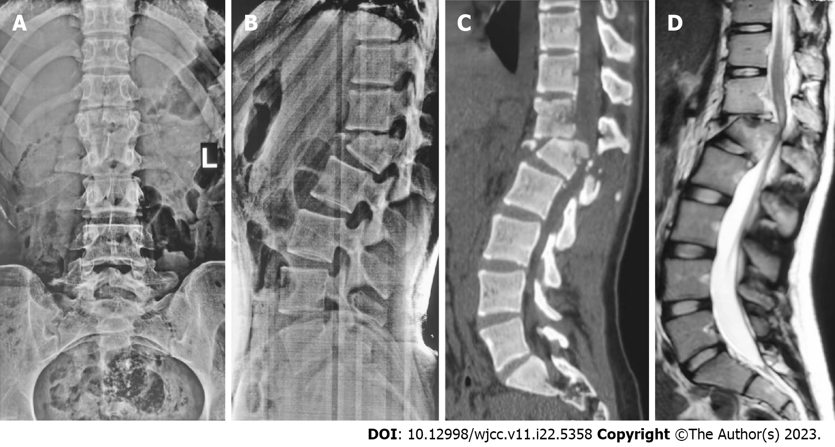
Figure 1 A 16-year-old female with thoracolumbar fracture dislocation of L1 (AO Type C, Load-sharing classification, leukemic stem cell = 8), ASIA B of spinal cord injury.
A and B: The pre-operative anteroposterior and lateral radiographs showed thoracolumbar kyphosis with a 26° Cobb’s angle; C: Pre-operative computed tomography scan reconstructions showed L1 fracture dislocation; D: Pre-operative sagittal T2-weighted magnetic resonance image revealed the spinal cord injury.
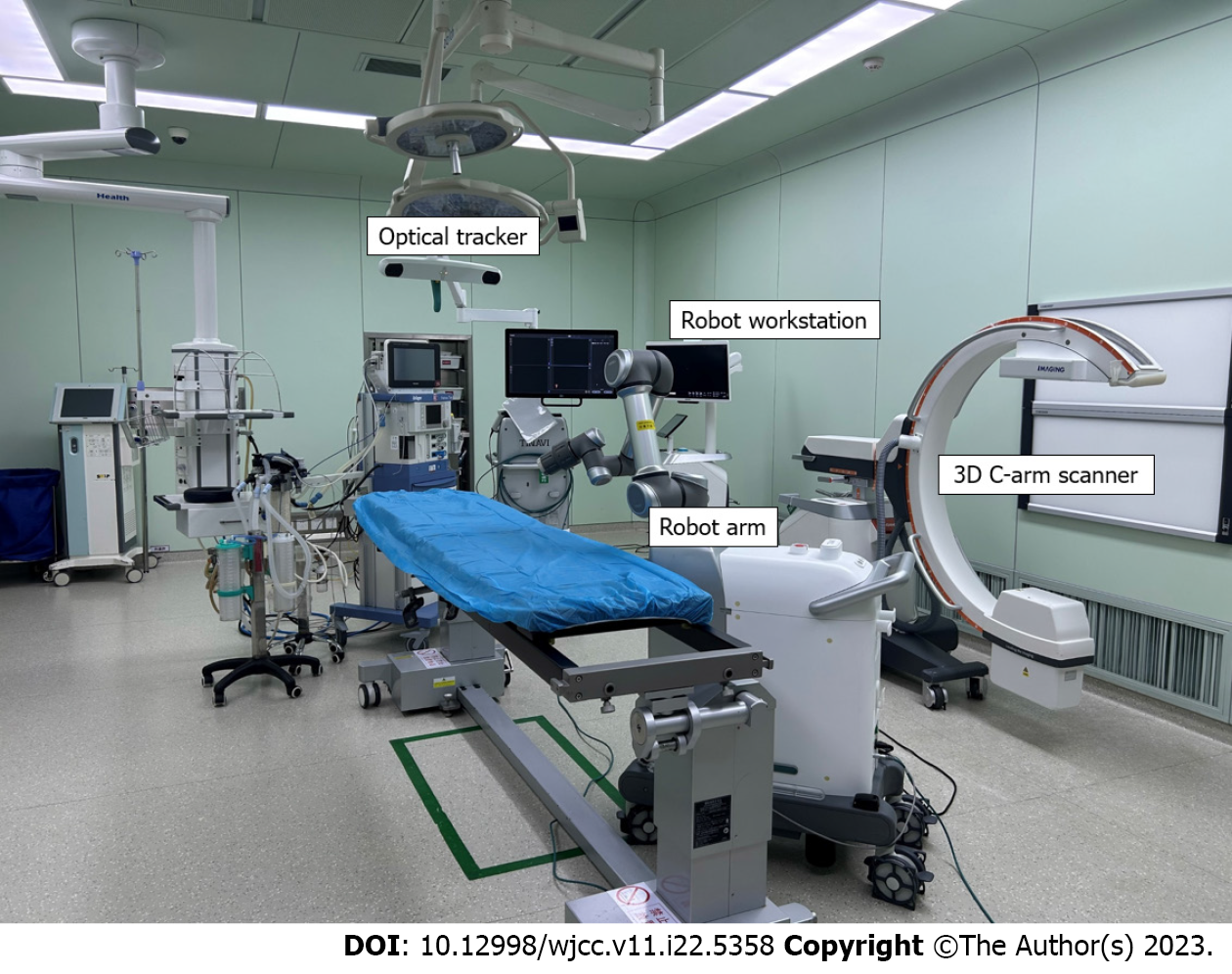
Figure 2 TINAVI orthopedic robotic system.
3D: Three dimensional.
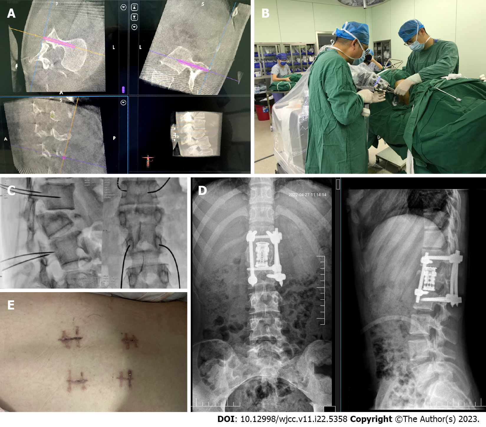
Figure 3 TINAVI robot-assisted one-stage anteroposterior surgery in lateral position.
A: Design the best virtual screw trajectory on 3D image; B: The electric drill is implanted with the guiding pin along the direction of the guiding cannula; C: C-arm fluoroscopy to determine the position of the guiding pin; D: The postoperative anteroposterior and lateral radiographs showed thoracolumbar kyphosis with a 5°Cobb’s angle; E: Postoperative wound.
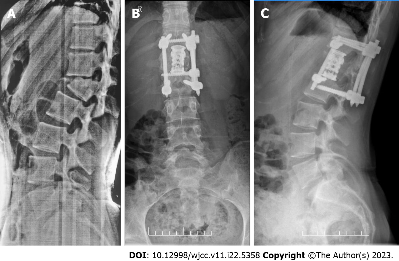
Figure 4 The anteroposterior and lateral radiographs of 5-mo follow-up showed no loss of corrected Cobb’s angle.
A: The pre-operative lateral radiographs showed thoracolumbar kyphosis with a 26° Cobb’s angle; B: The anteroposterior radiographs of 5-mo follow-up; C: The lateral radiographs of 5-mo follow-up showed thoracolumbar kyphosis with a 5.6° Cobb’s angle.
- Citation: Ye S, Chen YZ, Zhong LJ, Yu CZ, Zhang HK, Hong Y. TINAVI robot-assisted one-stage anteroposterior surgery in lateral position for severe thoracolumbar fracture dislocation: A case report. World J Clin Cases 2023; 11(22): 5358-5364
- URL: https://www.wjgnet.com/2307-8960/full/v11/i22/5358.htm
- DOI: https://dx.doi.org/10.12998/wjcc.v11.i22.5358


