©The Author(s) 2023.
World J Clin Cases. Jul 26, 2023; 11(21): 5167-5172
Published online Jul 26, 2023. doi: 10.12998/wjcc.v11.i21.5167
Published online Jul 26, 2023. doi: 10.12998/wjcc.v11.i21.5167
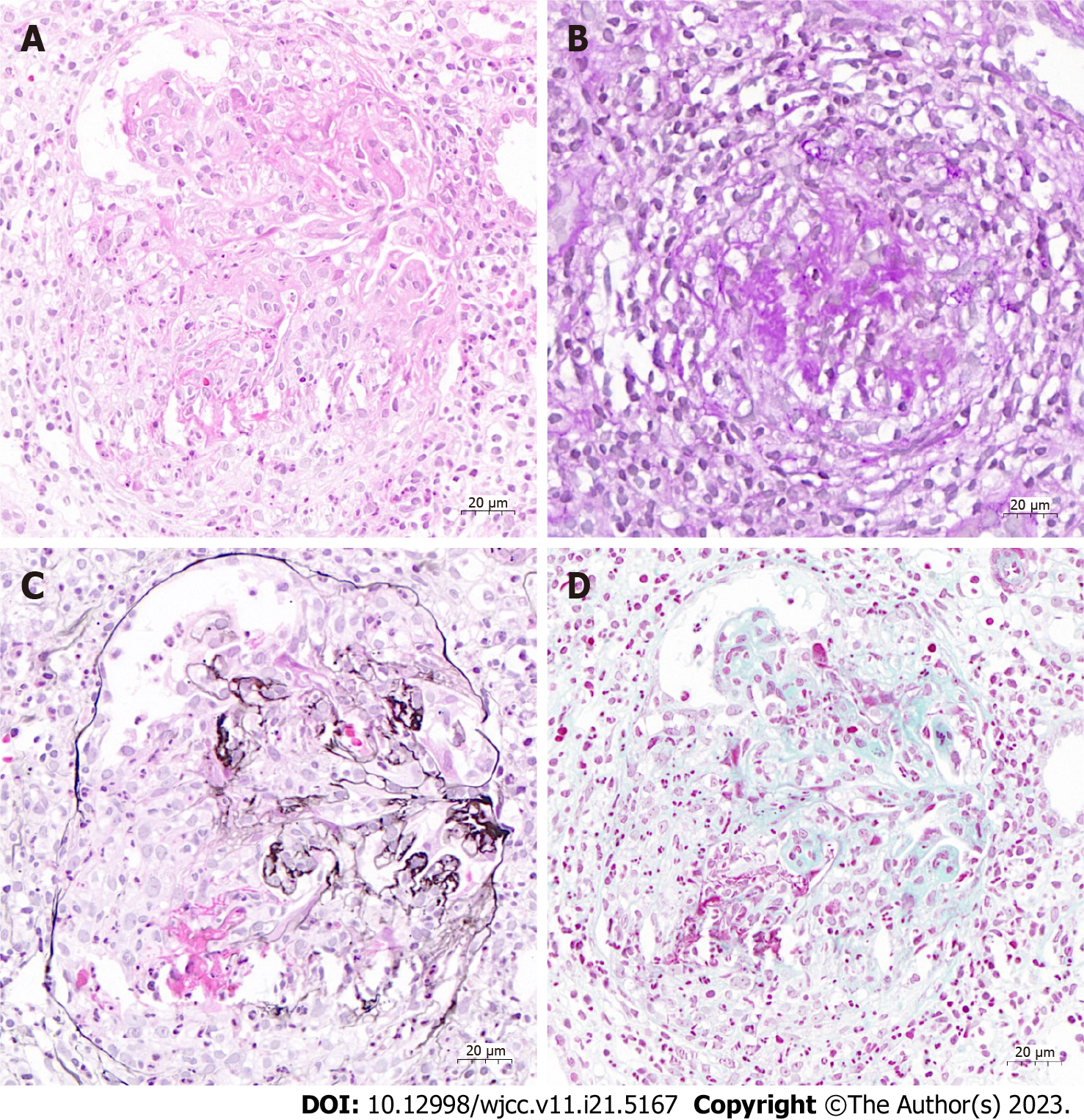
Figure 1 Kidney pathology showing crescentic glomerulonephritis and focal fibrinoid necrosis under a light microscope.
A: Hematoxylin and eosin staining, 400 × magnification; B: Periodic acid-Schiff staining, 400 × magnification; C: Periodic Schiff-methenamine silver staining, 400 × magnification; D: Masson trichrome staining, 400 × magnification.
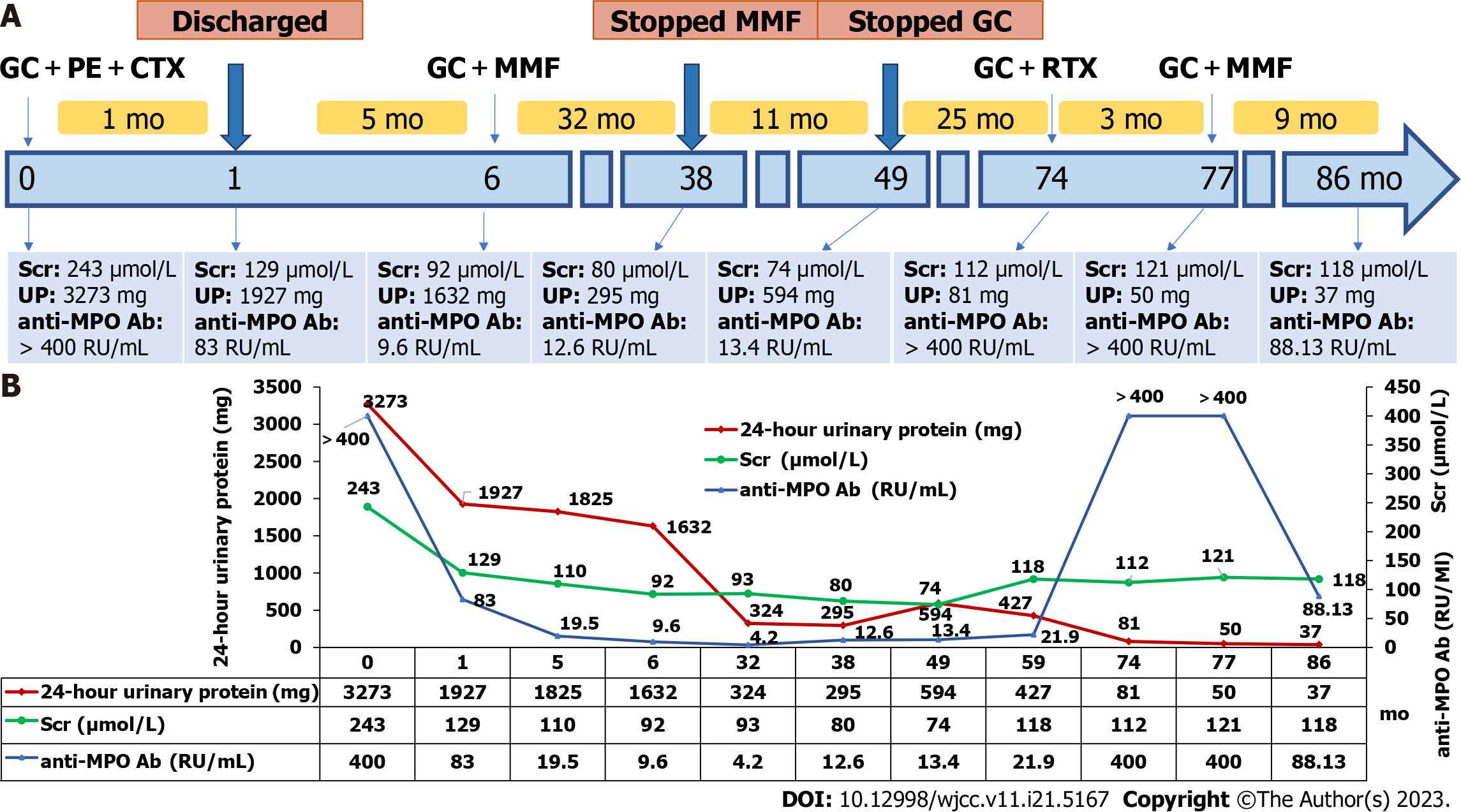
Figure 2 Clinical course, proteinuria, serum creatinine, and anti-myeloperoxidase antibody level in our patient.
A: Clinical course and laboratory test findings in our patient; B: Trends of 24-h urinary protein, serum creatinine, and anti-myeloperoxidase antibody titer. GC: Glucocorticoids; PE: Plasma exchange; CTX: Cyclophosphamide; MMF: Mycophenolate mofetil; RTX: Rituximab; Scr: Serum creatinine; UP: 24-h urinary protein or urinary protein; Ab: Antibody; MPO: Myeloperoxidase.
- Citation: Zhang X, Zhao GB, Li LK, Wang WD, Lin HL, Yang N. Myeloperoxidase-antineutrophil cytoplasmic antibody-associated vasculitis with headache and kidney involvement at presentation and with arthralgia at relapse: A case report. World J Clin Cases 2023; 11(21): 5167-5172
- URL: https://www.wjgnet.com/2307-8960/full/v11/i21/5167.htm
- DOI: https://dx.doi.org/10.12998/wjcc.v11.i21.5167


