Copyright
©The Author(s) 2023.
World J Clin Cases. May 6, 2023; 11(13): 3076-3085
Published online May 6, 2023. doi: 10.12998/wjcc.v11.i13.3076
Published online May 6, 2023. doi: 10.12998/wjcc.v11.i13.3076
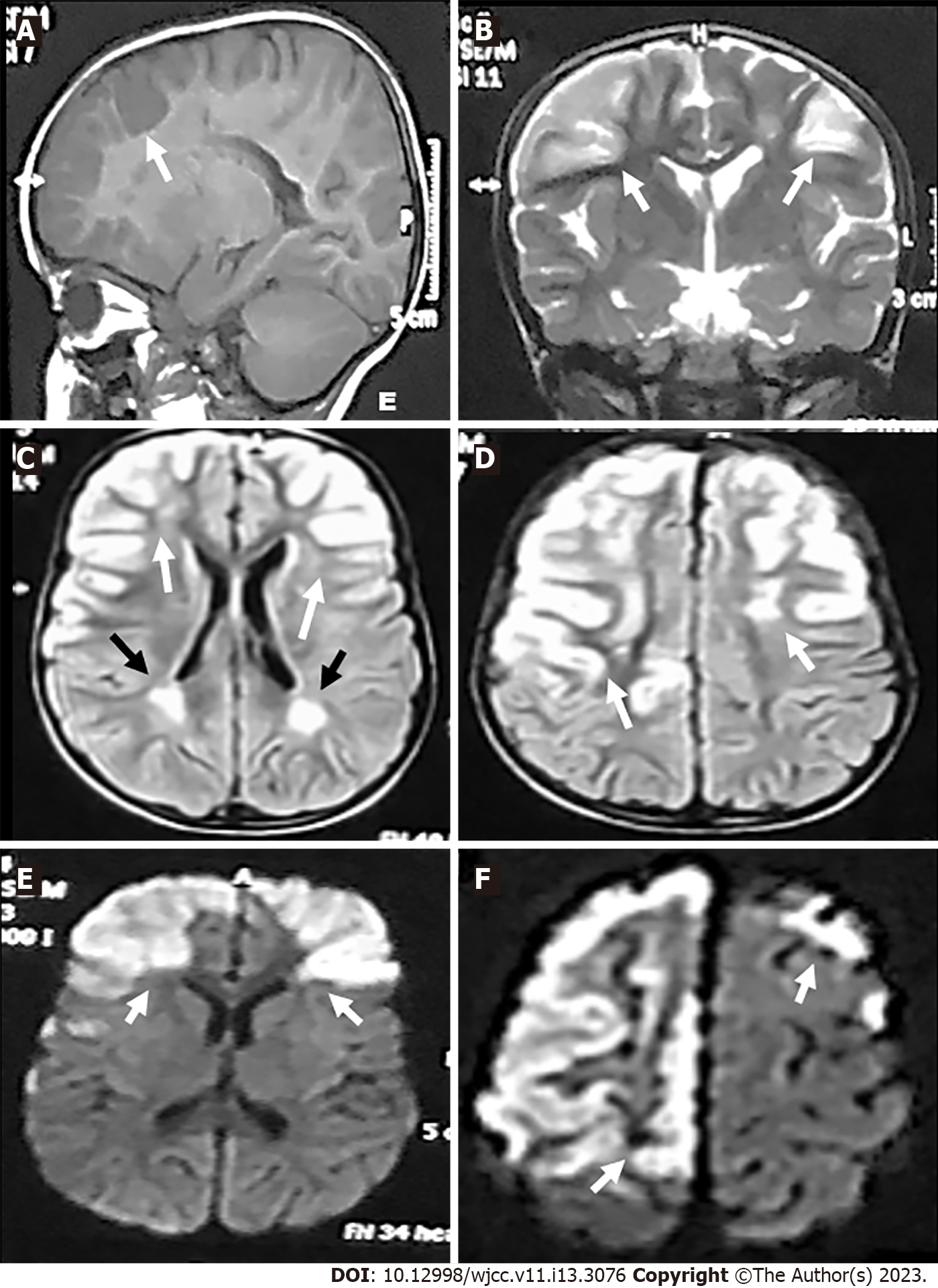
Figure 1 Magnetic resonance imaging-brain views (Date: July 2019).
A: Sagittal T1-weighted imaging revealed hypointense lesions in the fronto-parietal regions (white arrow); B: Coronal T2-weighted imaging; C and D: Axial fluid attenuation inversion recovery; E and F: Diffusion weighted imaging. Revealed hyperintense lesions in the right and left fronto-parietal areas (gyral pattern) (white arrows) and in the white matter adjacent to the lateral ventricle (C and D) (black arrows).
Figure 2 Magnetic resonance imaging-brain views (Date: October 2019).
A: Sagittal T1-weighted imaging; B: Coronal; C and D: Axial fluid attenuation inversion recovery; E and F: Diffusion weighted imaging; G and H: Diffusion/perfusion weighted. A, E and F revealed hypointense lesions in the fronto-parietal regions and areas of encephalomalacia (cerebrospinal fluid density) (white arrows). B-D, G, and H views revealed hyperintense lesions in the right and left fronto-parietal areas and areas of encephalomalacia (white arrows).
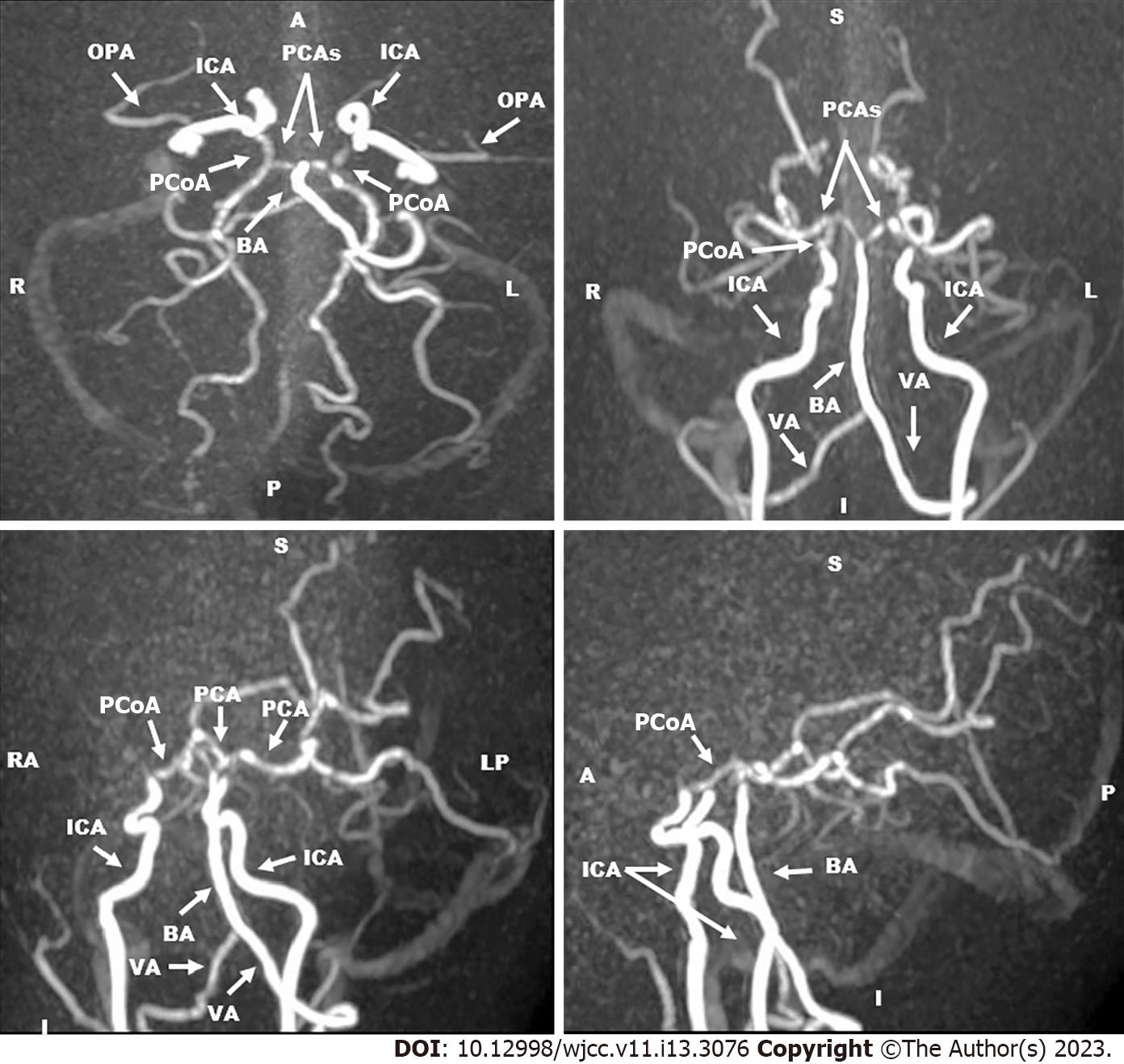
Figure 3 Magnetic resonance angiography of cerebral vessels three-dimension technique and maximum intensity projection images (Date: October 2019).
They revealed occlusion of the supraclinoid portion of internal carotid arteries. The right and left anterior cerebral arteries and middle cerebral arteries were not visualized. The basilar artery and the right and left posterior cerebral arteries had normal sizes and calibers. Collaterals were predominantly from the posterior circulation. OPA: The ophthalmic artery; ICA: The internal carotid artery; PCAs: The posterior cerebral arteries; PCoA: The posterior communicating artery; BA: The basilar artery; VA: The vertebral artery.
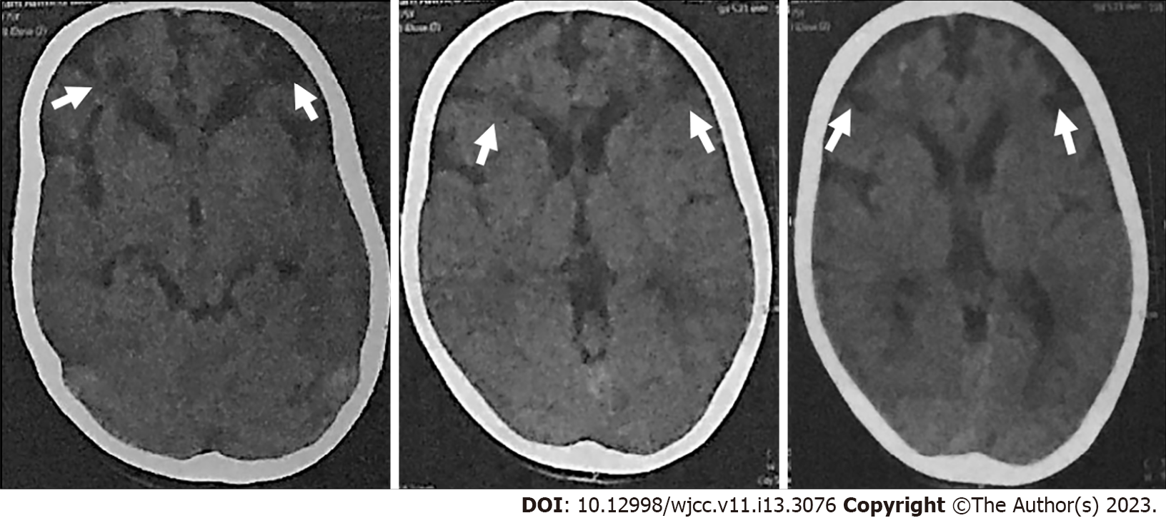
Figure 4 Axial computed tomography-brain views (Date: January 2021).
They revealed bilateral frontal subcortical and slightly cortical hypodense lesions (the cerebrospinal fluid density) (white arrows).
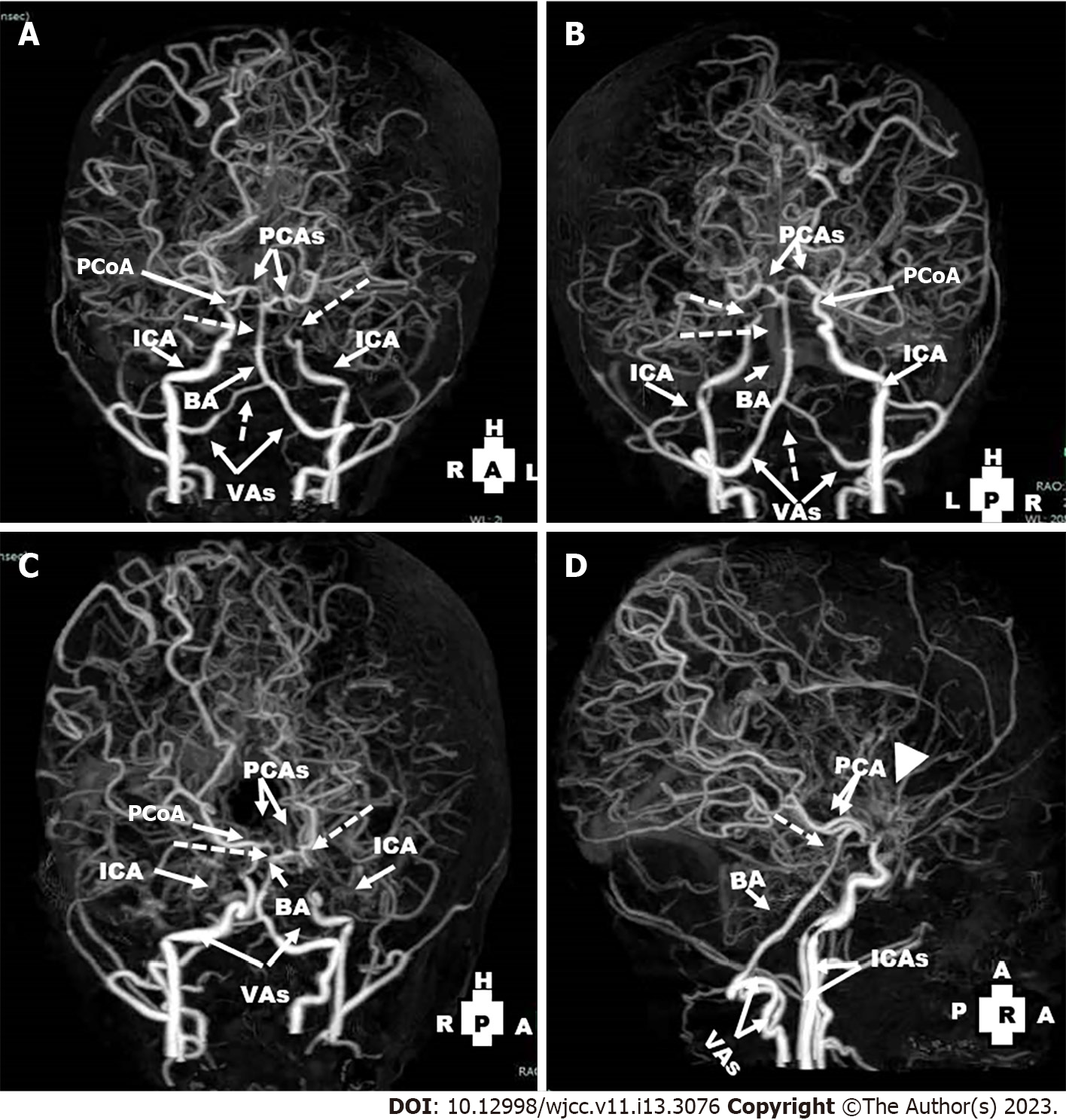
Figure 5 Computed tomography angiography views of the cerebral vessels (Date: January 2021): They revealed non-visualization of right and left anterior cerebral arteries and middle cerebral arteries.
A-C: The left internal carotid artery ended blindly before the siphon (dotted arrow), non-visualization of the left posterior communicating artery) and attenuation of the distal part of the right vertebral arteries and basilar artery (dotted arrows); D: There were extensive bilateral collaterals and prominence of moyamoya vessels (arrow head). PCoA: The posterior communicating artery; PCAs: The posterior cerebral arteries; ICA: The internal carotid artery; BA: The basilar artery; VAs: The vertebral arteries.
- Citation: Hamed SA, Yousef HA. Idiopathic steno-occlusive disease with bilateral internal carotid artery occlusion: A Case Report. World J Clin Cases 2023; 11(13): 3076-3085
- URL: https://www.wjgnet.com/2307-8960/full/v11/i13/3076.htm
- DOI: https://dx.doi.org/10.12998/wjcc.v11.i13.3076


