Copyright
©The Author(s) 2023.
World J Clin Cases. May 6, 2023; 11(13): 3052-3061
Published online May 6, 2023. doi: 10.12998/wjcc.v11.i13.3052
Published online May 6, 2023. doi: 10.12998/wjcc.v11.i13.3052
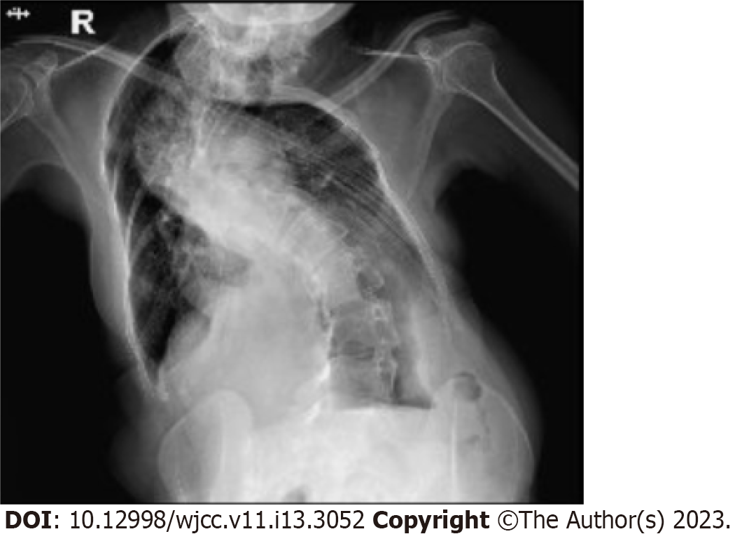
Figure 1 Chest X-ray examination after admission.
Chest X-ray reveals severe scoliosis, thoracic deformity, and thickened lung markings.
Figure 2 Ultrasound imaging before, during and after treatment.
A: B-mode ultrasound imaging (longitudinal section) demonstrates a homogeneous hypoechoic mass (bold arrow) prolapsing into the cervical canal; B: Color Doppler flow imaging reveals a large blood vessel (bold arrow) in the pedicle supplying the whole myoma; C: contrast-enhanced ultrasound imaging shows high enhancement in the pedicle, suggesting that it is the main blood supply source of the prolapsed lesion; D: Two days after UAE, both the uterus and myoma show slight atrophy; E: The upper segment of the pedicle shows enhancement on contrast-enhanced ultrasound imaging (bold arrow), indicating partial recanalization of the feeding arteries; F: During the PMWA procedure, the microwave antenna (white line) was inserted into the pedicle precisely under real-time TAUS guidance; G: After thorough ablation of both the pedicle stump and the endometrium, a hyperechoic cloud covering the whole uterine cavity (arrows) is observed; H: Intraoperative contrast-enhanced ultrasound imaging shows no enhancement in the pedicle stump or the inner myometrium (arrows). UAE: Uterine artery embolism; PMWA: Percutaneous microwave ablation.
Figure 3 Preoperative and postoperative magnetic resonance imaging demonstrations.
A: Preoperative T2WI demonstrates a well-defined pedunculated submucosal myoma prolapsed into the cervical canal; B: Preoperative CE-T1WI shows enhancement of the pedicle and the myoma; C: Postoperative T2WI shows slight edema in the ablated inner myometrium and no damage to the outer myometrium or the surrounding organs; D: Postoperative CE-T1WI confirms no enhancement in the inner myometrium or tumor pedicle stump.
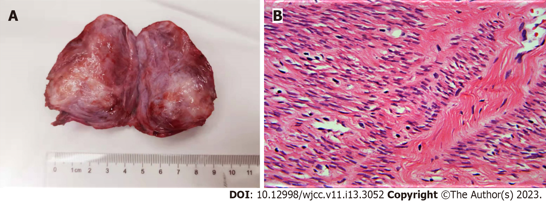
Figure 4 Gross specimen and hematoxylin and eosin-stained image.
A: Sectioned view of the resected specimen reveals a braided parenchymal mass without any significant degeneration; B: Hematoxylin and eosin-stained image shows abundant spindle smooth muscle cells and fibrous connective tissue with cellular heteromorphism.
Figure 5 Findings for the blood supply of the myoma before and after uterine artery embolism.
A: Aortography demonstrates a dilated left arcuate artery and two downstream radial arteries supplying the pedicle of the myoma; B: Bilateral uterine arteries contribute to the blood supply of the myoma; C: After complete occlusion of the radial arteries, no perfusion of the pedicle or myometrium is observed.
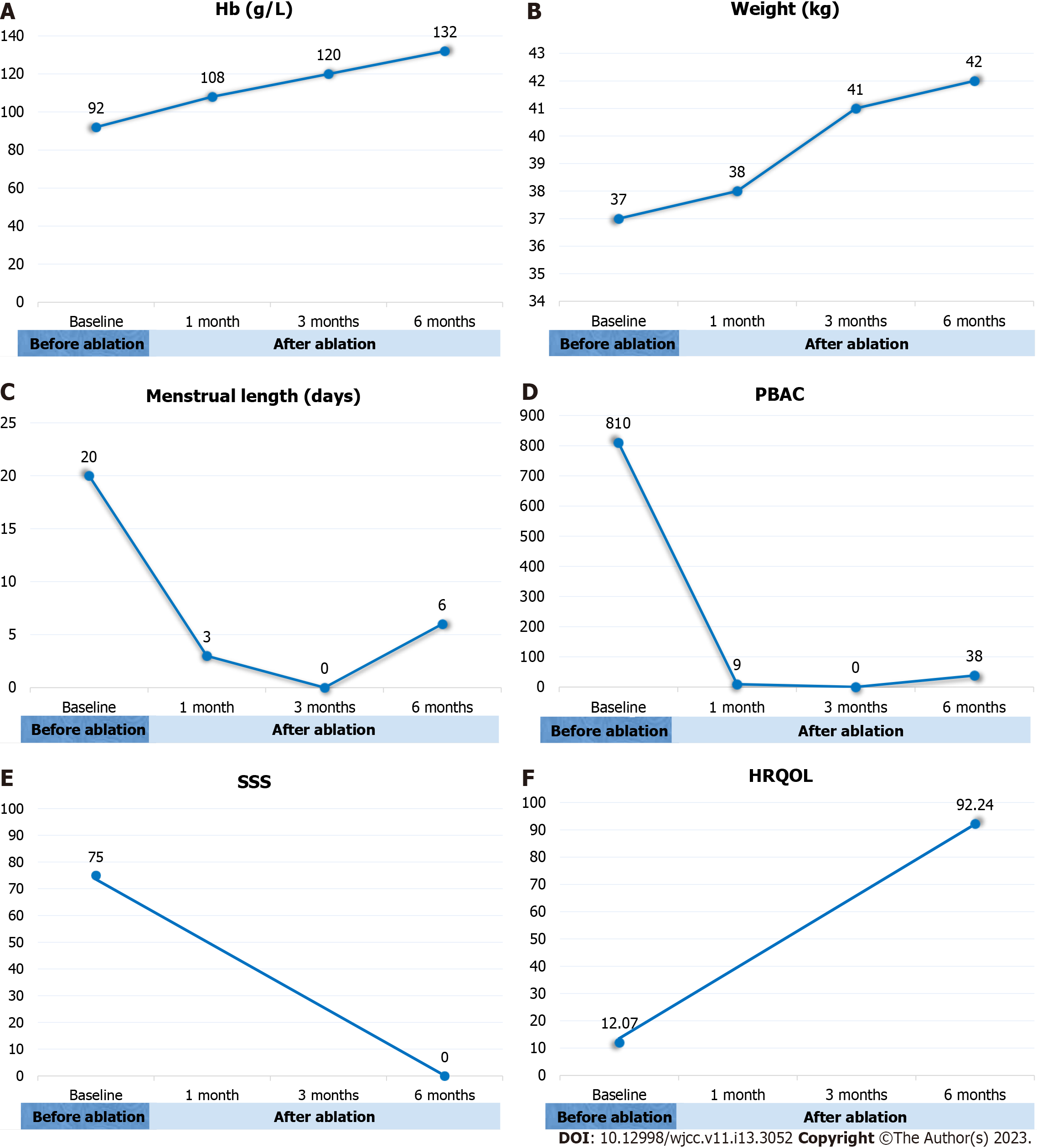
Figure 6 Changes in weight, hemoglobin level and clinical scores.
A: The Hb level increased to normal range 3 mo after treatment; B: The patient had gained weight gradually after ablation; C: Menstrual length was 6 mo for each cycle 6 mo after treatment; D: The patient had a transient amenorrhea from 3 to 5 mo after treatment, and returned to normal mense 6 mo after treatment with a PBAC score < 100; E: SSS score decreased from 75 to 0 after treatment, indicating complete relief of symptoms; F: HRQOL score increased from 12.07 to 92.24 at 6 mo after treatment, indicating huge improvement of the life quality. Hb: Hemoglobin; PBAC: Pictorial blood-loss assessment chart; SSS: Symptom severity scale; HRQOL: Health-related quality of life.
- Citation: Zhang HL, Yu SY, Cao CW, Zhu JE, Li JX, Sun LP, Xu HX. Uterine artery embolization combined with percutaneous microwave ablation for the treatment of prolapsed uterine submucosal leiomyoma: A case report. World J Clin Cases 2023; 11(13): 3052-3061
- URL: https://www.wjgnet.com/2307-8960/full/v11/i13/3052.htm
- DOI: https://dx.doi.org/10.12998/wjcc.v11.i13.3052


