Copyright
©The Author(s) 2023.
World J Clin Cases. Apr 16, 2023; 11(11): 2457-2463
Published online Apr 16, 2023. doi: 10.12998/wjcc.v11.i11.2457
Published online Apr 16, 2023. doi: 10.12998/wjcc.v11.i11.2457
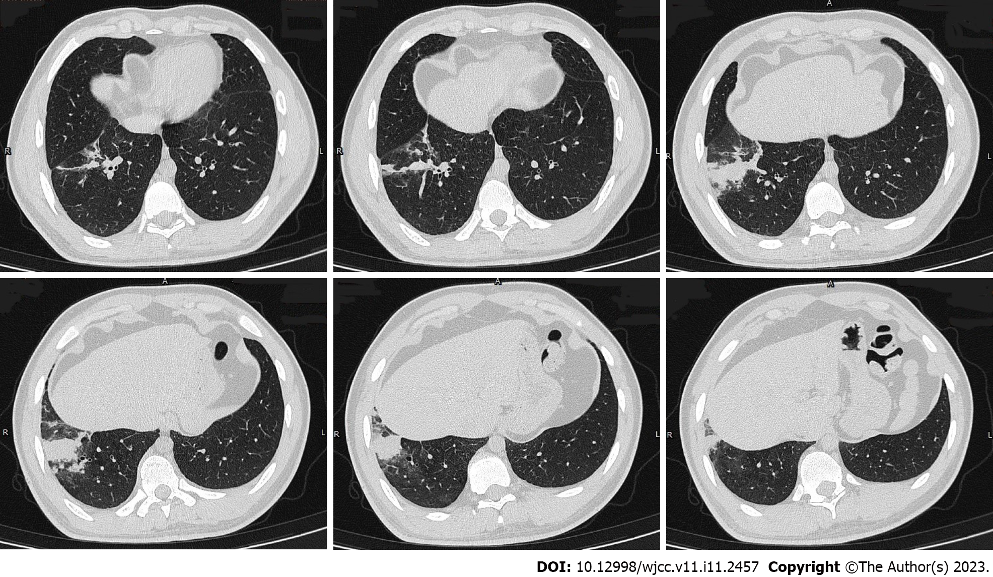
Figure 1 Chest computed tomography images (February 3, 2021) showing consolidation in the anterior basal segment of the right lower lobe, with multiple small nodules surrounded by blurred margins, some of which merged into small patchy consolidations.
Figure 2 Hematoxylin-eosin-stained sections of lung tissue.
A: Magnification × 20; B: Magnification × 40. The alveolar structure can be identified, the alveolar septum is slightly widened, and there are many eosinophils and a small amount of scattered lymphoplasmacytic infiltration in the alveolar septum and interstitium. A small amount of eosinophilic exudation can be seen in some alveolar spaces with no signs of necrosis. No typical granulomas or vasculitis is observed.
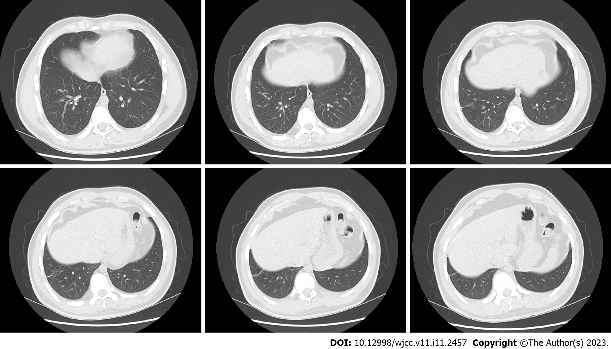
Figure 3 Repeat chest computed tomography performed after 4 wk of glucocorticoid treatment showing resorption of the shadow in the right lower lobe; only a fibrous cord shadow is seen, and the adjacent bronchus is stretched and dilated.
- Citation: Zhang XX, Zhou R, Liu C, Yang J, Pan ZH, Wu CC, Li QY. Allergic bronchopulmonary aspergillosis with marked peripheral blood eosinophilia and pulmonary eosinophilia: A case report. World J Clin Cases 2023; 11(11): 2457-2463
- URL: https://www.wjgnet.com/2307-8960/full/v11/i11/2457.htm
- DOI: https://dx.doi.org/10.12998/wjcc.v11.i11.2457


