Copyright
©The Author(s) 2023.
World J Clin Cases. Apr 6, 2023; 11(10): 2123-2139
Published online Apr 6, 2023. doi: 10.12998/wjcc.v11.i10.2123
Published online Apr 6, 2023. doi: 10.12998/wjcc.v11.i10.2123
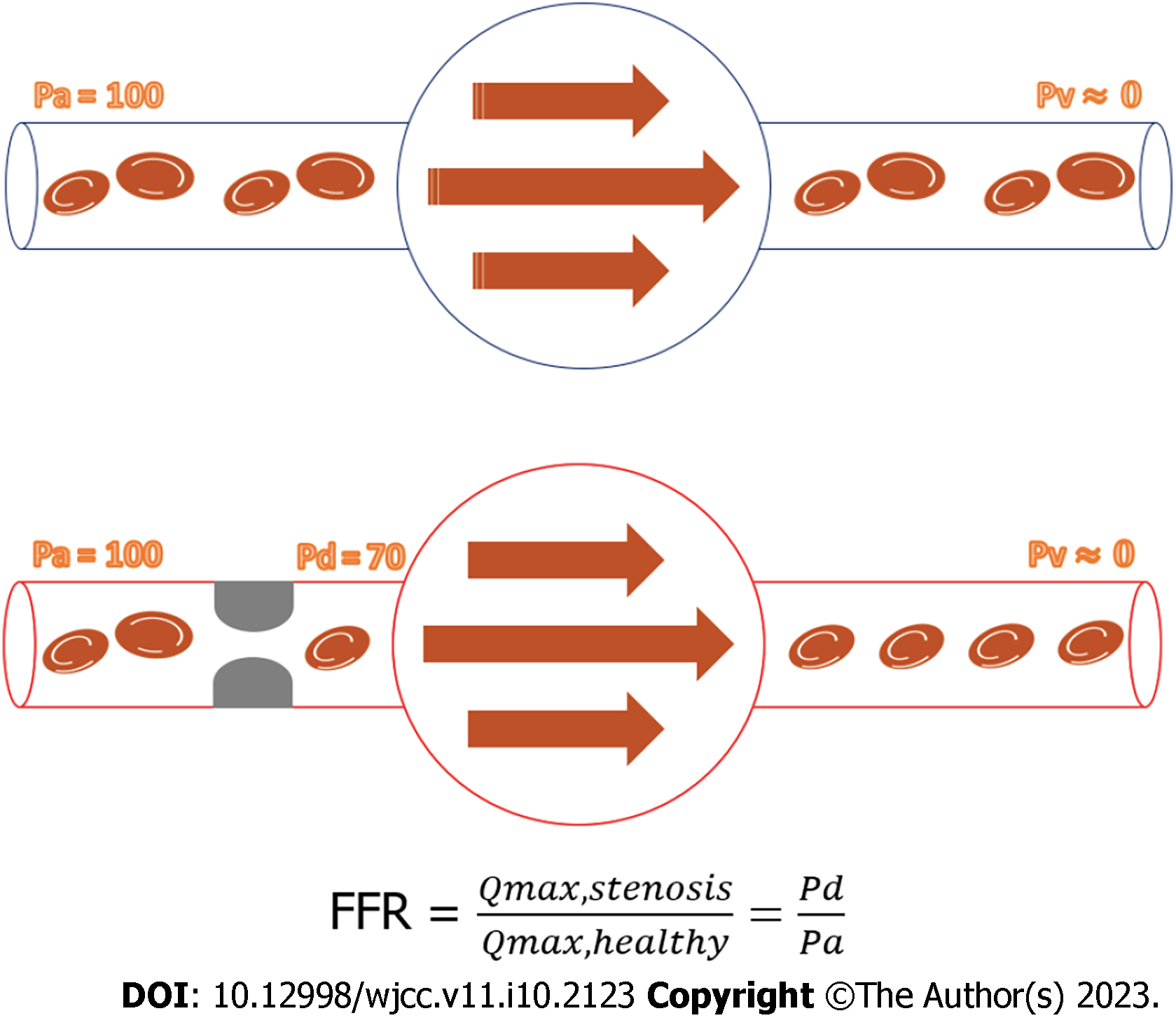
Figure 1 Invasive measurement of fractional flow reserve.
FFR: Fractional flow reserve.
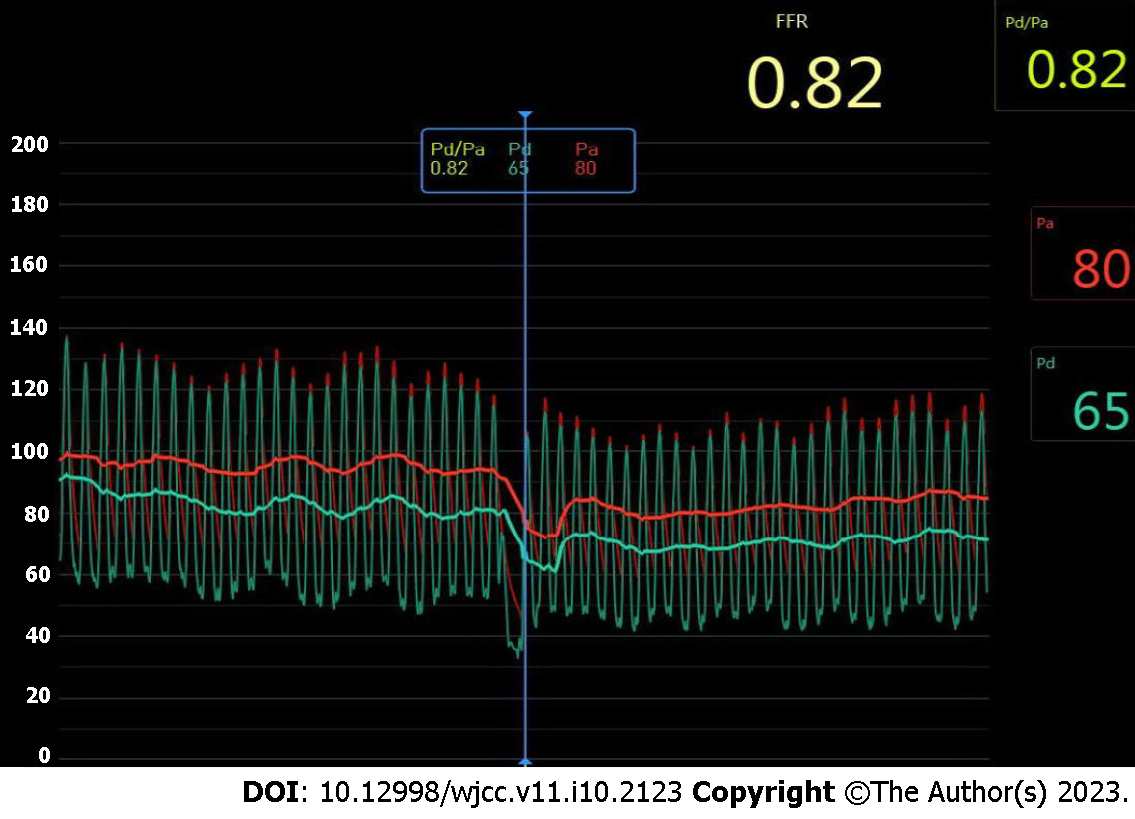
Figure 2
POLARIS display screen showing negative fractional flow reserve value.
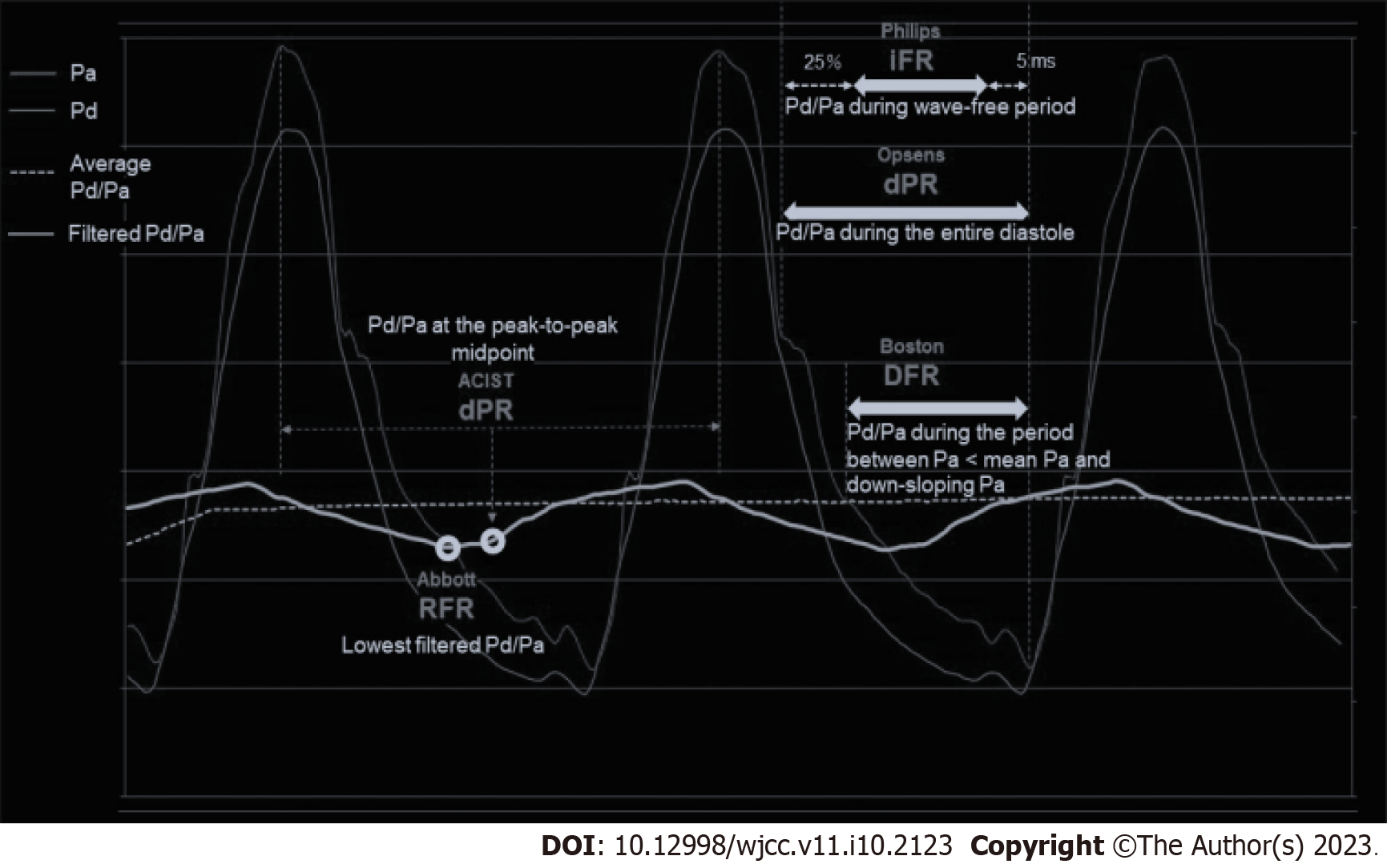
Figure 3 Non-Hyperemic pressure ratios calculation over cardiac cycle.
RFR: Resting full-cycle ratio; DFR: Diastolic hyperemia-free ratio; dPR: Diastolic pressure ratio.
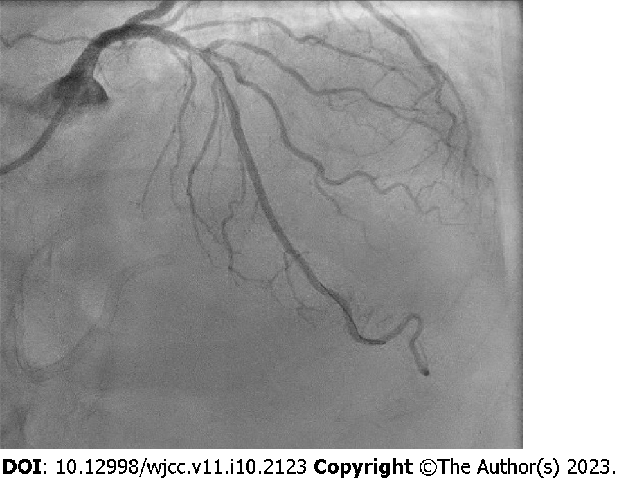
Figure 4
Left anterior oblique cranial view demonstrating a moderate proximal left anterior descending stenosis.
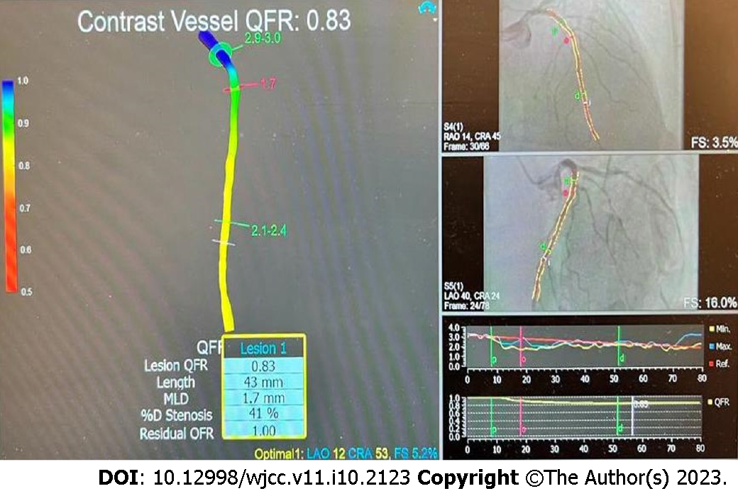
Figure 5 Quantitative flow ratio of the left anterior descending.
QFR: Quantitative flow ratio.
Figure 6
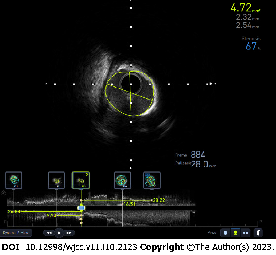
Intravascular ultrasound imaging showing the minimal luminal area of 4.
72 mm², diameters and percentage stenosis of the left anterior descending.
Figure 7
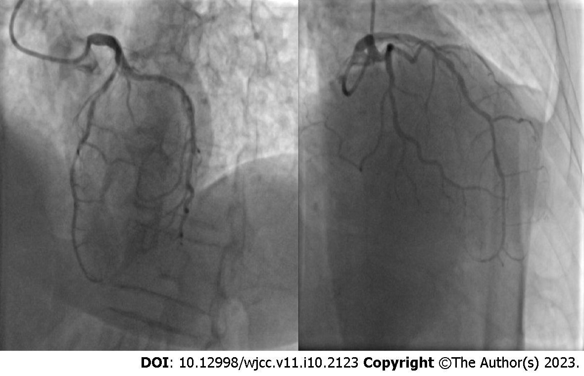
Cranial left and right anterior oblique view demonstrating serial stenoses of the left anterior descending.
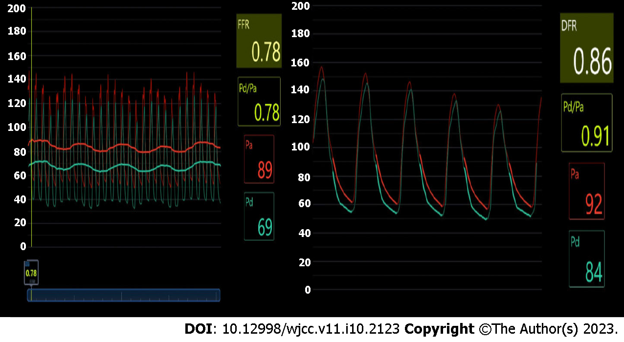
Figure 8 Distal left anterior descending diastolic flow ratio and fractional flow reserve.
FFR: Fractional flow reserve; DFR: Diastolic hyperemia-free ratio.
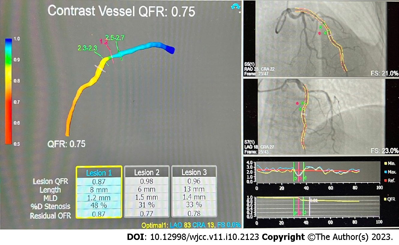
Figure 9 Quantitative flow ratio of left anterior descending evaluating each stenosis residual quantitative flow ratio and final result.
QFR: Quantitative flow ratio.
- Citation: Boutaleb AM, Ghafari C, Ungureanu C, Carlier S. Fractional flow reserve and non-hyperemic indices: Essential tools for percutaneous coronary interventions. World J Clin Cases 2023; 11(10): 2123-2139
- URL: https://www.wjgnet.com/2307-8960/full/v11/i10/2123.htm
- DOI: https://dx.doi.org/10.12998/wjcc.v11.i10.2123


