Copyright
©The Author(s) 2022.
World J Clin Cases. Nov 6, 2022; 10(31): 11646-11651
Published online Nov 6, 2022. doi: 10.12998/wjcc.v10.i31.11646
Published online Nov 6, 2022. doi: 10.12998/wjcc.v10.i31.11646
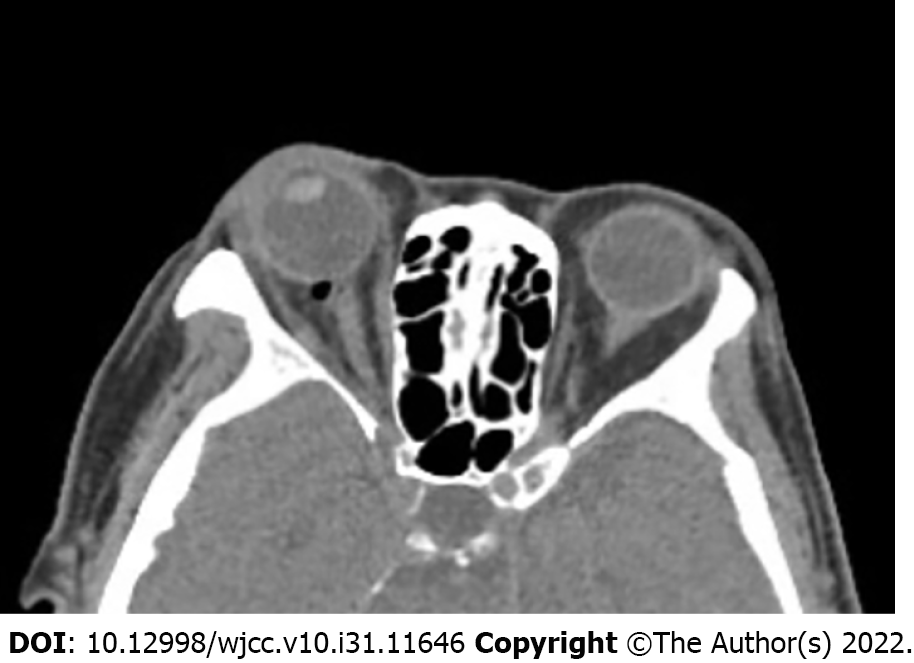
Figure 1 Postoperative head computed tomography.
Two hours after retrobulbar anesthesia, head computed tomography showed bubbles at the posterior of the patient’s right eye, partially compressing the optic nerve.
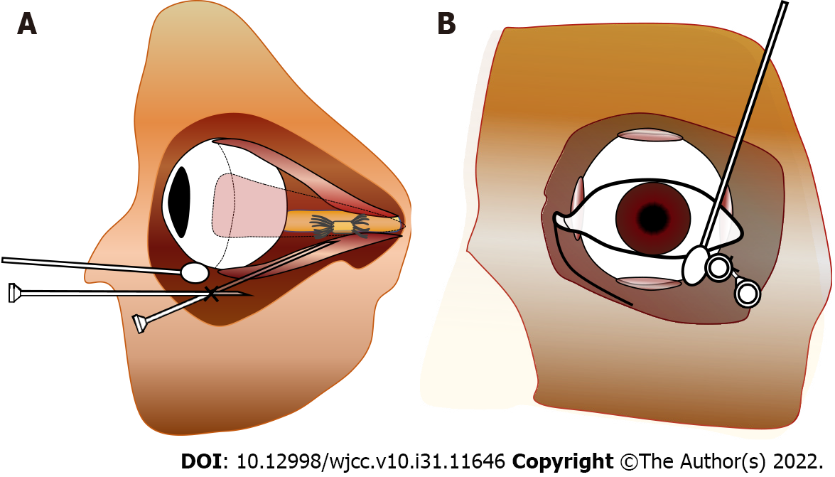
Figure 2 Schematic diagram of retrobulbar anesthesia.
A: Side view, the trick of using a cotton bud was applied to assist the operation, as it lifts the eyeball but with the eyes staring at the front, helping to prevent penetration of the eyeball and decreasing risk of injury to the optic nerve; B: Front view.
- Citation: Wang YL, Lan GR, Zou X, Wang EQ, Dai RP, Chen YX. Apnea caused by retrobulbar anesthesia: A case report. World J Clin Cases 2022; 10(31): 11646-11651
- URL: https://www.wjgnet.com/2307-8960/full/v10/i31/11646.htm
- DOI: https://dx.doi.org/10.12998/wjcc.v10.i31.11646


