Copyright
©The Author(s) 2022.
World J Clin Cases. Nov 6, 2022; 10(31): 11561-11566
Published online Nov 6, 2022. doi: 10.12998/wjcc.v10.i31.11561
Published online Nov 6, 2022. doi: 10.12998/wjcc.v10.i31.11561
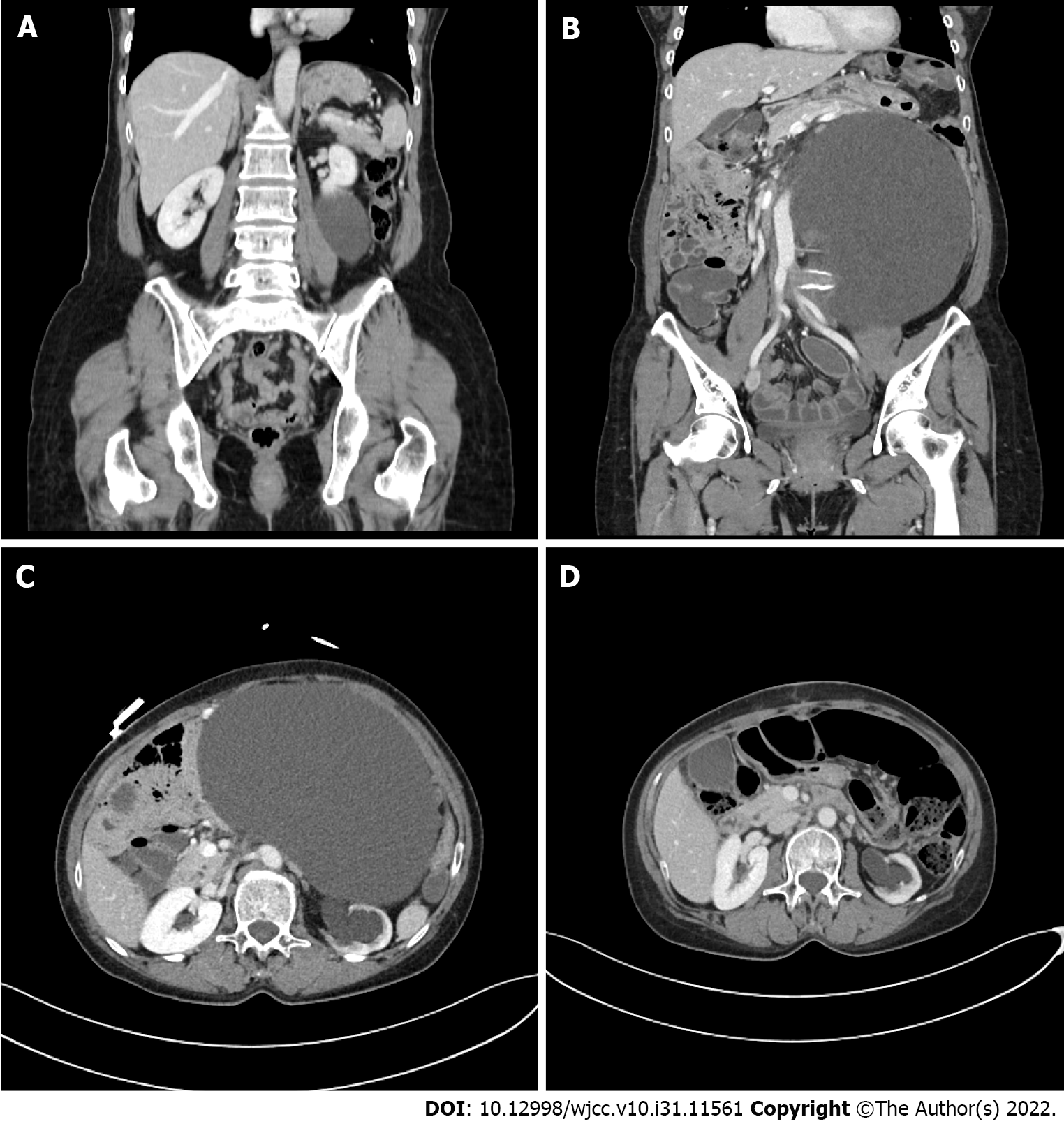
Figure 1 The pre- and post-operative computed tomography scan of the patient.
A: A retroperitoneal cystic mass on abdominal computed tomography (CT) scan; B and C: Follow-up CT scan in seven years from initial diagnosis showed significant increase in mass size (from 11 cm to 24 cm) with left hydronephrosis caused by external compression of cystic mass; D: Follow-up CT scan in one month after surgical excision showed complete removal of mass and decreased left hydronephrosis.
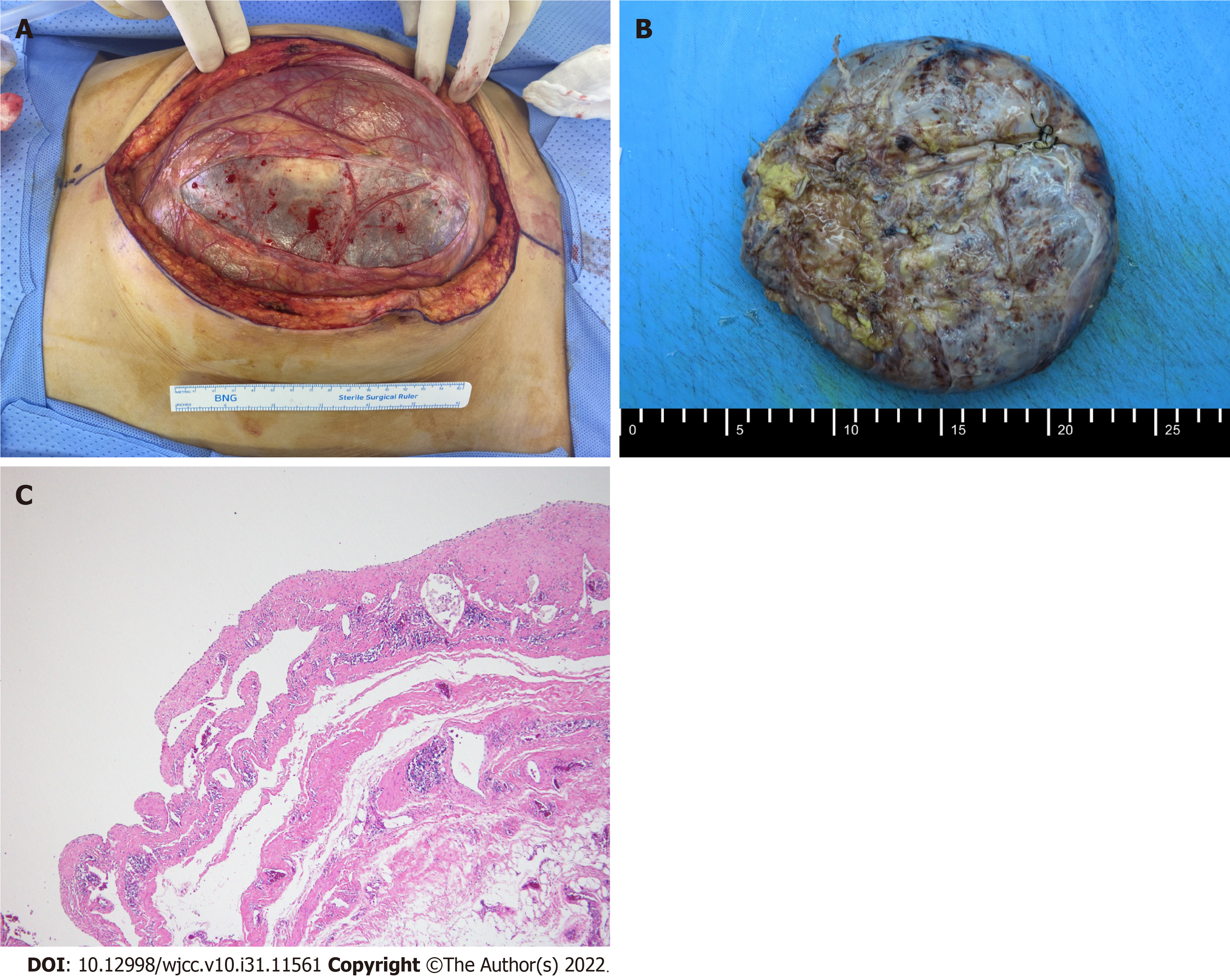
Figure 2 The gross and microscopic findings of the mass.
A: Huge cystic mass before excision; B: The mass after excision; C: The microscopic feature which shows the cystic space and dilated lymphatic vessels lined by flattened endothelium consistent with lymphangioma.
- Citation: Park JH, Lee D, Maeng YH, Chang WB. Surgical excision of a large retroperitoneal lymphangioma: A case report. World J Clin Cases 2022; 10(31): 11561-11566
- URL: https://www.wjgnet.com/2307-8960/full/v10/i31/11561.htm
- DOI: https://dx.doi.org/10.12998/wjcc.v10.i31.11561


