Copyright
©The Author(s) 2022.
World J Clin Cases. Oct 16, 2022; 10(29): 10647-10654
Published online Oct 16, 2022. doi: 10.12998/wjcc.v10.i29.10647
Published online Oct 16, 2022. doi: 10.12998/wjcc.v10.i29.10647
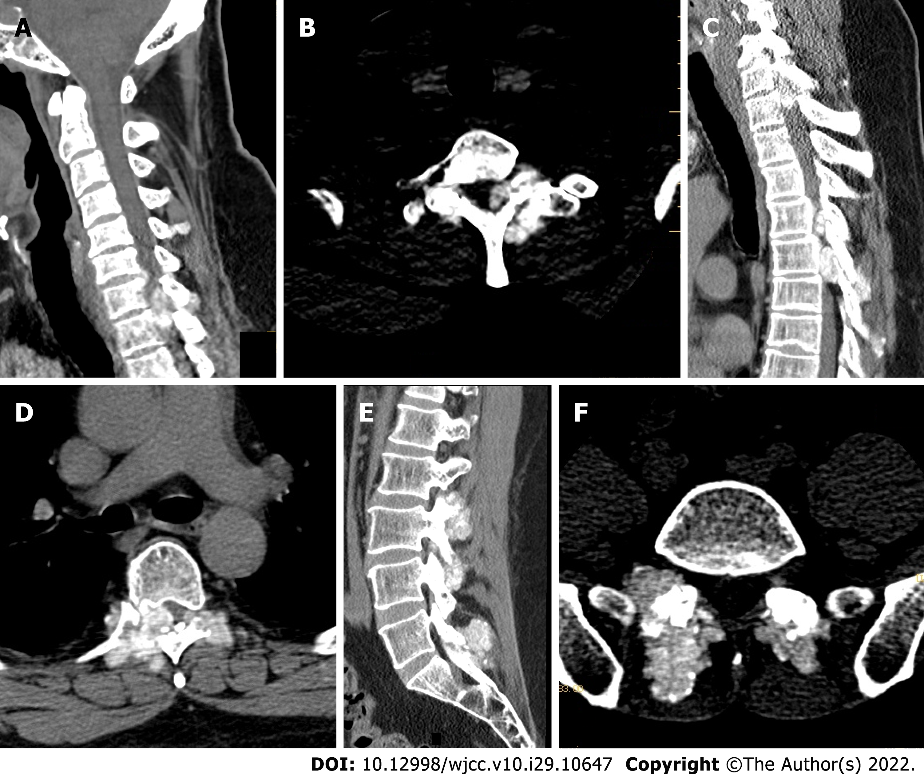
Figure 1 Preoperative computed tomography of the cervical, thoracic, lumbar, and sacral spine.
A-D: Multiple tophi in the whole spine, with spinal cord compression and degeneration at C7/T1, T5, and T6 Levels; E and F: Multiple abnormal high signals that tophi in the lumbar and sacral vertebral subtalar joints, and tophus deposits.
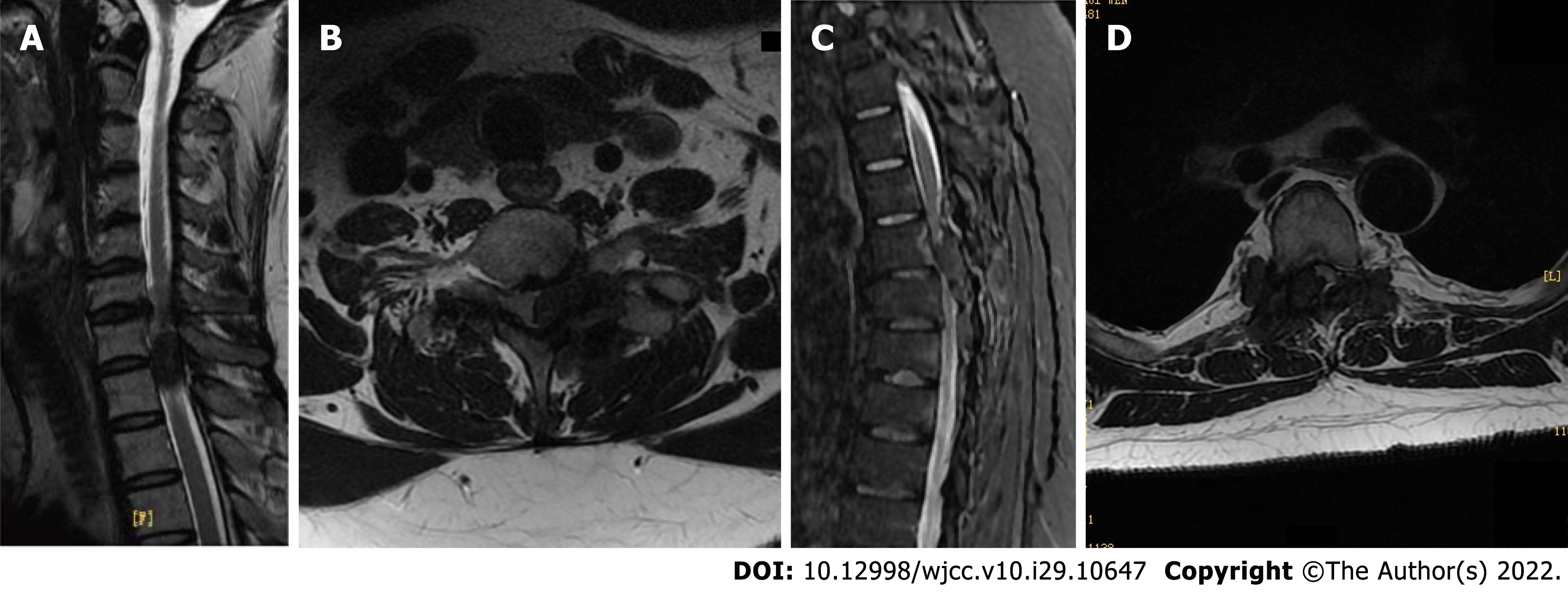
Figure 2 Preoperative cervical and thoracic magnetic resonance imaging.
A and B: Multiple tophi in the compressed and degenerated spine and spinal cord at C7/T1; C and D: Multiple tophi in the compressed and degenerated spine and spinal cord at T5, T6 Levels.
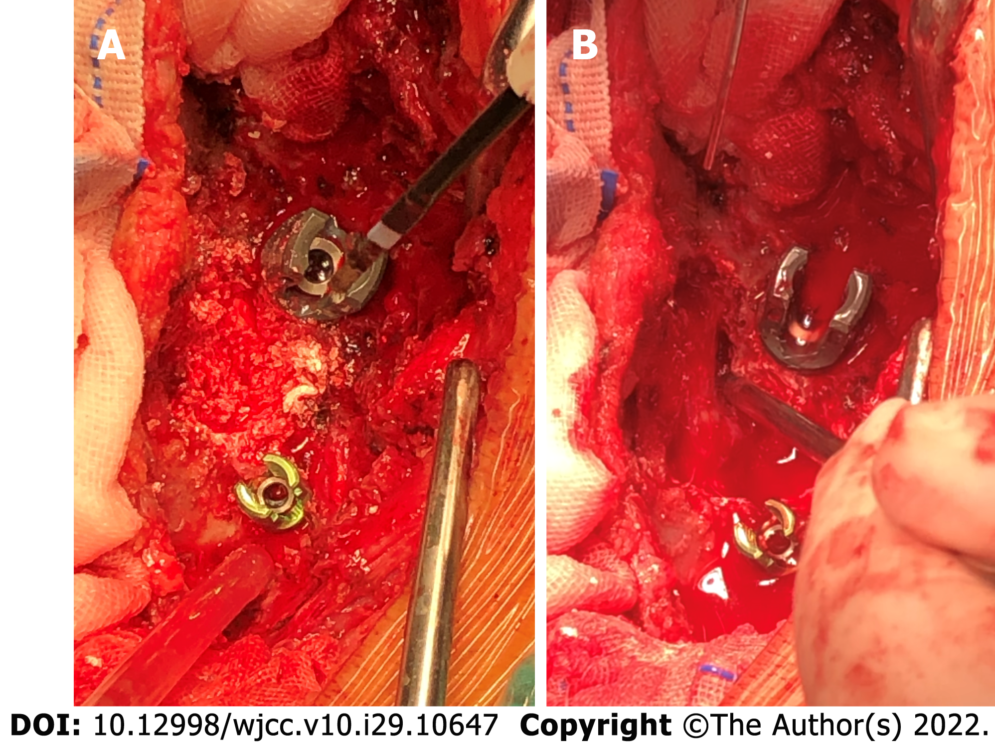
Figure 3 Intraoperative findings.
A: Intraoperative lime-like changes before decompression of the thoracic vertebral plate; B: Intraoperative decompression of the thoracic vertebral plate.
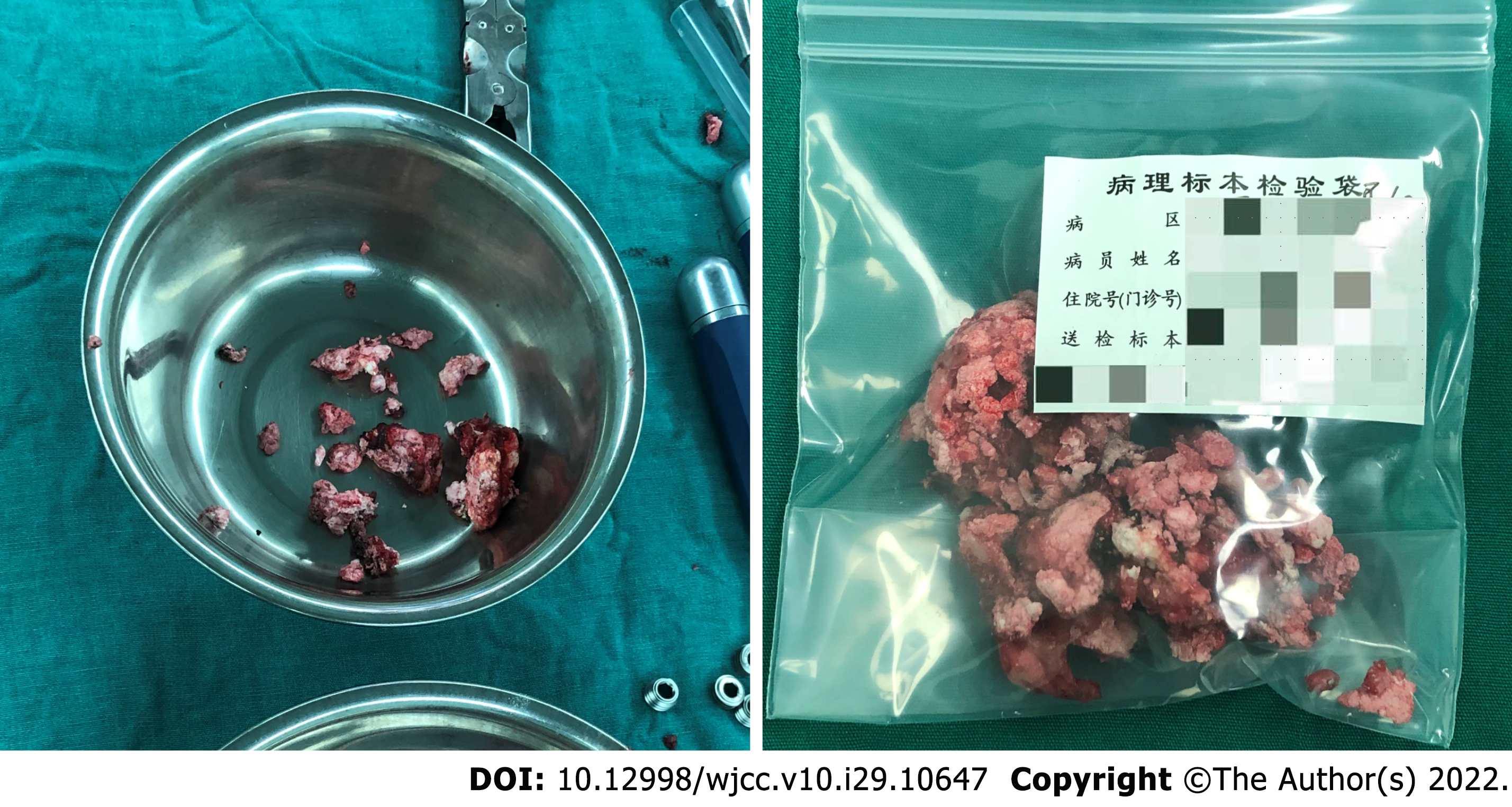
Figure 4 Intraoperative resection of spinal tophus sample.
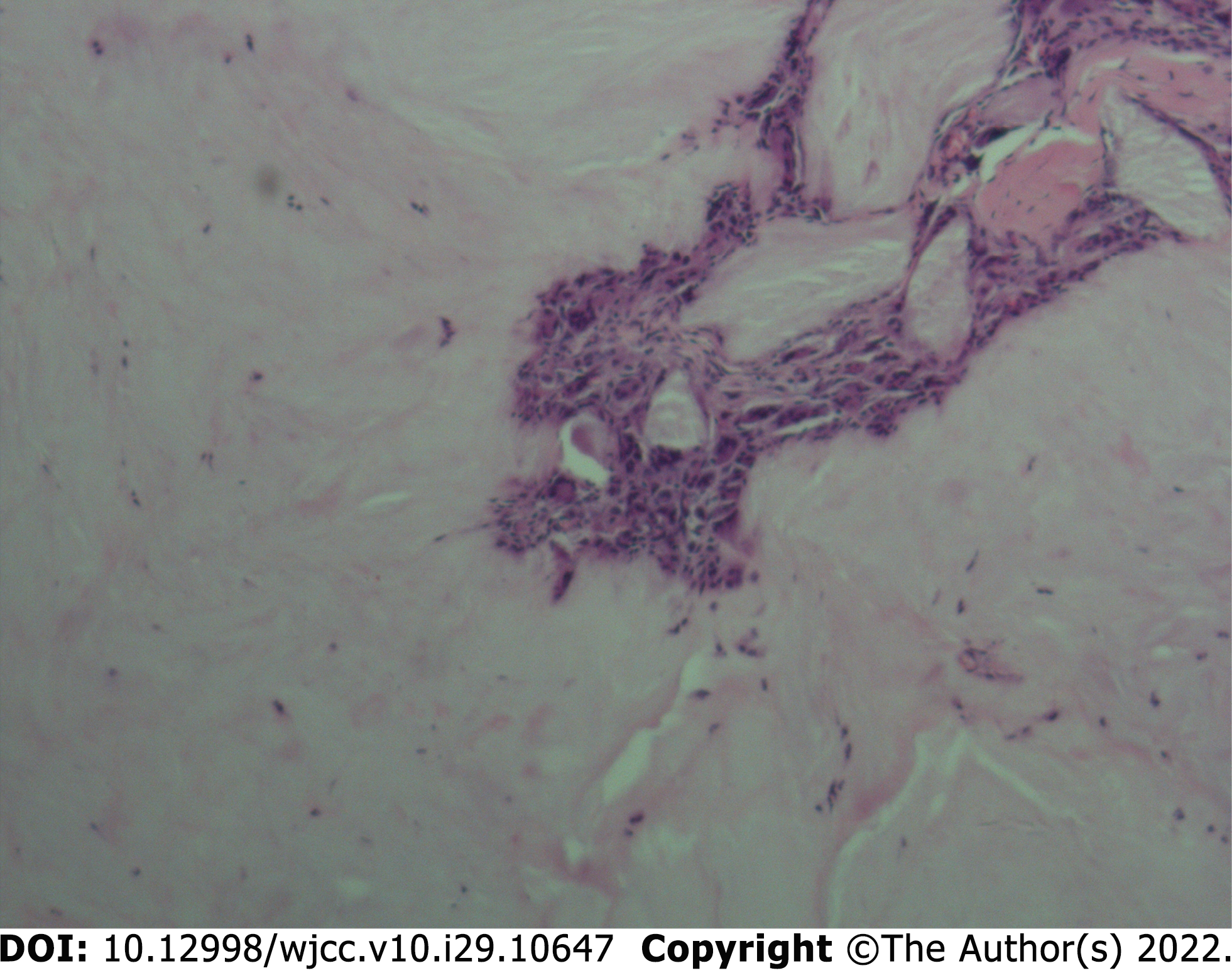
Figure 5 Histopathological sections (hematoxylin-eosin staining) of C7/T1, T5, T6 epidural, vertebral tuberosities, and surrounding soft tissues show urate crystals arranged in needle-like, parallel, or radial patterns, surrounded by foreign body giant cell reaction and fibroblast hyperplasia.
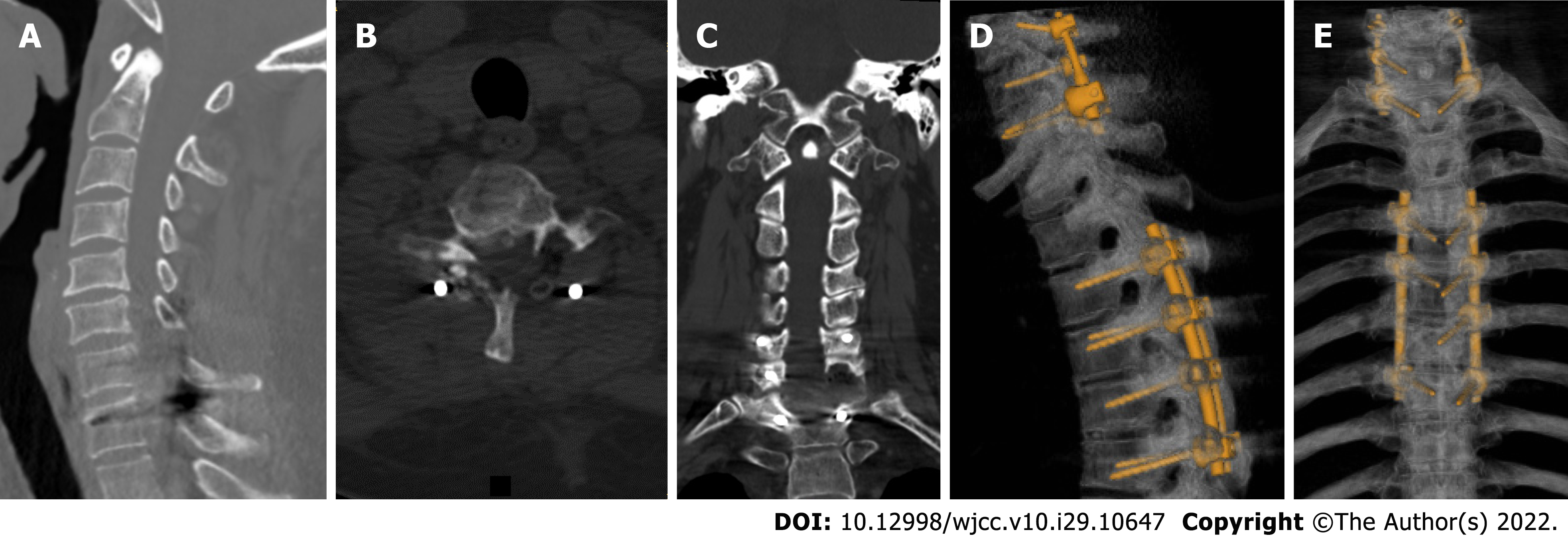
Figure 6 Postoperative computed tomography shows no loosening of internal fixation of the C6-T1 and T4-7 vertebrae.
A-C: The tophi around the C6-T1 vertebral facet joint and attachment have been removed, the compression of the spinal cord and the left spinal nerve root has been relieved, and there is no spinal canal stenosis; D and E: Part of the tophi around the facet joints of T4-7 vertebrae has been removed, the spinal stenosis is better than before, and the compression of the spinal cord is relieved; the tophi around the facet joints of T10-12 vertebrae is the same as before. And no loosening of internal fixation of the C6-T1 and T4-7 vertebrae.
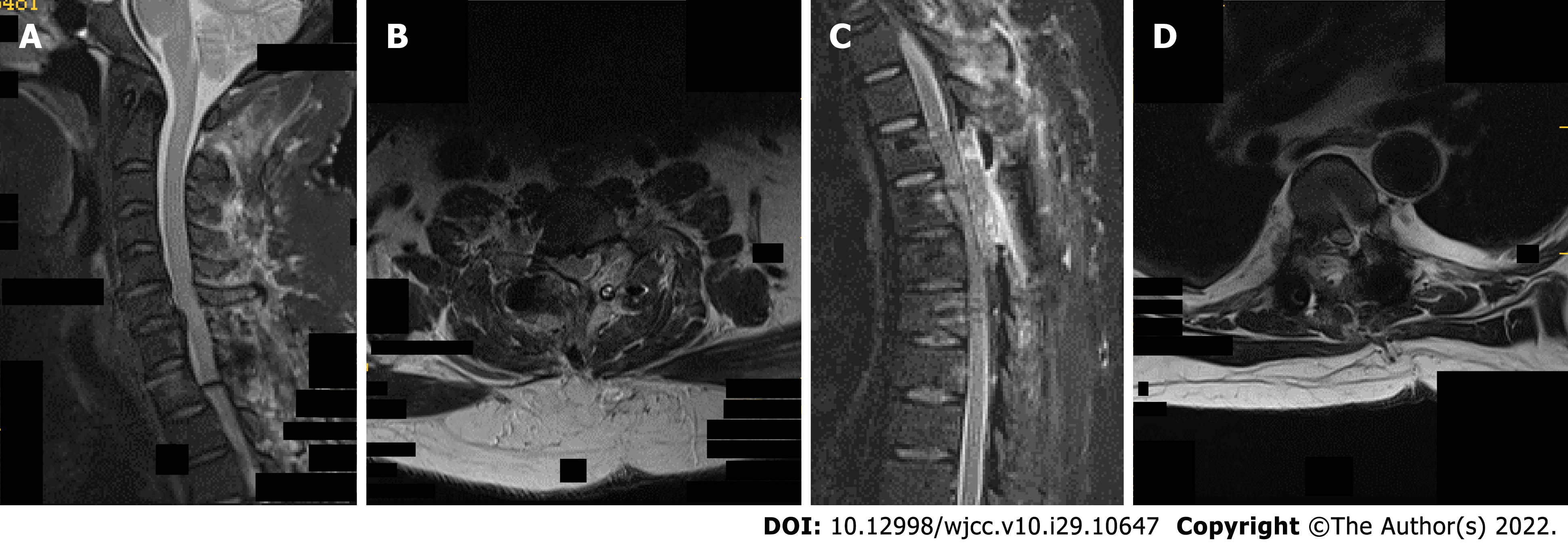
Figure 7 Postoperative cervicothoracic magnetic resonance imaging shows postoperative changes in the C6-T1 and T4-7 vertebral bodies, with patency in the spinal canal and release of spinal cord compression.
A and B: C7/T1 left vertebral facet joint multiple tophi have been cleared; C and D: T4/5 right side, T5/6 bilateral vertebral facet joint multiple tophi have been cleared, T4/5-T5/6 spinal canal stenosis returned to patency, C7/T1 spinal canal stenosis has returned to normal, spinal cord compression has been relieved; multiple exudative postoperative changes at the back of the cervicothoracic spinous process.
- Citation: Chen HJ, Chen DY, Zhou SZ, Chi KD, Wu JZ, Huang FL. Multiple tophi deposits in the spine: A case report. World J Clin Cases 2022; 10(29): 10647-10654
- URL: https://www.wjgnet.com/2307-8960/full/v10/i29/10647.htm
- DOI: https://dx.doi.org/10.12998/wjcc.v10.i29.10647


