Copyright
©The Author(s) 2022.
World J Clin Cases. Sep 6, 2022; 10(25): 9148-9155
Published online Sep 6, 2022. doi: 10.12998/wjcc.v10.i25.9148
Published online Sep 6, 2022. doi: 10.12998/wjcc.v10.i25.9148
Figure 1 Computed tomography image of the chest showing bronchiectasis with multiple miliary nodules.
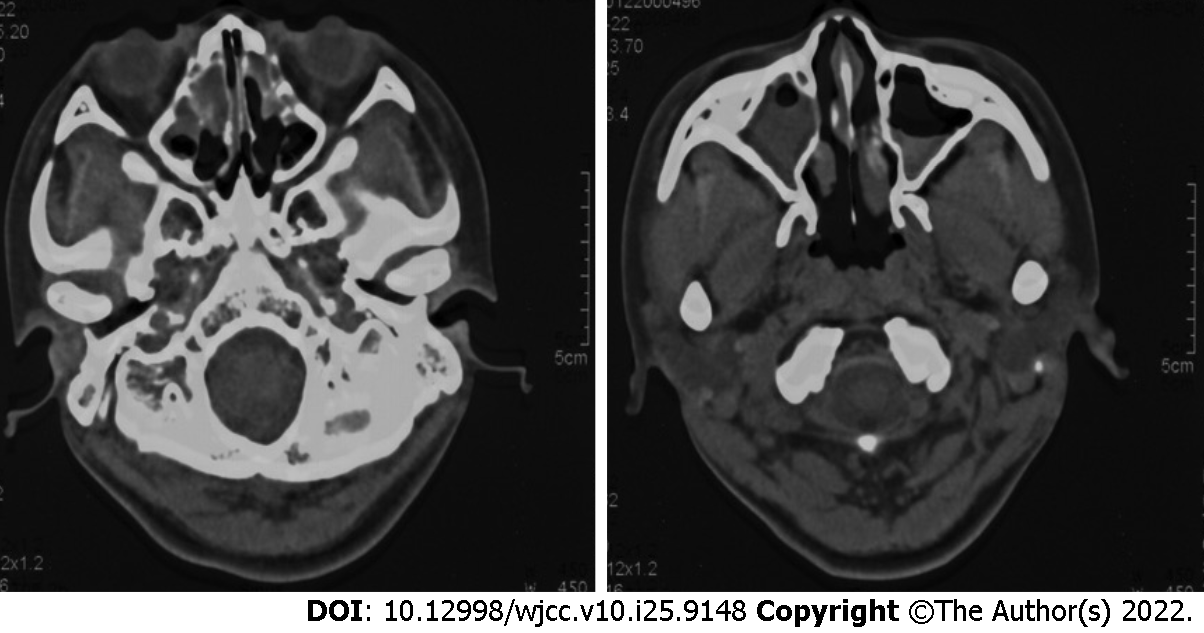
Figure 2 Computed tomography image of the sinus showing the mucosa of bilateral ethmoid sinus and maxillary sinus was thickened and edematous, and the lesion of the right sinus cavity was more serious than that of the left.
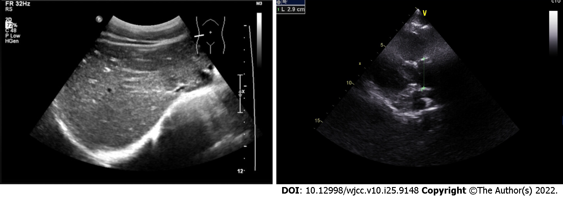
Figure 3 Abdominal ultrasound and cardiac ultrasound images showing that the position of all organs and all atrial ventricles was normal.
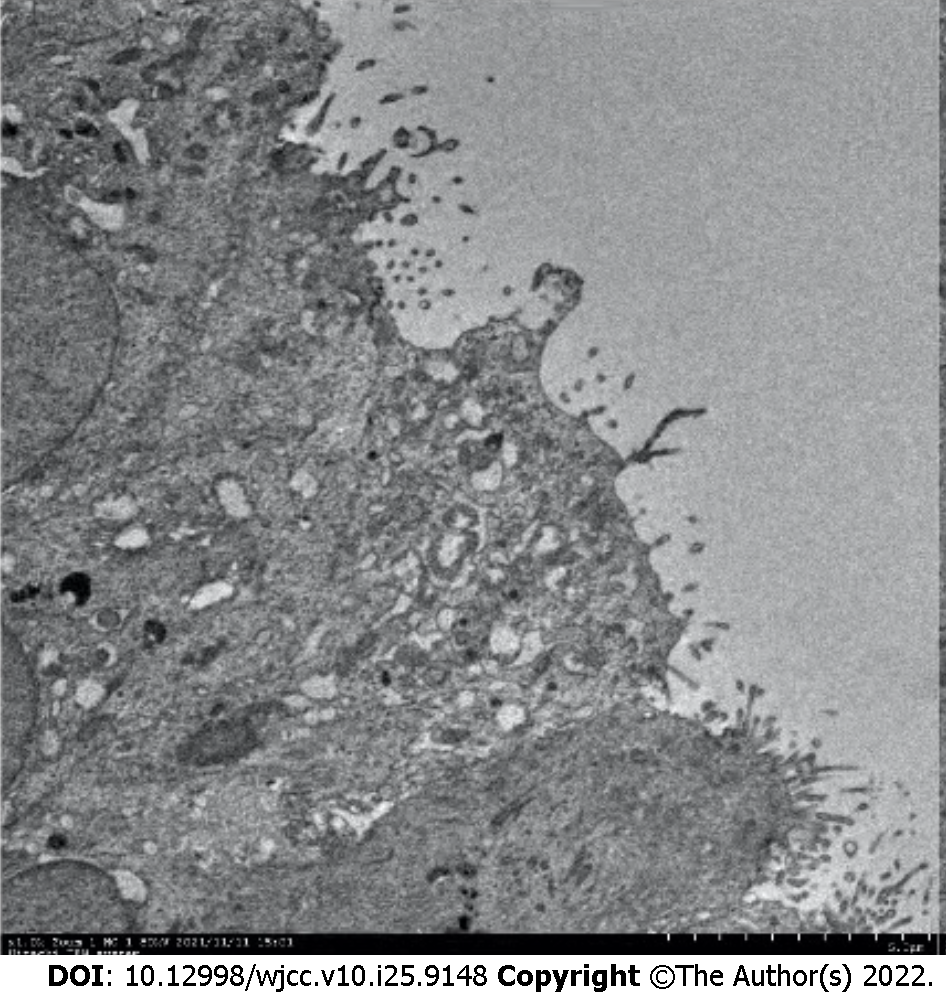
Figure 4 Electron microscopic examination of tracheoscopic biopsy showed that cilia were short and no cilia power arm was found.
Transmission electron microscope, 1000 × magnification.
Figure 5 Computed tomography image of the chest 2 mo after azithromycin treatment.
Nodular shadow was obviously weakened, but signs of bronchiectasis didn’t change.
- Citation: Zhang YY, Lou Y, Yan H, Tang H. CCNO mutation as a cause of primary ciliary dyskinesia: A case report. World J Clin Cases 2022; 10(25): 9148-9155
- URL: https://www.wjgnet.com/2307-8960/full/v10/i25/9148.htm
- DOI: https://dx.doi.org/10.12998/wjcc.v10.i25.9148


