Copyright
©The Author(s) 2022.
World J Clin Cases. Sep 6, 2022; 10(25): 9036-9043
Published online Sep 6, 2022. doi: 10.12998/wjcc.v10.i25.9036
Published online Sep 6, 2022. doi: 10.12998/wjcc.v10.i25.9036
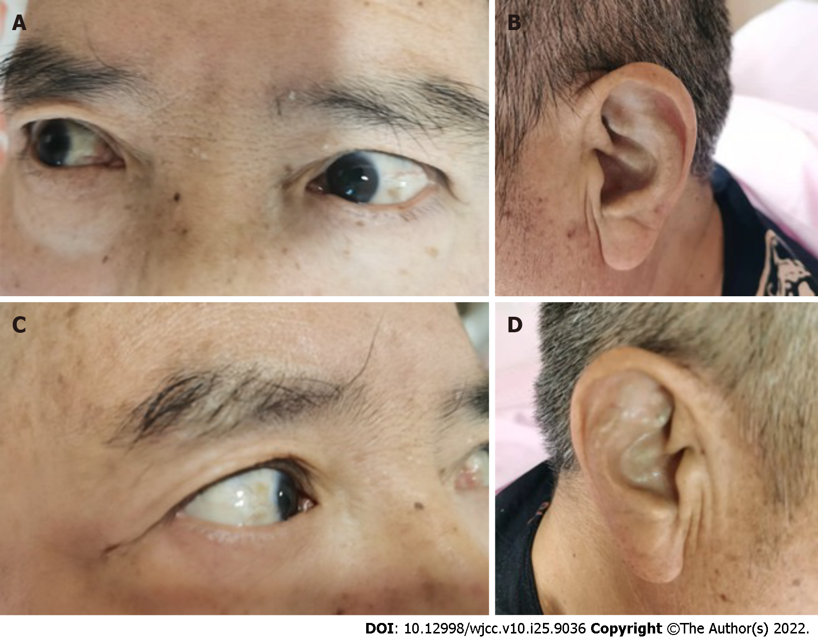
Figure 1 Brown-black pigment can be seen on both sides of the auricle and sclera.
A: Left eye; B: Left ear; C: Right eye; D: Right ear.
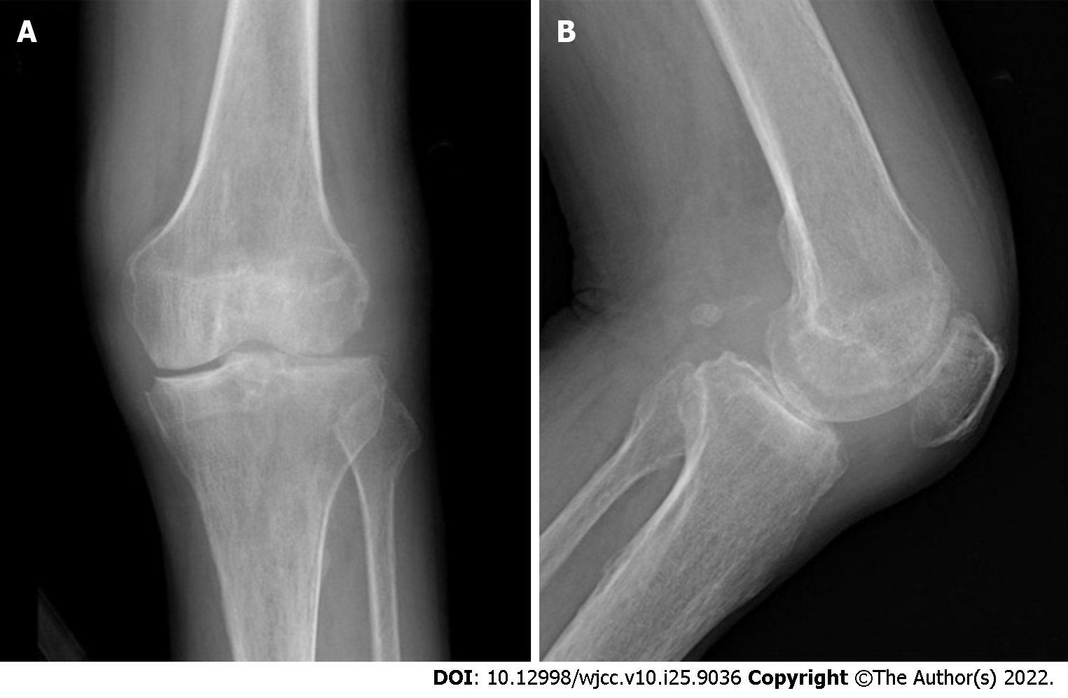
Figure 2 Radiographs of the patient’s knee showed joint space narrowing and osteophyte formation.
A: Coronal plane; B: Sagittal plane.
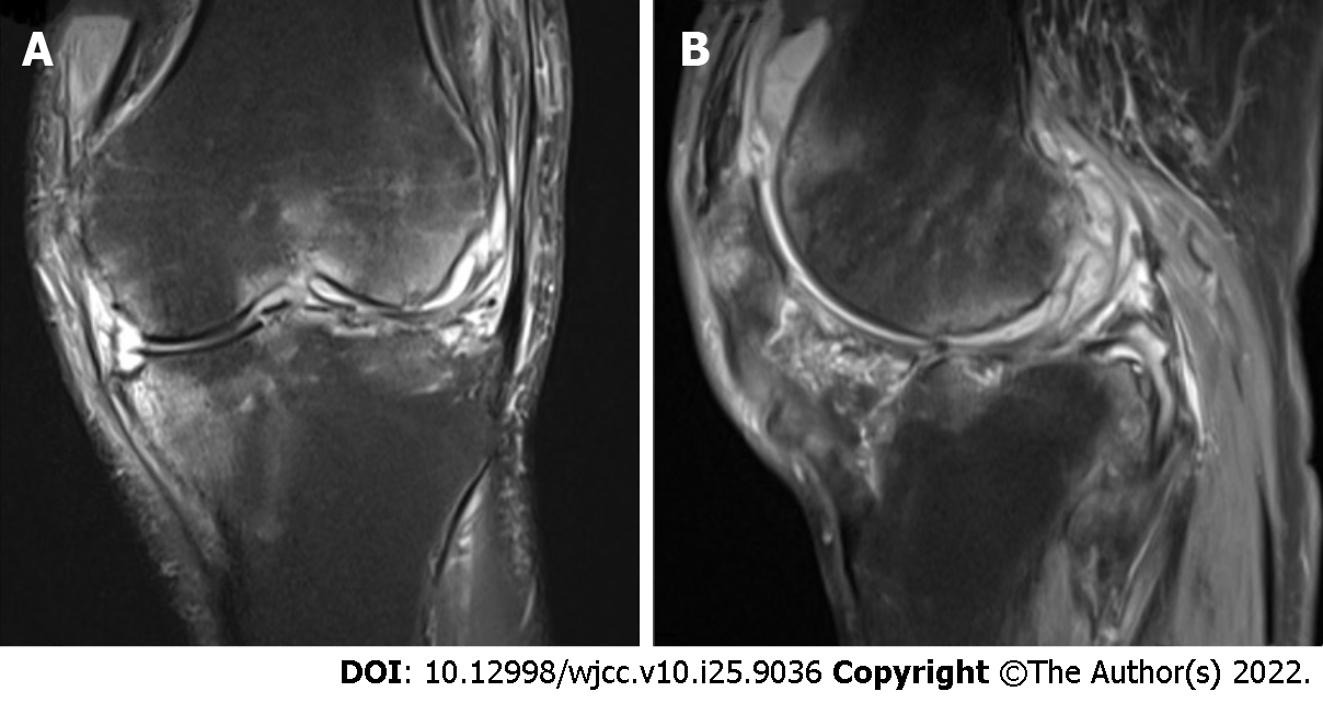
Figure 3 Magnetic resonance image of the patient's left knee revealed bone marrow edema.
A: Coronal plane; B: Sagittal plane.
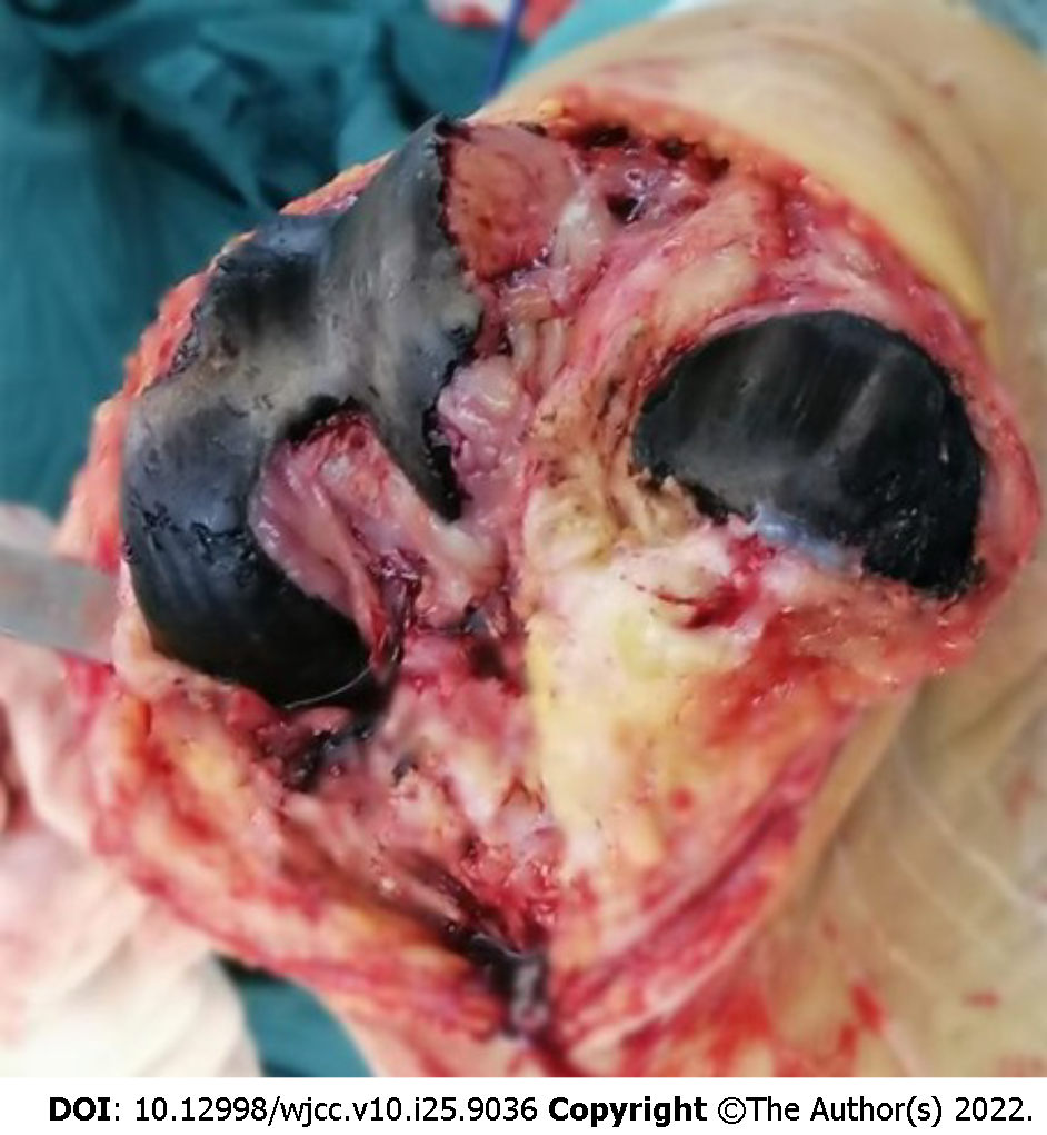
Figure 4 Intraoperative images show that the surface of the knee cartilage was dark brown.
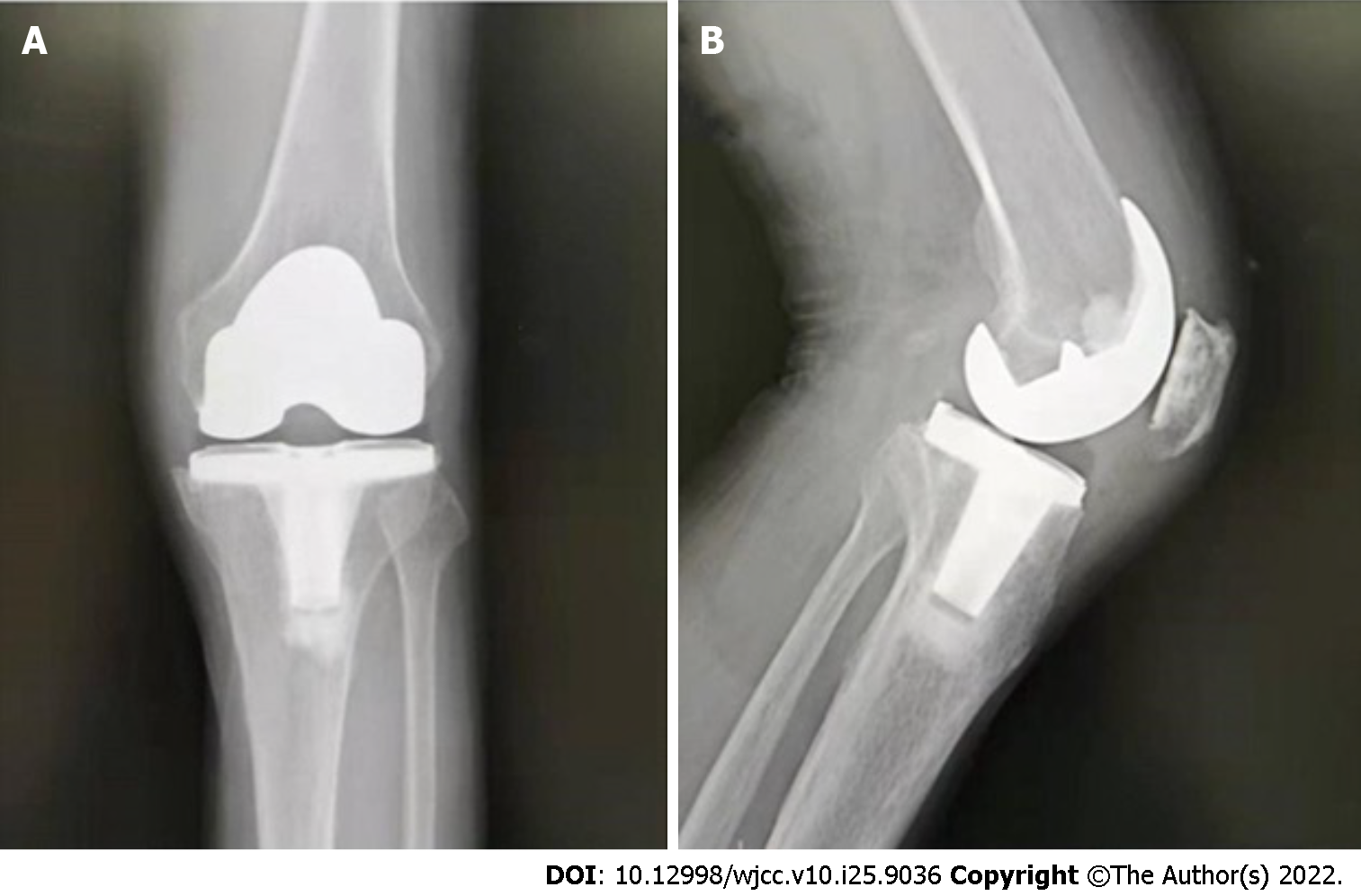
Figure 5 One week postoperatively, the knee X-ray showed that the prosthesis was in a good position.
A: Coronal plane; B: Sagittal plane.
- Citation: Wang XC, Zhang XM, Cai WL, Li Z, Ma C, Liu YH, He QL, Yan TS, Cao XW. One-stage revision arthroplasty in a patient with ochronotic arthropathy accompanied by joint infection: A case report. World J Clin Cases 2022; 10(25): 9036-9043
- URL: https://www.wjgnet.com/2307-8960/full/v10/i25/9036.htm
- DOI: https://dx.doi.org/10.12998/wjcc.v10.i25.9036


