Copyright
©The Author(s) 2022.
World J Clin Cases. Sep 6, 2022; 10(25): 8998-9003
Published online Sep 6, 2022. doi: 10.12998/wjcc.v10.i25.8998
Published online Sep 6, 2022. doi: 10.12998/wjcc.v10.i25.8998
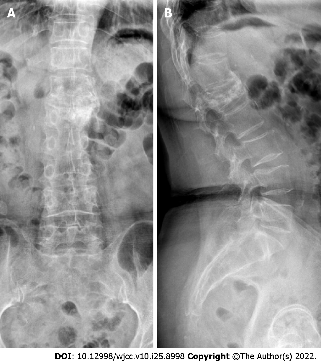
Figure 1 Postoperative X-ray of the first vertebroplasty.
A: Cement diffused only in the left side of L1 vertebral body; B: Lateral view showing compressed vertebral body with cement.
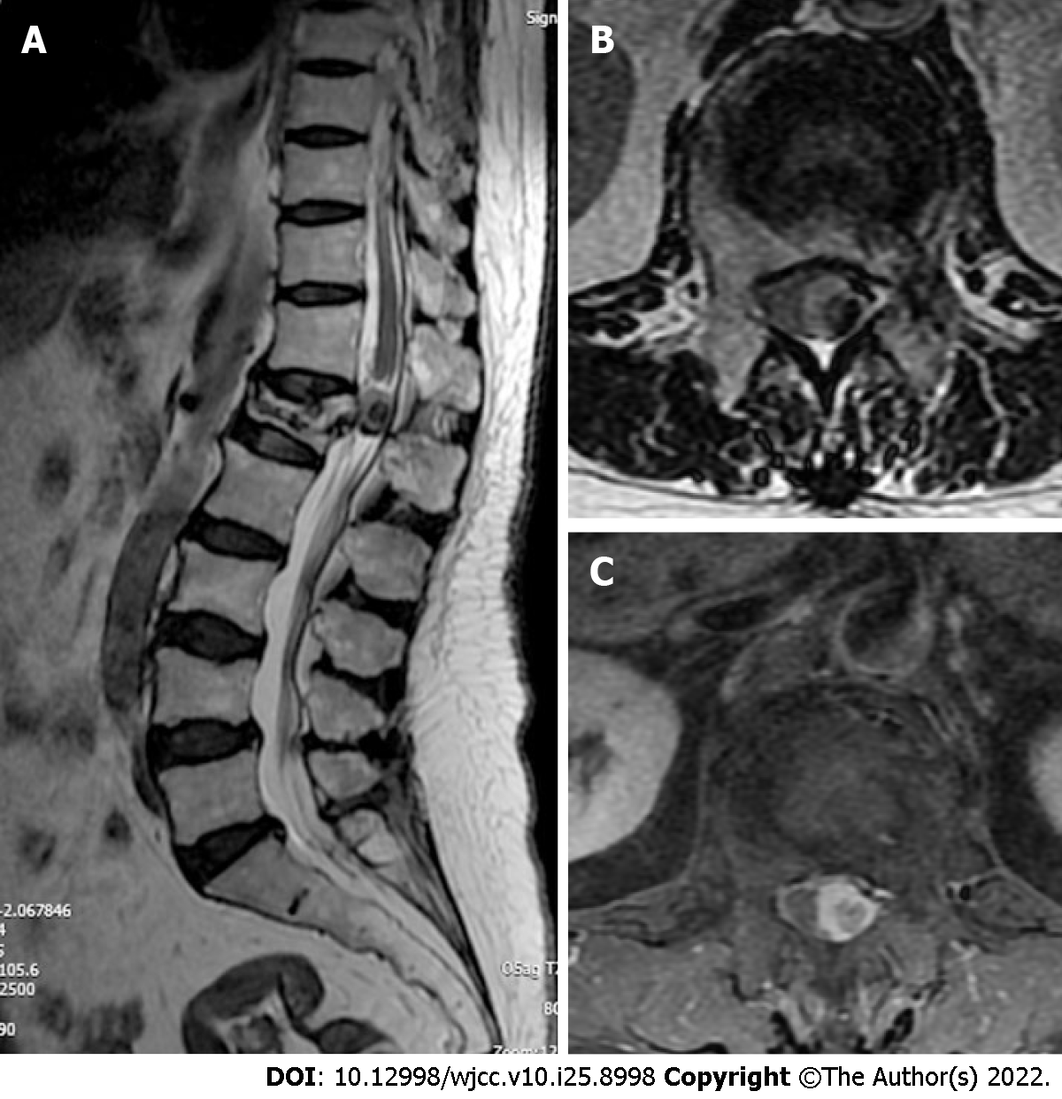
Figure 2 Lumbar magnetic resonance imaging before the second operation.
A: Intradural mass in L1 Level; B and C: The cauda equina was deviated to the right.
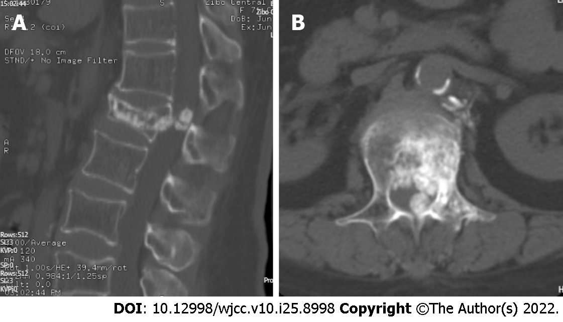
Figure 3 Reconstruction computed tomography scan level before the second operation.
A: Intradural calcification mass in L1 Level; B: The mass connected with cement extravasated into the anterior space of spine canal.
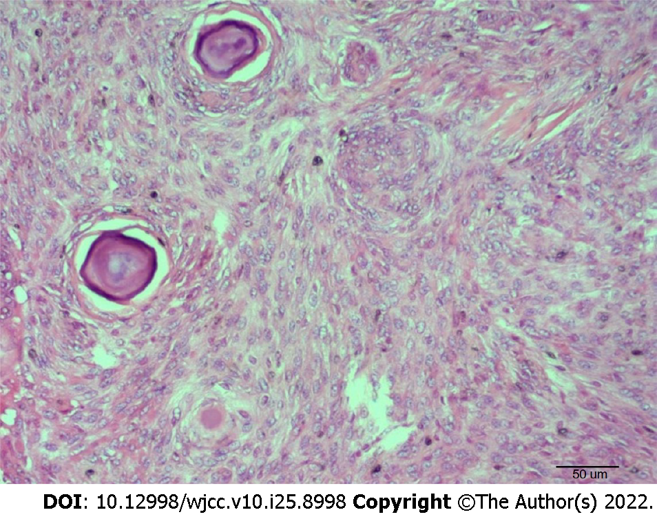
Figure 4 Result of postoperative histopathology imaging showing granulomatous formation with focal chronic inflammatory cell infiltration.
Magnification, × 200.
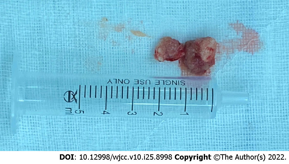
Figure 5 Postoperative image of two pieces of the mass covered with fibrosis and thickening.
- Citation: Ma QH, Liu GP, Sun Q, Li JG. Delayed complications of intradural cement leakage after percutaneous vertebroplasty: A case report . World J Clin Cases 2022; 10(25): 8998-9003
- URL: https://www.wjgnet.com/2307-8960/full/v10/i25/8998.htm
- DOI: https://dx.doi.org/10.12998/wjcc.v10.i25.8998


