Copyright
©The Author(s) 2022.
World J Clin Cases. Jun 26, 2022; 10(18): 6234-6240
Published online Jun 26, 2022. doi: 10.12998/wjcc.v10.i18.6234
Published online Jun 26, 2022. doi: 10.12998/wjcc.v10.i18.6234
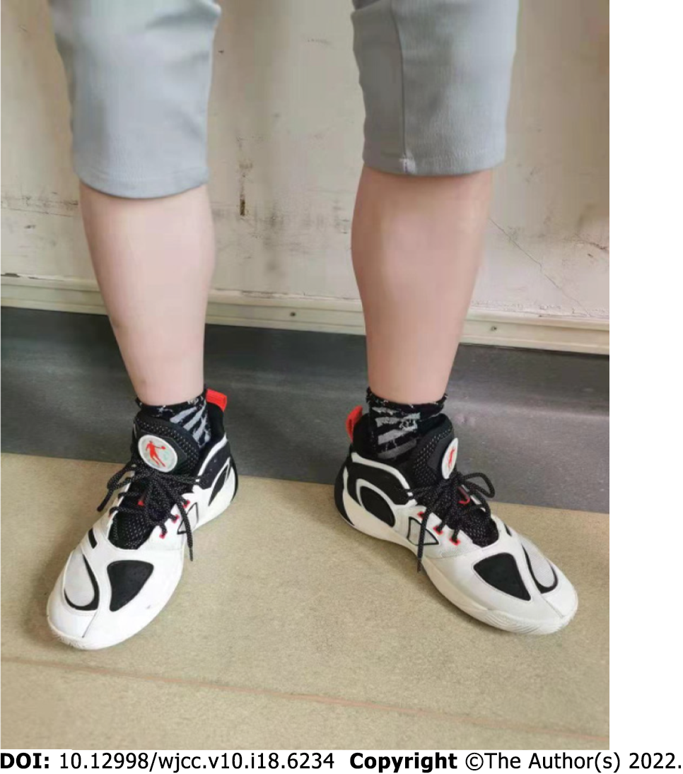
Figure 1 Normal lower limbs.
No edema of the lower limbs was observed in the 19-year-old patient in this case.
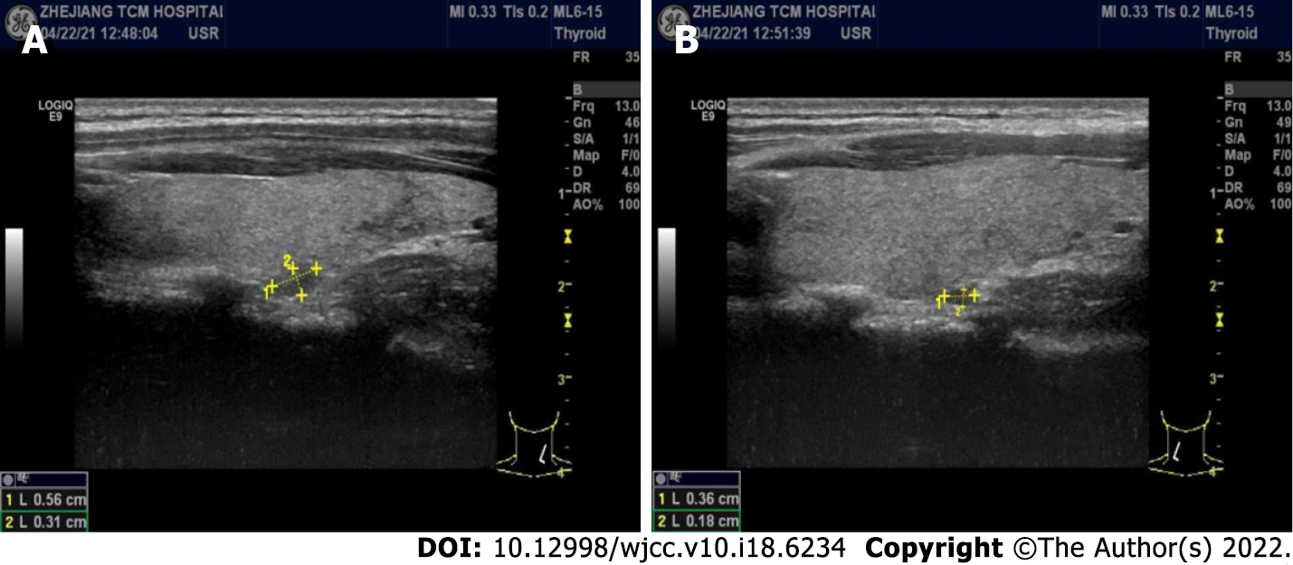
Figure 2 Parathyroid gland ultrasound images.
Hypoechoic nodules can be seen in the parathyroid gland, 0.56 cm x 0.31 cm on the right (A) and 0.36 cm x 0.18 cm on the left (B), with a clear boundary, regular shape, and few blood flow signals.
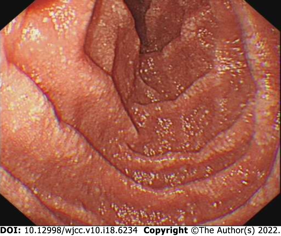
Figure 3 Gastroscopic image.
Multiple white spots and patches can be clearly seen in the duodenum.
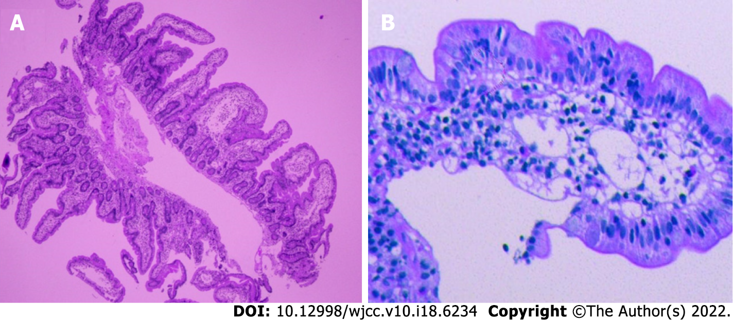
Figure 4 Gastroscopic pathological images.
A: Moderate chronic inflammatory cell infiltration distributed around a small mucosal patch in the descending duodenum (25 × magnification); B: The descending duodenum followed by lymphatic dilatation in the mucosal lamina propria (400 × magnification).
- Citation: Cao Y, Feng XH, Ni HX. Primary intestinal lymphangiectasia presenting as limb convulsions: A case report. World J Clin Cases 2022; 10(18): 6234-6240
- URL: https://www.wjgnet.com/2307-8960/full/v10/i18/6234.htm
- DOI: https://dx.doi.org/10.12998/wjcc.v10.i18.6234


