©The Author(s) 2022.
World J Clin Cases. May 26, 2022; 10(15): 5005-5011
Published online May 26, 2022. doi: 10.12998/wjcc.v10.i15.5005
Published online May 26, 2022. doi: 10.12998/wjcc.v10.i15.5005
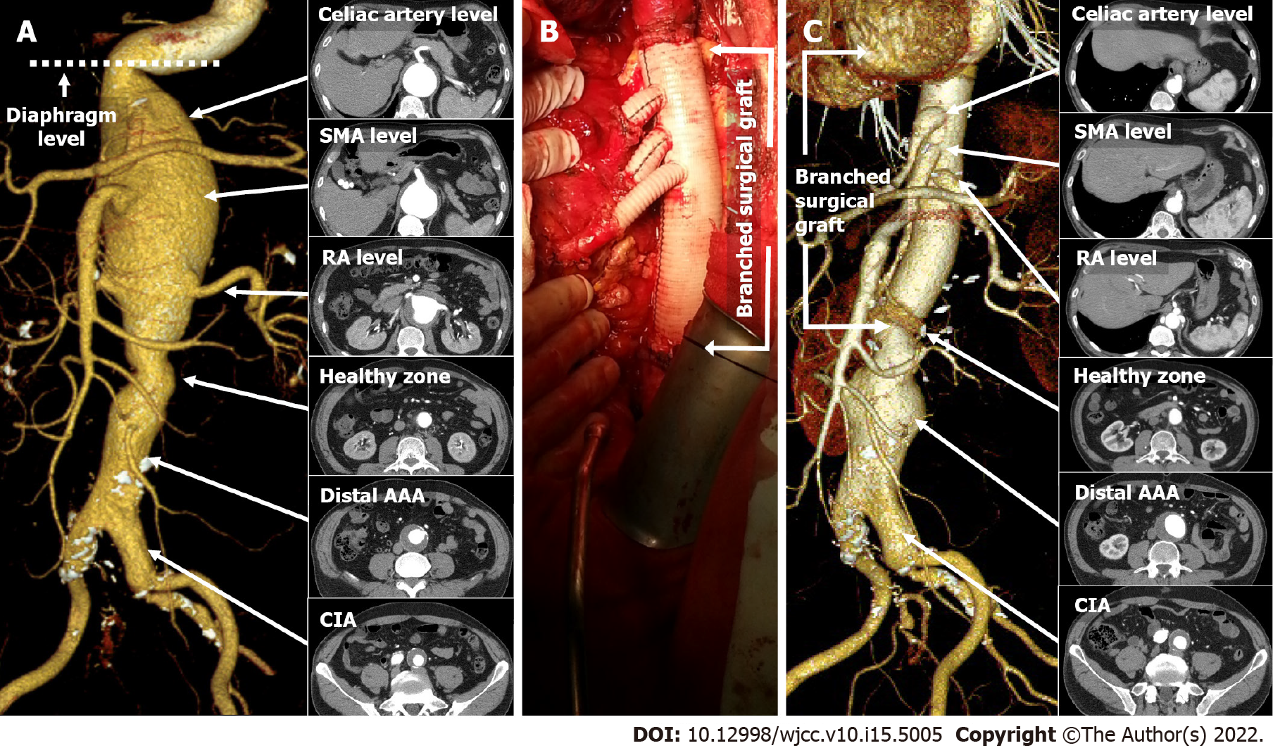
Figure 1 Computed tomographic aortography and surgical images of complex abdominal aortic aneurysm with bilateral involvement of the common iliac artery pre- and post-surgery and endovascular aortic repair.
A: The computed tomographic aortography image before surgery shows complex thoracoabdominal aortic aneurysm initiating directly distal to the diaphragm level and extending to both common iliac arteries, while the extensive lesion is separated by a short healthy zone in the infrarenal level; B: After surgery, the visceral and renal arteries were anastomosed to the Coselli branched graft and the distal end of the surgical graft was sutured to the 1 cm healthy zone; C: The main body of the stent graft was sealed to the infrarenal landing zone facilitated by the surgical graft. SMA: Superior mesenteric artery; RA: Renal artery; AAA: Abdominal aortic aneurysm; CIA: Common iliac artery.
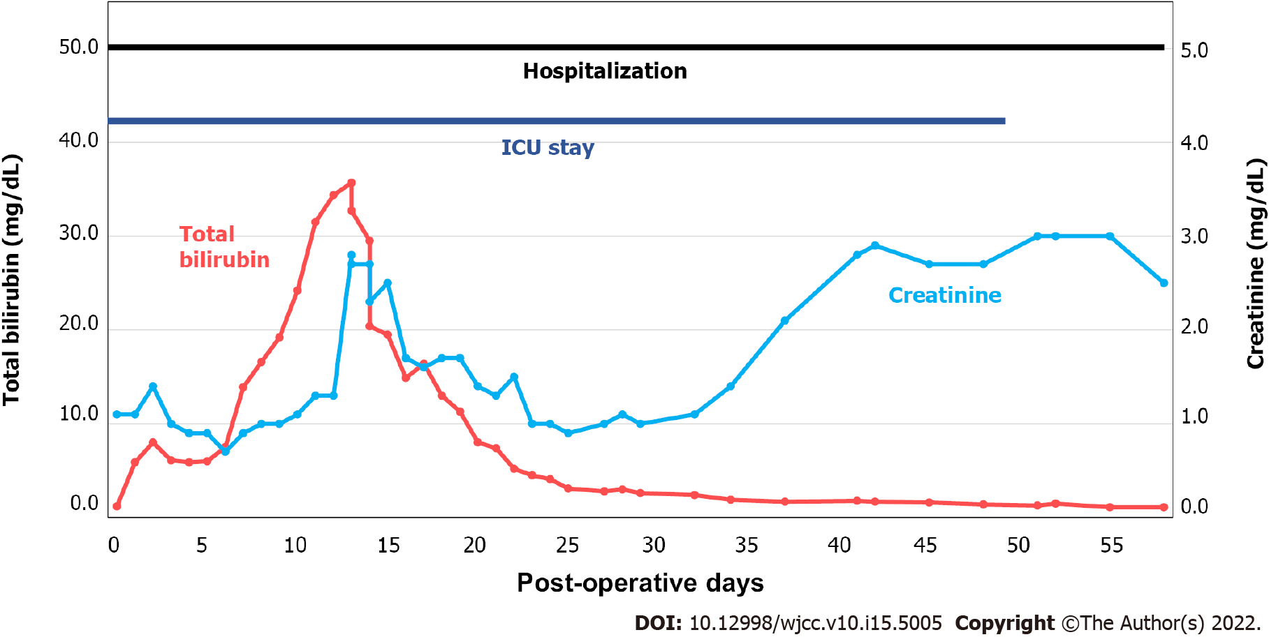
Figure 2 Diagram of hospital course after the surgery.
The patient was discharged after 60 d post-surgery. His stay in the intensive care unit was prolonged for 49 d due to difficulty in weaning from ventilator support, initiated due to pneumonia. He also developed liver and kidney failure. Total bilirubin peaked at post-operative day 13 up to 35.7 mg/dL and serum creatinine was not recovered until discharge.
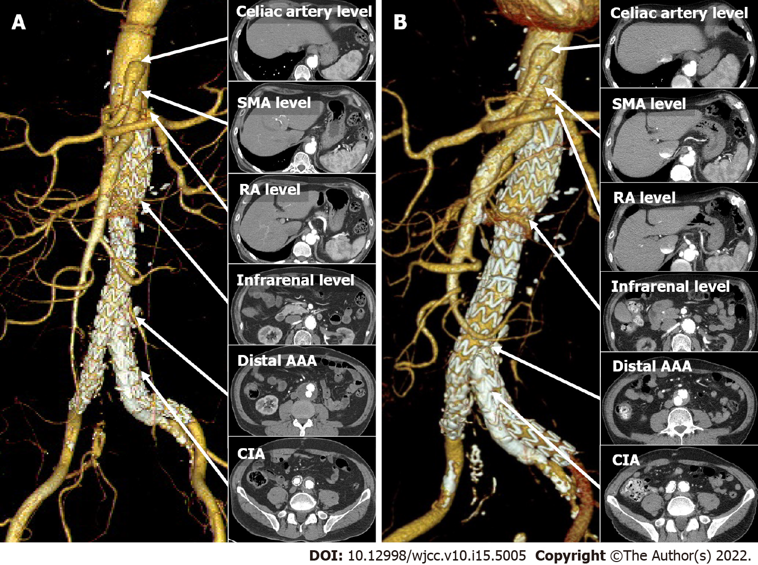
Figure 3 Computed tomographic angiography images after endovascular aortic repair.
A: The computed tomographic angiography (CTA) images 5 mo after endovascular aortic repair (EVAR); B: The CTA image of the 5-year post-EVAR showing reduced maximal diameter of the distal abdominal aortic aneurysm and common iliac artery aneurysm. SMA: Superior mesenteric artery; RA: Renal artery; AAA: Abdominal aortic aneurysm; CIA: Common iliac artery.
- Citation: Jang AY, Oh PC, Kang JM, Park CH, Kang WC. Extensive complex thoracoabdominal aortic aneurysm salvaged by surgical graft providing landing zone for endovascular graft: A case report. World J Clin Cases 2022; 10(15): 5005-5011
- URL: https://www.wjgnet.com/2307-8960/full/v10/i15/5005.htm
- DOI: https://dx.doi.org/10.12998/wjcc.v10.i15.5005


