©The Author(s) 2022.
World J Clin Cases. Apr 26, 2022; 10(12): 3951-3958
Published online Apr 26, 2022. doi: 10.12998/wjcc.v10.i12.3951
Published online Apr 26, 2022. doi: 10.12998/wjcc.v10.i12.3951
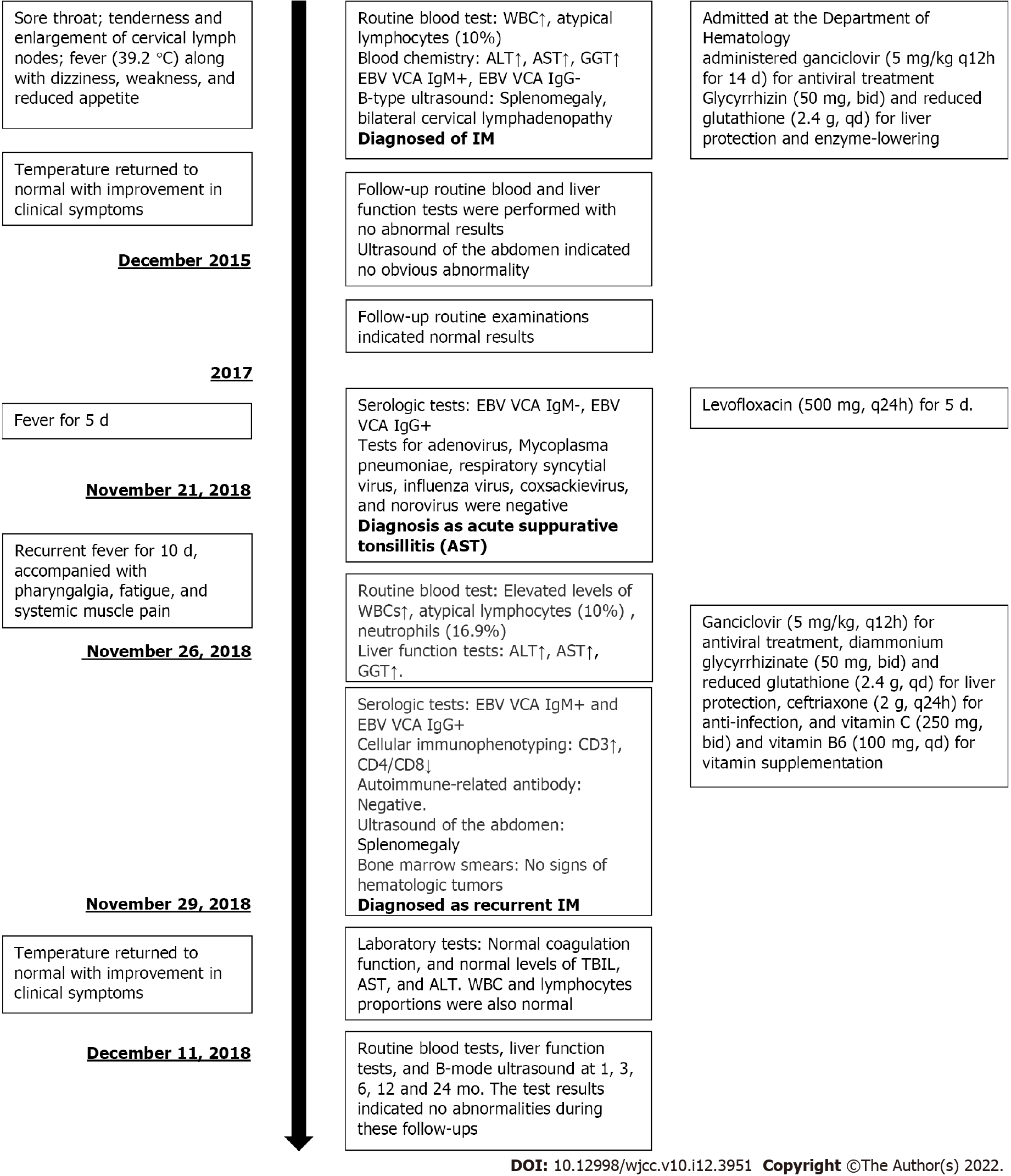
Figure 1 Timeline of symptoms, diagnosis and interventions.
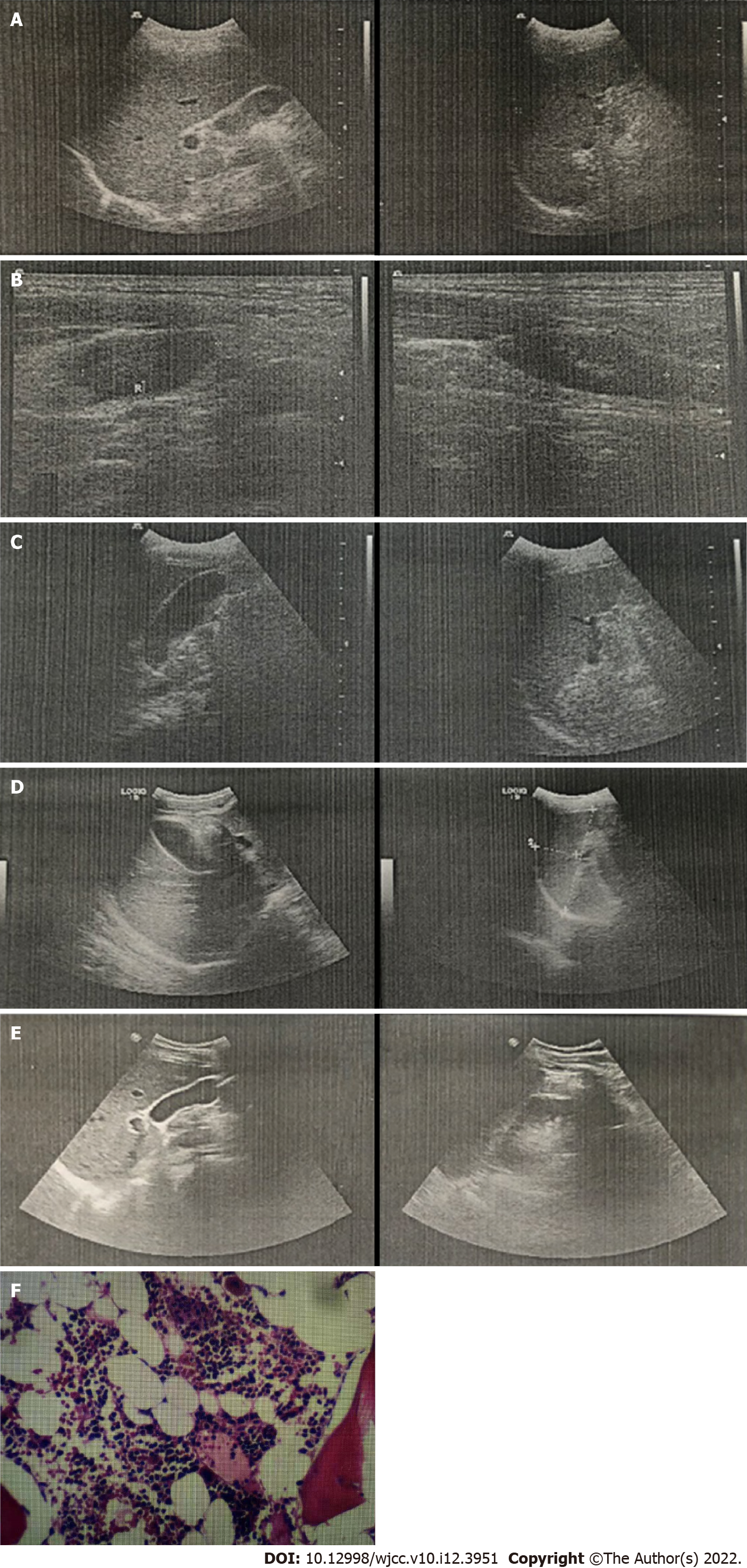
Figure 2 Results of imaging examinations and biopsy.
A: The liver is normal in size, smooth in contour, and heterogeneous in echogenicity. The gallbladder is normal in size. The spleen is about 10.9 × 4.3 cm in size and smooth in contour; B: Echogenic areas seen within cervical lymph nodes. Maximum size of the echogenic areas is 2.2 × 0.9 cm at the left side and 2.1 × 1.1 cm at the right side; C: The liver is normal in size, smooth in contour, and heterogeneous in echogenicity. The gallbladder is 7.2 × 2.5 cm in size. The spleen is about 11.0 × 4.3 cm in size and smooth in contour; D: The liver is normal in size, smooth in contour, and heterogeneous in echogenicity. The gallbladder is normal in size. The spleen is about 10.8 × 3.8 cm in size and smooth in contour. E: The liver is normal in size, smooth in contour, and heterogeneous in echogenicity. The gallbladder is normal in size. The spleen is about 11.4 × 3.6 cm in size and smooth in contour; F: Bone marrow smears indicating active proliferative myeloid series with granulocytes at all stages of development and active proliferative erythroid series with discretely distributed nucleated cells.
- Citation: Zhang XY, Teng QB. Recurrence of infectious mononucleosis in adults after remission for 3 years: A case report. World J Clin Cases 2022; 10(12): 3951-3958
- URL: https://www.wjgnet.com/2307-8960/full/v10/i12/3951.htm
- DOI: https://dx.doi.org/10.12998/wjcc.v10.i12.3951


