©The Author(s) 2022.
World J Clin Cases. Apr 26, 2022; 10(12): 3893-3898
Published online Apr 26, 2022. doi: 10.12998/wjcc.v10.i12.3893
Published online Apr 26, 2022. doi: 10.12998/wjcc.v10.i12.3893
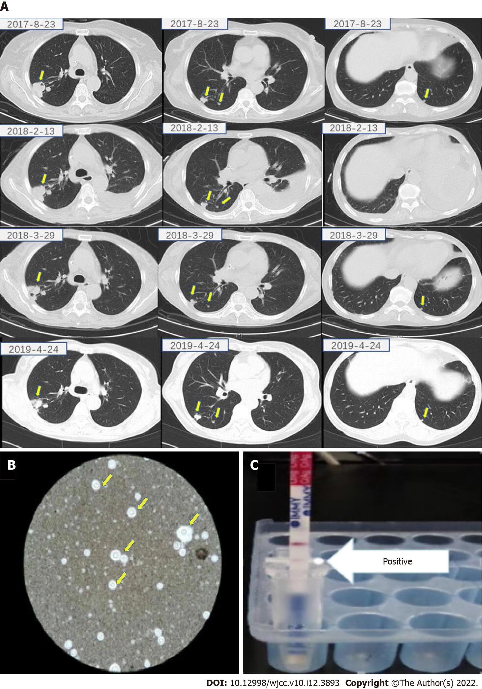
Figure 1 Imaging, pathology and cryptococcal antigen testing of lung tissue homogenate of case 1.
A: Computed tomography scan of the chest showing multiple nodules in both lungs (arrow); B: Ink staining of lung biopsy specimen showing polysaccharide capsule that surrounds the cell body; C: Results of cryptococcal antigen lateral flow immunoassay of lung tissue homogenate.
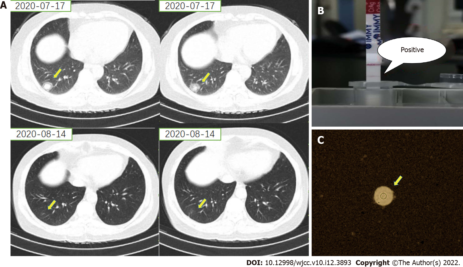
Figure 2 Imaging, pathology and cryptococcal antigen testing of lung tissue homogenate of case 2.
A: Computed tomography scan of the chest showing a nodule in the right lung (arrow); B: Positive result for cryptococcal antigen in lateral flow immunoassay of lung tissue homogenate; C: Ink staining of lung biopsy specimen showing polysaccharide capsule that surrounds the cell body.
- Citation: Wang WY, Zheng YL, Jiang LB. Cryptococcal antigen testing of lung tissue homogenate improves pulmonary cryptococcosis diagnosis: Two case reports. World J Clin Cases 2022; 10(12): 3893-3898
- URL: https://www.wjgnet.com/2307-8960/full/v10/i12/3893.htm
- DOI: https://dx.doi.org/10.12998/wjcc.v10.i12.3893


