©The Author(s) 2022.
World J Clin Cases. Apr 26, 2022; 10(12): 3872-3878
Published online Apr 26, 2022. doi: 10.12998/wjcc.v10.i12.3872
Published online Apr 26, 2022. doi: 10.12998/wjcc.v10.i12.3872
Figure 1 Reconstruction of pulmonary vein and left atrial appendage by cardiac computed tomography.
A: Reconstruction of pulmonary vein; B-C: Reconstruction and measurement of left atrial appendage (LAA); D: LAA occluder was released. Orange arrow shows LAA occluder.
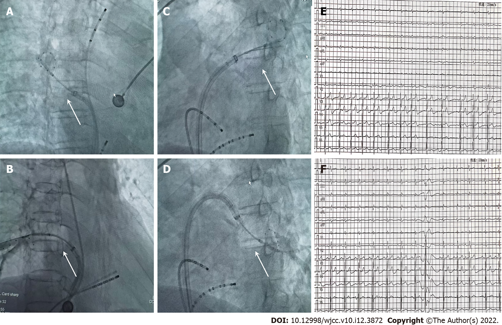
Figure 2 Cryoballoon ablation.
A-D: Cryoballoon ablation of all four pulmonary veins with good balloon occlusion; E-F: Preoperative and postoperative electrocardiogram. White arrows show balloon occlusion.
Figure 3 Outcome and follow-up.
A: Preoperative echocardiography for atrial septal defect (ASD); B: Final X-ray image after left atrial appendage occlusion and ASD occlusion; C: Postoperative echocardiography for ASD; D: Follow-up at 3 mo by transesophageal echocardiography. Orange arrow shows ASD occluder; white arrow shows left atrial appendage occluder.
- Citation: Wu YC, Wang MX, Chen GC, Ruan ZB, Zhang QQ. Cryoballoon pulmonary vein isolation and left atrial appendage occlusion prior to atrial septal defect closure: A case report. World J Clin Cases 2022; 10(12): 3872-3878
- URL: https://www.wjgnet.com/2307-8960/full/v10/i12/3872.htm
- DOI: https://dx.doi.org/10.12998/wjcc.v10.i12.3872


