Copyright
©The Author(s) 2022.
World J Clin Cases. Apr 16, 2022; 10(11): 3593-3600
Published online Apr 16, 2022. doi: 10.12998/wjcc.v10.i11.3593
Published online Apr 16, 2022. doi: 10.12998/wjcc.v10.i11.3593
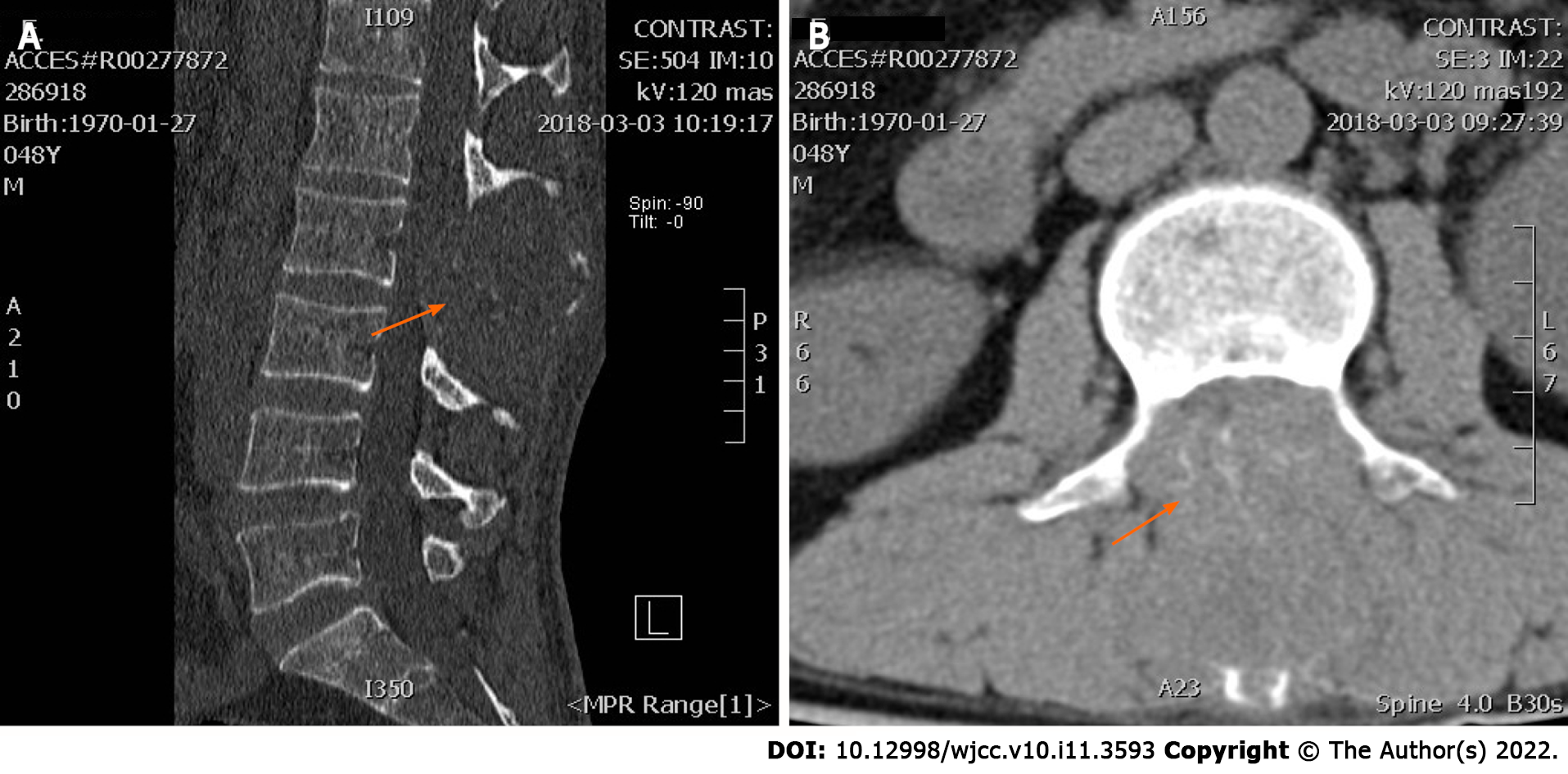
Figure 1 Lumbar computed tomography was taken on March 5, 2018, which showed lumbar spinous process level occupied space with destruction and absorption of lumbar 2 appendages and invasion of the spinal canal.
A: Sagittal sections of the masses; B: Coronal sections of the masses (red arrow).
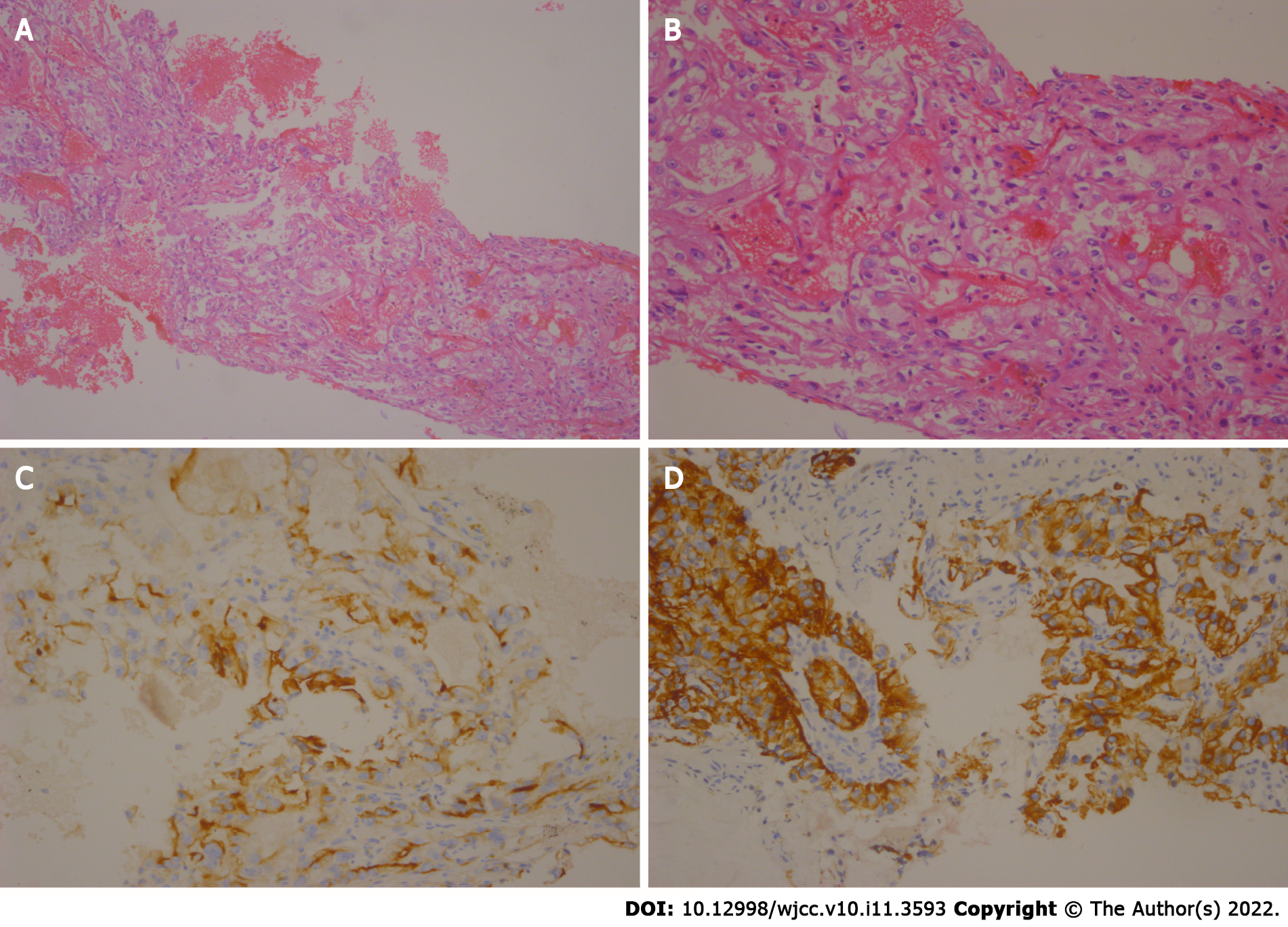
Figure 2 Haematoxylin and eosin staining of a tumour section.
A: Under × 10 times microscope; B: Under × 40 times microscope. Immunohistochemical staining of a tumour section; C: It showed that CD10 was postive; D: It showed that CD20 was negtive.
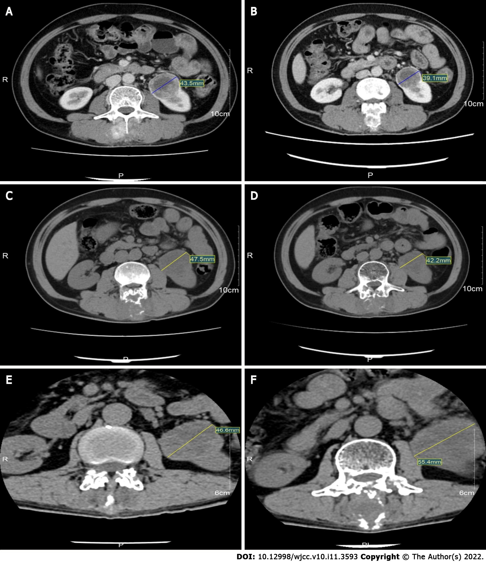
Figure 3 Computed tomography images during treatment showing the changes of the mass.
A: Computed tomography (CT) images obtained on July 26, 2018; B: CT images obtained on October 20, 2018; C: CT images obtained on March 5, 2019; D: CT images obtained on December 24, 2019; E: CT images obtained on October 5, 2020; F: CT images obtained on December 14, 2020.
- Citation: Wei HP, Mao J, Hu ZL. Successful apatinib treatment for advanced clear cell renal carcinoma as a first-line palliative treatment: A case report. World J Clin Cases 2022; 10(11): 3593-3600
- URL: https://www.wjgnet.com/2307-8960/full/v10/i11/3593.htm
- DOI: https://dx.doi.org/10.12998/wjcc.v10.i11.3593


