Copyright
©2013 Baishideng Publishing Group Co.
World J Clin Cases. Oct 16, 2013; 1(7): 217-219
Published online Oct 16, 2013. doi: 10.12998/wjcc.v1.i7.217
Published online Oct 16, 2013. doi: 10.12998/wjcc.v1.i7.217
Figure 1 Abdominal computed tomography scan revealing a homogeneous round splenic mass with rim enhancement.
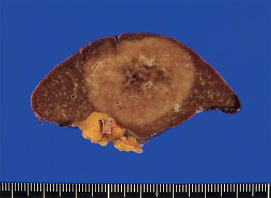
Figure 2 A well-defined homogeneous red-tan mass can be seen on the cut surface compressing the adjacent normal splenic parenchyma.
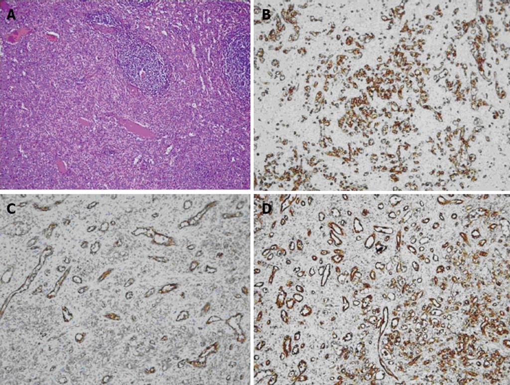
Figure 3 Pathological findings: Hematoxylin-eosin staining and immunohistochemical stainings.
A: The tumor contains haphazardly arranged small slit-like vascular spaces lined by plump endothelial cells; B: The endothelial cells lining the vascular channels are positive for CD8; C: CD31; D: Vimentin.
- Citation: Sim J, Ahn HI, Han H, Jun YJ, Rehman A, Jang SM, Jang K, Paik SS. Splenic hamartoma: A case report and review of the literature. World J Clin Cases 2013; 1(7): 217-219
- URL: https://www.wjgnet.com/2307-8960/full/v1/i7/217.htm
- DOI: https://dx.doi.org/10.12998/wjcc.v1.i7.217


