Published online Oct 16, 2013. doi: 10.12998/wjcc.v1.i7.212
Revised: July 29, 2013
Accepted: August 12, 2013
Published online: October 16, 2013
Processing time: 187 Days and 8.6 Hours
Generally, ingested foreign bodies are excreted from the digestive tract without any complications or morbidity. In adults, ingestion of foreign bodies frequently occurs in alcoholics and elderly individuals with dentures. The most commonly ingested foreign bodies are food stuffs or their parts, such as fish bones or fragments of bone and phytobezoars. Sharp foreign bodies like fish and chicken bones can lead to intestinal perforation and peritonitis. We report herein two cases, one of bowel perforation and another of anal impaction, both caused by ingested bone fragments. Complications due to ingested bone fragments are not common and preoperative diagnosis remains a challenge and therefore it must be considered in susceptible cases.
Core tip: The ingested bone fragment may cause bowel perforation at any site from the jejunum to anal margin, obstruction and fistula formation. An experienced clinician should suspect such conditions in the presence of some predisposing factors, such as rapid eating and the use of dentures in the elderly, and should consider various surgical options. We report herein two cases, one of bowel perforation and another of anal impaction, both caused by ingested bone fragments. Complications due to ingested bone fragments are not common and preoperative diagnosis remains a challenge and therefore it must be considered in susceptible cases.
- Citation: Emir S, Özkan Z, Altınsoy HB, Yazar FM, Sözen S, Bali İ. Ingested bone fragment in the bowel: Two cases and a review of the literature. World J Clin Cases 2013; 1(7): 212-216
- URL: https://www.wjgnet.com/2307-8960/full/v1/i7/212.htm
- DOI: https://dx.doi.org/10.12998/wjcc.v1.i7.212
The majority of ingested foreign bodies (IFB) are excreted from the digestive tract without any complications or morbidity; however, occasionally they may lead to serious clinical problems, such as obstruction, perforation or bleeding[1-3]. Although IFB are a common problem in children, they are infrequently encountered in adults but are seen in elderly people wearing dentures, alcoholics and/or patients with learning difficulties[4]. IFB, such as chicken bones, fish bones, toothpicks and dentures, rarely require surgical intervention (5%). Patients are not usually aware of the IFB which is usually detected either during laparotomy or at the time of pathology examination of the surgical specimen[5]. Less than 1% of IFB, especially large, sharp and/or pointed objects, cause bowel perforation. Perforation usually occurs at the narrowest parts of the bowel, either at the ileocecal valve or at the rectosigmoid junction[6]. In the literature, there are reports of ingested bone causing intestinal perforation, enterovesical fistula and perianal abscesses[4-7].
We report herein two patients who presented with different complications caused by an ingested bone fragment; we also review the existing literature on IFB in the gastrointestinal (GI) tract.
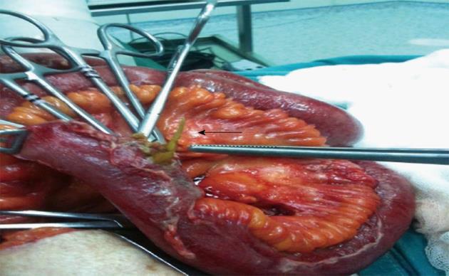
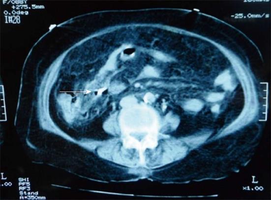
An 87-year-old woman was admitted to the emergency department with complaints of abdominal pain and vomiting for 2 d. Her past medical history included chronic obstructive pulmonary disease, cardiac failure and renal failure. On physical examination, she was conscious and alert, with a mild pyrexia. Abdominal examination revealed generalized rebound tenderness. Her white blood cell count, BUN and creatinine levels were out of the normal range; 13.500/μL, 79.5 mg/dL and 2.5 mg/dL respectively. Apart from free intra-abdominal fluid, no other abnormality was detected on abdominal X-ray, abdominal ultrasound (US) and computed tomography (CT). As the reason of the acute abdominal pain was not clear, a laparotomy was performed. At laparotomy, 500 cc of a purulent fluid collection in the right paracolic region and a perforation caused by a protruding sharp-pointed bone fragment, 15 cm proximal of the ileocecal valve, were noted (Figure 1). Partial ileal resection and end ileostomy were performed. She was discharged on the postoperative 8th day. Ileostomy closure was successfully performed after three months. After surgery, her abdominal CT scan was re-evaluated by the radiologists and a lesion with bone density was identified at the terminal ileal region (Figure 2).
A 27-year-old female was admitted to the general surgery outpatient clinic complaining of severe anal pain for 3 d. Previous medical history revealed no significant pathology. Anal inspection in the knee-chest position was normal; at anal digital examination a hard, flat object lodged in the anal canal 4 cm above the anal margin was identified. Abdominal CT confirmed the presence of the foreign body in bone density. In the operation room under sedation and analgesia, a 2 cm bone fragment that was lodged at the lateral rectal wall was removed by a Kelly clamp with anoscopy. The patient was discharged 6 h after the intervention.
Accidentally ingested foreign bodies are a common problem. Most of the IFB pass uneventfully through the gastrointestinal tract and are excreted in the stool within 1 wk[4]. Foreign body ingestion generally occurs in childhood but may also be seen in adults. In adults, IFB are usually seen in alcoholics, elderly individuals with dentures, drug abusers, prisoners, individuals with mental disorders or learning difficulties, people with fast eating habits and workers such as carpenters and dressmakers who tend to hold small sharp objects in their mouths[4,8]. Elderly people may have trouble using dentures and as the sense of feeling in the palate is decreased, they may become prone to FB ingestion. Generally patients do not recall ingesting a foreign body and this is usually detected on radiological imaging studies, during surgery or in the pathological examination of the surgical specimens[5,7]. Both patients presented herein did not recall any FB ingestion. Although the first case was an elderly individual who wore dentures and had comorbidities, the second case was a young woman with no prior mental or physical disorders. However, she eventually admitted to being a fast eater.
The American Society for Gastrointestinal Endoscopy classifies IFB as: (1) food bolus impactions, usually meat; (2) blunt objects, such as coins; (3) long objects, longer than 6-10 cm such as toothpicks; (4) sharp-pointed objects, such as fish bones or small bones; (5) disk batteries; and (6) narcotic packets, wrapped in plastic or latex. The types of IFB vary according to regional differences and feeding habits. For example, fish bone ingestion is more common in eastern countries while meat bolus impactions are mostly seen in western countries[4,9]. The most common IFB are food stuffs or their parts, such as fish bones, bone fragments or vegetable bezoars and toothpicks[4]. Although generally the ingested bones are digested or uneventfully pass through the gastrointestinal tract within 1 wk, complications such as impaction, perforation or obstruction may rarely occur[7,10-13]. Gastrointestinal perforation occurs in less than 1% of all patients. The possibility of perforation is associated with the length and sharpness of the swallowed object[14]. Ingested sharp bones, fish and chicken bones can lead to intestinal perforation and peritonitis[15]. Goh et al[16] state that most of the foreign bodies causing gastrointestinal tract perforation were of a food origin, such as fish bones, chicken bones, bone fragments or shells. Another study on IFB found that a fish bone was the most frequently encountered foreign body causing GI tract perforation[17]. In some cultures or religions, people prefer to eat all parts of the fish and thus fish bone ingestion and related complications are common in these populations[18]. GI perforations caused by a chicken bone are less frequently reported. Also, different GI complications were defined as caused by poultry bones, duck bones, rabbit bones and meat bone fragments in the literature[19,20].
Although bowel perforations may occur at any part of the intestinal tract, the most common site is at an acute angulation or physiological narrowing, such as the ileocecal region and rectosigmoid junction[7,11]. It is reported that the ileum was the site of perforation in 83% of cases[21]. Goh et al[16] recorded the most common site of intra abdominal perforation as the terminal ileum in 38.6%. Perforation of the jejunum is less frequent and its incidence is approximately 14.3%[4]. Predisposing factors for perforation or other complications are bowel disease, adhesions, diverticular disease, inflammatory bowel disease, bowel tumors, abdominal wall hernias and a blind loop of bowel[16]. Glasson et al[11] reported a case with perforated sigmoid diverticulum caused by a chicken bone. Akhtar et al[12] reported 3 cases with bowel perforation caused by chicken bones, two cases had a hernia and the other case had diverticulitis. In their case report and review of literature, McGregor et al[13] presented a case in whom clinical diagnosis of previously undiagnosed carcinoma was established based on colonic perforation resulting from ingested chicken/poultry bone and they also reported 3 such cases in the literature.
There were no intestinal or abdominal disorders in our cases but they said they experienced constipation from time to time. Bowel perforations can present with different clinical manifestations, such as intra abdominal abscesses, anal fistula or rectal abscess, coloenteric, colovesical or rectocutaneous fistulas, and acute abdomen. A very interesting clinical presentation reported in the literature is an aortocolic fistula[8,22-24]. Although generally anal or rectal FB engage transanally and are possible causes of anal pain, ingested fish bone has been reported to lead to perianal abscesses, anal fistulae and severe anal pain. Adufull[10] reported that ingested chicken bones and fragments of meat bone can also cause anal pain, abscess formation and anal fistula[10]. Cash et al[8] also reported anorectal abscess and fistula caused by ingested chicken bones; they stated that a partially closed anal sphincter against rectal contractions might lead to these disorders.
IFB usually present with non-specific symptoms and different clinical symptoms may occur in patients. Abdominal pain is the most common complaint (95%), followed by fever (81%) and localized peritonitis (39%). The other symptoms that may occur are nausea, vomiting, hematochezia and melena. Bowel perforation and acute surgical abdomen can lead to misdiagnosis with other conditions causing surgical abdominal diseases, such as acute appendicitis, diverticulitis or perforated peptic ulcer[7,25]. The most common preoperative diagnosis is acute abdomen of uncertain origin[18]. Gastric, duodenal or colonic perforations can present as more chronic events, such as abdominal mass or abscess[14]. Usually physicians cannot establish a preoperative diagnosis as the patient cannot recall a foreign body ingestion.
Our first case presented with acute abdominal pain and the second with severe anal pain, particularly during defecation. Generally, no specific image is detected by imaging methods. Free abdominal gas due to pneumoperitoneum, abdominal fluid collection, gas-fluid levels due to bowel obstruction, or a chicken bone image facilitates the preoperative diagnosis[15].
Free gas is rarely detected on abdominal X-rays; it was present in only 20% of cases with perforation[7]. According to the studies, the degree of radiopacity of ingested fish bones varies according to the species of the fish[16]. A prospective study of 358 patients with fish bone ingestion revealed that a plain radiography had a sensitivity of only 32%[26]. Ingested fish bones are overlooked on plain films as they are minimally radiopaque and adjacent inflammatory tissue or fluids interfere with the image of the fish bone[22].
Ultrasound can detect even non radiopaque FB, such as fish bones and toothpicks, based on their high reflectivity and variable posterior shadowing. There are reported cases of ingested FB defined by US[17]. Intra abdominal fluid and adjacent tissue changes can be seen using US.
Abdominal CT scan can detect even more details, such as intestinal obstruction, pneumoperitoneum, a thickened intestinal wall or foreign body[16]. Goh et al[17] reported, in their study of seven patients with fish bone perforations, that a correct diagnosis was made in five of the seven original radiology reports. However, on retrospective review of the scans, the fish bones could be identified in all cases, typically appearing as a linear calcified lesion surrounded by an area of inflammation.
In this report, the abdominal X-ray of the first case was evaluated as normal, ultrasound and CT scan only showed intra abdominal fluid and therefore the preoperative evaluation was non diagnostic. However, in the postoperative evaluation of the CT scan, the radiologists detected a radiopaque lesion at the terminal ileum. In the second case, no X-ray examination was performed as X-ray examination was insufficient in revealing non radiopaque FBs and other intra abdominal complications. We preferred only CT scan for imaging methods. The CT scan revealed no pathology except a bone density lesion in the rectum.
Only 1% of the complicated ingested FB in the gastrointestinal tract requires a surgical operation; 10% to 20% of them are successfully removed by non operative methods such as endoscopy[11]. If the foreign body is at the anorectal region, it is easily removed via proctosigmoidoscopy or digitally[27]. Watanabe et al[28] detected a fish bone stuck in the sigmoid colon wall by sigmoidoscopy and removed it with a sigmoidoscopy snare[28].
In recent years, laparoscopy has been used for intraperitoneal and intraluminal foreign body removal with success. Laparoscopy is less invasive than laparotomy and thus it can be a good choice for FB removal[29]. Hur et al[15] reported two cases of peritonitis caused by sharp bones perforating the intestinal tract and the bones were successfully removed by laparoscopy. Surgery treatment is based on removal of FB and peritoneal lavage. The appropriate surgical intervention is decided according to the anatomical location of the perforation or other clinical pathological findings, such as primary suture of a perforated bowel segment, bowel resection and a Hartman procedure. Antibiotic treatment should also be added to the surgical treatment[7,27]. Generally, surgeons prefer resection; the use of primary sutures is rare in the literature. In the presence of any accompanied surgical disorders, such as abdominal wall hernias, diverticulitis or sigmoid tumor, appropriate treatment is performed[13,21].
When a perianal fistula or abscess or a colovesical fistula is caused by a FB, after removal of the FB, abscess may be drained and the fistula can be operated on. Aduful[10] also reported two cases of swallowed bones that caused anal pain and anal fistula.
Our first case was an acute surgical abdomen; she was an elderly patient with cardiac, respiratory and renal disorders. The anesthesiologists evaluated the patient using the American Society of Anesthesiologists (ASA) classification, ASA-4, and thus a laparotomy was preferred, which was a fast surgery and a well-known method. Laparoscopy was not preferred because of technical conditions. During the operation, ileum perforation by a bone fragment and intra abdominal diffuse purulent fluid was observed. The case was not suitable for primary repair, we considered anastomosis may pose a risk and therefore resection and end ileostomy was performed. In the second case, as a FB was suspected and a careful rectoscopic examination under sedation was done, the impacted bone fragment was seen and removed.
Complications due to ingested bone fragments are not common and preoperative diagnosis remains a challenge. The patient’s medical history can be misleading and the clinical symptoms are not specific. They can present with different clinical manifestations in the bowel. The ingested bone fragment may cause bowel perforation at any site from the jejunum to anal margin, obstruction and fistula formation. An experienced clinician should suspect such conditions in the presence of some predisposing factors, such as rapid eating and the use of dentures in the elderly, and should consider various surgical options.
P- Reviewers Ingle SB, Jia HG, Koulaouzidis A, Yen HH S- Editor Zhai HH L- Editor Roemmele A E- Editor Wang CH
| 1. | Paul RI, Christoffel KK, Binns HJ, Jaffe DM. Foreign body ingestions in children: risk of complication varies with site of initial health care contact. Pediatric Practice Research Group. Pediatrics. 1993;91:121-127. [PubMed] |
| 2. | Hashmonai M, Kaufman T, Schramek A. Silent perforations of the stomach and duodenum by needles. Arch Surg. 1978;113:1406-1409. [RCA] [PubMed] [DOI] [Full Text] [Cited by in Crossref: 28] [Cited by in RCA: 34] [Article Influence: 0.7] [Reference Citation Analysis (0)] |
| 3. | Cheng W, Tam PK. Foreign-body ingestion in children: experience with 1,265 cases. J Pediatr Surg. 1999;34:1472-1476. [RCA] [PubMed] [DOI] [Full Text] [Cited by in Crossref: 224] [Cited by in RCA: 204] [Article Influence: 7.6] [Reference Citation Analysis (0)] |
| 4. | Rodríguez-Hermosa JI, Codina-Cazador A, Sirvent JM, Martín A, Gironès J, Garsot E. Surgically treated perforations of the gastrointestinal tract caused by ingested foreign bodies. Colorectal Dis. 2008;10:701-707. [RCA] [PubMed] [DOI] [Full Text] [Cited by in Crossref: 85] [Cited by in RCA: 87] [Article Influence: 4.8] [Reference Citation Analysis (0)] |
| 5. | Yilmaz M, Akbulut S, Ozdemir F, Gozeneli O, Baskiran A, Yilmaz S. A swallowed dental prosthesis causing duodenal obstruction in a patient with schizophrenia: Description of a new technique. Int J Surg Case Rep. 2012;3:308-310. [RCA] [PubMed] [DOI] [Full Text] [Cited by in Crossref: 9] [Cited by in RCA: 8] [Article Influence: 0.6] [Reference Citation Analysis (0)] |
| 6. | Kornprat P, Langner C, Mohadjer D, J Mischinger H. Chicken-bone perforation of a sigmoid colon diverticulum into the right groin and subsequent phlegmonous inflammation of the abdominal wall. Wien Klin Wochenschr. 2009;121:220-222. [PubMed] |
| 7. | Joglekar S, Rajput I, Kamat S, Downey S. Sigmoid perforation caused by an ingested chicken bone presenting as right iliac fossa pain mimicking appendicitis: a case report. J Med Case Rep. 2009;3:7385. [RCA] [PubMed] [DOI] [Full Text] [Full Text (PDF)] [Cited by in Crossref: 15] [Cited by in RCA: 19] [Article Influence: 1.1] [Reference Citation Analysis (0)] |
| 8. | Cash DJ, Sadat MM, Abu-Own AS. Anorectal abscess and fistula caused by an ingested chicken bone. Am J Gastroenterol. 2004;99:1617-1618. [RCA] [PubMed] [DOI] [Full Text] [Cited by in RCA: 1] [Reference Citation Analysis (0)] |
| 9. | Guideline for the management of ingested foreign bodies. American Society for Gastrointestinal Endoscopy. Gastrointest Endosc. 1995;42:622-625. [RCA] [PubMed] [DOI] [Full Text] [Cited by in Crossref: 40] [Cited by in RCA: 45] [Article Influence: 1.5] [Reference Citation Analysis (0)] |
| 10. | Aduful HK. Anal pain secondary to swallowed bone. Ghana Med J. 2006;40:31-32. [PubMed] |
| 11. | Glasson R, Haghighi KS, Richardson G. Chicken bone perforation of a sigmoid diverticulum. ANZ J Surg. 2002;72:448-449. [PubMed] |
| 12. | Akhtar S, McElvanna N, Gardiner KR, Irwin ST. Bowel perforation caused by swallowed chicken bones--a case series. Ulster Med J. 2007;76:37-38. [PubMed] |
| 13. | McGregor DH, Liu X, Ulusarac O, Ponnuru KD, Schnepp SL. Colonic perforation resulting from ingested chicken bone revealing previously undiagnosed colonic adenocarcinoma: report of a case and review of literature. World J Surg Oncol. 2011;9:24. [RCA] [DOI] [Full Text] [Full Text (PDF)] [Cited by in Crossref: 9] [Cited by in RCA: 11] [Article Influence: 0.7] [Reference Citation Analysis (0)] |
| 14. | Hoxha FT, Hashani SI, Komoni DS, Gashi-Luci LH, Kurshumliu FI, Hashimi MSh, Krasniqi AS. Acute abdomen caused by ingested chicken wishbone: a case report. Cases J. 2009;2:64. [RCA] [PubMed] [DOI] [Full Text] [Full Text (PDF)] [Cited by in Crossref: 3] [Cited by in RCA: 4] [Article Influence: 0.2] [Reference Citation Analysis (0)] |
| 15. | Hur H, Song KY, Jung SE, Jeon HM, Park CH. Laparoscopic removal of bone fragment causing localized peritonitis by intestinal perforation: a report of 2 cases. Surg Laparosc Endosc Percutan Tech. 2009;19:e241-e243. [RCA] [PubMed] [DOI] [Full Text] [Cited by in Crossref: 4] [Cited by in RCA: 5] [Article Influence: 0.3] [Reference Citation Analysis (0)] |
| 16. | Goh BK, Chow PK, Quah HM, Ong HS, Eu KW, Ooi LL, Wong WK. Perforation of the gastrointestinal tract secondary to ingestion of foreign bodies. World J Surg. 2006;30:372-377. [RCA] [PubMed] [DOI] [Full Text] [Cited by in Crossref: 243] [Cited by in RCA: 220] [Article Influence: 11.0] [Reference Citation Analysis (0)] |
| 17. | Goh BK, Tan YM, Lin SE, Chow PK, Cheah FK, Ooi LL, Wong WK. CT in the preoperative diagnosis of fish bone perforation of the gastrointestinal tract. AJR Am J Roentgenol. 2006;187:710-714. [RCA] [PubMed] [DOI] [Full Text] [Cited by in Crossref: 162] [Cited by in RCA: 138] [Article Influence: 6.9] [Reference Citation Analysis (0)] |
| 18. | Yamamoto T, Hirohashi K, Iwasaki H, Kubo S, Tanaka Y, Yamasaki K, Koh M, Uenishi T, Ogawa M, Sakabe K. Pseudotumor of the omentum with a fishbone nucleus. J Gastroenterol Hepatol. 2007;22:597-600. [RCA] [PubMed] [DOI] [Full Text] [Cited by in Crossref: 8] [Cited by in RCA: 9] [Article Influence: 0.5] [Reference Citation Analysis (0)] |
| 19. | WARD-McQUAID JN. Perforation of the intestine of intestine by swallowed foreign bodies, with a report of two cases of perforation by rabbit bones. Br J Surg. 1952;39:349-351. [RCA] [PubMed] [DOI] [Full Text] [Cited by in Crossref: 15] [Cited by in RCA: 16] [Article Influence: 0.7] [Reference Citation Analysis (1)] |
| 20. | Yıldız F, Terzi A, Coban S, Cece H, Uzunkoy A. Perforation of the terminal ileum secondary to ingestion of duck bone. Acta Medica Academica. 2009;3:35-38. |
| 21. | Singh RP, Gardner JA. Perforation of the sigmoid colon by swallowed chicken bone: case reports and review of literature. Int Surg. 1981;66:181-183. [PubMed] |
| 22. | Drakonaki E, Chatzioannou M, Spiridakis K, Panagiotakis G. Acute abdomen caused by a small bowel perforation due to a clinically unsuspected fish bone. Diagn Interv Radiol. 2011;17:160-162. [PubMed] |
| 23. | Caes F, Vierendeels T, Welch W, Willems G. Aortocolic fistula caused by an ingested chicken bone. Surgery. 1988;103:481-483. [PubMed] |
| 24. | Khan MS, Bryson C, O’Brien A, Mackle EJ. Colovesical fistula caused by chronic chicken bone perforation. Ir J Med Sci. 1996;165:51-52. [RCA] [PubMed] [DOI] [Full Text] [Cited by in Crossref: 10] [Cited by in RCA: 10] [Article Influence: 0.3] [Reference Citation Analysis (0)] |
| 25. | Cho HJ, Kim SJ, Lee SW, Moon SW, Park JH. Pseudotumor of the omentum associated with migration of the ingested crab-leg. J Korean Med Sci. 2012;27:569-571. [RCA] [PubMed] [DOI] [Full Text] [Full Text (PDF)] [Cited by in Crossref: 5] [Cited by in RCA: 5] [Article Influence: 0.4] [Reference Citation Analysis (0)] |
| 26. | Ngan JH, Fok PJ, Lai EC, Branicki FJ, Wong J. A prospective study on fish bone ingestion. Experience of 358 patients. Ann Surg. 1990;211:459-462. [RCA] [PubMed] [DOI] [Full Text] [Cited by in Crossref: 163] [Cited by in RCA: 172] [Article Influence: 4.8] [Reference Citation Analysis (0)] |
| 27. | Davies DH. A chicken bone in the rectum. Arch Emerg Med. 1991;8:62-64. [RCA] [PubMed] [DOI] [Full Text] [Cited by in Crossref: 14] [Cited by in RCA: 15] [Article Influence: 0.4] [Reference Citation Analysis (0)] |
| 28. | Watanabe M, Kou T, Nishikawa Y, Sakuma Y, Kumagai N, Oda Y, Kato Y, Kudo Y, Yamauchi A, Sugiura Y. Perforation of the sigmoid colon by an ingested fish bone. Intern Med. 2010;49:1041-1042. [RCA] [PubMed] [DOI] [Full Text] [Cited by in Crossref: 6] [Cited by in RCA: 6] [Article Influence: 0.4] [Reference Citation Analysis (0)] |
| 29. | Chin EH, Hazzan D, Herron DM, Salky B. Laparoscopic retrieval of intraabdominal foreign bodies. Surg Endosc. 2007;21:1457. [RCA] [PubMed] [DOI] [Full Text] [Cited by in Crossref: 15] [Cited by in RCA: 16] [Article Influence: 0.8] [Reference Citation Analysis (0)] |


