BPG is committed to discovery and dissemination of knowledge
Letter to the Editor Open Access
Copyright ©The Author(s) 2025. Published by Baishideng Publishing Group Inc. All rights reserved.
World J Psychiatry. Nov 19, 2025; 15(11): 111985
Published online Nov 19, 2025. doi: 10.5498/wjp.v15.i11.111985
Potential role of meningeal lymphatic drainage in repetitive transcranial magnetic stimulation-induced cognitive improvement: A call for further research
Ying-Pei Li, Yin Niu, Zhi Chen, Department of Neurosurgery, Southwest Hospital, Army Medical University (Third Military Medical University), Chongqing 400038, China
Hong Ding, Qiang Zhang, Department of Neurosurgery, The 961st Hospital of the Chinese People’s Liberation Army Joint Logistic Support Force, Qiqihar 161000, Heilongjiang Province, China
Qiang Zhang, Department of Neurosurgery, Qiqihar Medical University, Qiqihar 161000, Heilongjiang Province, China
Qiang Zhang, Department of Neurosurgery, The First Affiliated Hospital, Jiamusi University, Jiamusi 15400, Heilongjiang Province, China
ORCID number: Yin Niu (0000-0003-0789-2403); Zhi Chen (0000-0002-8404-4937); Qiang Zhang (0009-0001-9418-8520).
Co-first authors: Ying-Pei Li and Yin Niu.
Co-corresponding authors: Zhi Chen and Qiang Zhang.
Author contributions: Li YP, Niu Y, and Ding H wrote the original draft; Li YP and Niu Y contributed equally to this article, they are the co-first authors of this manuscript; Chen Z and Zhang Q participated in drafting the manuscript, they contributed equally to this article, they are the co-corresponding authors of this manuscript; and all authors have read and approved the final version of the manuscript.
Supported by the Heilongjiang Provincial Health Commission Scientific Research Project, No. 20230404040027; and the Scientific Technology Project of Qiqihar City, No. CSFGG-2025030.
Conflict-of-interest statement: All the authors report no relevant conflicts of interest for this article.
Open Access: This article is an open-access article that was selected by an in-house editor and fully peer-reviewed by external reviewers. It is distributed in accordance with the Creative Commons Attribution NonCommercial (CC BY-NC 4.0) license, which permits others to distribute, remix, adapt, build upon this work non-commercially, and license their derivative works on different terms, provided the original work is properly cited and the use is non-commercial. See: https://creativecommons.org/Licenses/by-nc/4.0/
Corresponding author: Qiang Zhang, Professor, Department of Neurosurgery, The 961st Hospital of the Chinese People’s Liberation Army Joint Logistic Support Force, No. 71 Postal Road, Qiqihar 161000, Heilongjiang Province, China. qiangzhang1982@163.com
Received: July 16, 2025
Revised: August 2, 2025
Accepted: August 22, 2025
Published online: November 19, 2025
Processing time: 110 Days and 12 Hours

Abstract

Mild cognitive impairment (MCI), which is a high-risk transitional phase leading to Alzheimer’s disease, is characterized by mild memory deficits and specific cognitive dysfunctions. Without effective intervention, a significant proportion of patients with MCI progress to dementia. However, current pharmacological treatments are characterized by side effects and poor patient compliance. Therefore, it is necessary to develop effective, noninvasive alternative treatments. Repetitive transcranial magnetic stimulation (rTMS) is becoming a widely studied noninvasive treatment for central nervous system disease. The therapeutic effects of rTMS on patients with MCI and its underlying mechanism are noteworthy issues. Recently, a growing number of studies have shown that meningeal lymphatic vessel damage may be related to cognitive dysfunction. Whether the improvement of the meningeal lymphatic system is an important mechanism through which rTMS improves the clinical manifestations of MCI is worthy of further study.

Key Words: Mild cognitive impairment; Repetitive transcranial magnetic stimulation; Memory function; Cognitive function; Meningeal lymphatic system

Core Tip: The treatment group demonstrated improved long-term memory performance, short-term memory performance, and cognitive function after treatment compared with baseline measurements, although no significant changes were observed in immediate memory performance. In contrast, the control group showed no statistically significant differences in any parameters before or after treatment. Further investigations are warranted regarding the mechanism of action of repetitive transcranial magnetic stimulation. Future studies should explore its functional effects on the meningeal lymphatic system to elucidate its underlying therapeutic mechanisms.


TO THE EDITOR

In aging societies, the socioeconomic burden imposed by Alzheimer’s disease (AD) continues to increase annually. Mild cognitive impairment (MCI), which represents the prodromal stage of AD, makes effective early intervention crucial for delaying disease progression. However, current pharmacological interventions for MCI and AD, such as donepezil and rivastigmine, face challenges, including side effects and compliance issues, creating an urgent need for noninvasive alternatives[1]. In recent years, the potential therapeutic effects of repetitive transcranial magnetic stimulation (rTMS) on cognitive dysfunction have been widely reported. The advantages of rTMS include its noninvasive nature, repeatability, precise targeting capability (e.g., prefrontal stimulation), and high levels of biosafety[2]. Compared with pharmacological interventions, rTMS avoids systemic side effects and is suitable for long-term therapy[3]. Up to 74% of patients with heart failure experience cognitive impairment[4]. However, current antidementia drugs may increase the risk of cardiovascular events, making rTMS a novel option for medication-intolerant patients, particularly elderly individuals with comorbidities[5].

In this context, the study by Fu[6] merits particular attention, as rTMS offers a noninvasive and safe physical modulation approach for MCI. In this study, researchers evaluated the effects of rTMS treatment on the improvement in memory function between the research group and the control group, with specific types of memory improvements discussed. The electroencephalograms of the two groups of patients were also studied. The key findings revealed improved short-term and long-term memory in the research group, while immediate memory showed no significant changes. Additionally, rTMS therapy improved cognitive dysfunction in patients with MCI. With a certain sample size, this study provides compelling clinical evidence for the potential of rTMS to improve cognitive and memory impairments, highlighting the significant therapeutic potential of noninvasive methods in treating cognitive function disorders. Although the efficacy of rTMS has been preliminarily established, this study has several limitations, such as its small cohort, small number of observation indicators, and unaddressed issues of tolerability. Future research should improve the study design, expand the sample size, and introduce healthy controls to enhance the scientific rigor and persuasiveness of the findings. More importantly, the mechanism of action of rTMS remains incompletely elucidated, which partially constrains broader clinical adoption.

Recent studies have revealed that meningeal lymphatic vessels (mLVs) play a pivotal role in clearing neurotoxic proteins (e.g., Aβ and tau)[7]. Reports have shown that cerebrospinal fluid (CSF)-interstitial fluid circulation is impaired in patients with MCI[8]. However, it remains unclear whether noninvasive interventions such as rTMS increase cognitive improvement through modulating mLV function. While Fus study[6] did not explore the underlying mechanisms - particularly whether rTMS influences cerebral waste clearance - emerging evidence has demonstrated that in intracerebral hemorrhage mouse models, rTMS accelerates the clearance of CSF tracers by increasing meningeal lymphatic drainage, concomitantly improving neurological function[9]. Additionally, studies have reported that cognitive improvement in 5xFAD transgenic mouse models occurs through rTMS-mediated enhancement of meningeal lymphatic function[10]. rTMS has also been reported to reduce clinical symptoms by promoting CSF-interstitial fluid drainage in patients with neurological disorders[11,12]. These findings suggest that rTMS may indirectly improve cognition by modulating mLV activity. However, differences in the anatomy of mLVs, their role in draining CSF, and their response to physical and chemical factors may exist between mice and humans; therefore, further investigations using CSF biomarkers or dynamic contrast-enhanced magnetic resonance imaging (MRI) are warranted to assess rTMS-mediated regulation of mLV function in patients with MCI[13].

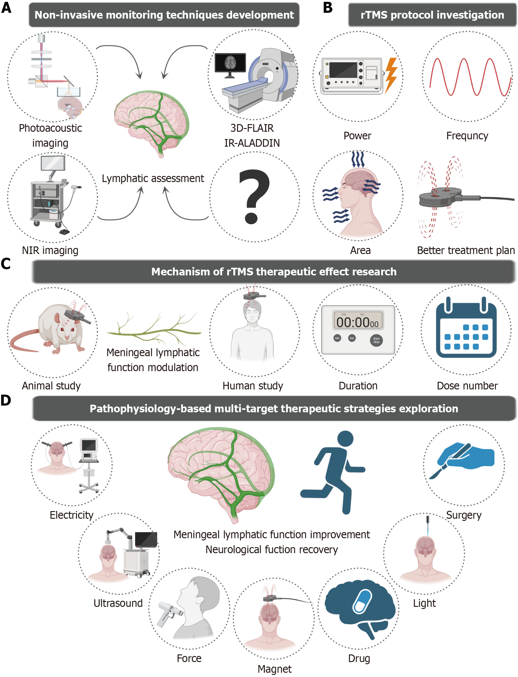
Therefore, future research should focus on investigating the modulatory effects of rTMS on mLVs and elucidating its underlying mechanisms. First, the development of noninvasive imaging techniques that specifically target meningeal lymphatic function is essential. Through multimodal imaging technology - such as dynamic contrast-enhanced MRI, near-infrared fluorescence imaging (NIR), and photoacoustic microscopy - it is feasible to monitor rTMS-induced dynamic changes in human mLV drainage efficiency in real time. Current assessments of human mLV drainage efficiency rely mainly on gadolinium-enhanced MRI sequences (e.g., T1 black-blood imaging and T2 fluid attenuated inversion recovery) for quantification[14,15]. However, owing to the uncertain biosafety of gadolinium contrast media, it is difficult for most researchers to conduct large-scale clinical trials on human mLV function. Therefore, it is necessary to develop noninvasive functional assessment tools for the meningeal lymphatic system. Emerging contrast-free techniques have substantially expanded the functional evaluation capabilities of noninvasive human mLVs. The MRI signals of a specific T2-fluid attenuated inversion recovery sequence of liquids with different protein contents are different. Based on the characteristic difference between the protein content of lymphatic fluid and the surrounding tissue fluid, the lymphatic vessels can be visualized without the injection of contrast agent by using 3D fluid attenuated inversion recovery magnetic resonance methods[13]. Additionally, the use of preceding imaging slices as marker planes along with long reversion times makes the inversion–recovery alternate ascending/descending directional navigation technique highly sensitive to components such as mLV slow-flow components, thereby enabling the visualization of lymphatic vessels[16]. These advanced approaches provide critical technical support for large-scale clinical investigations of meningeal lymphatic dysfunction under pathological conditions and meningeal lymphatic remodeling effects after treatment. However, the specificity of MRI for mLV imaging and sensitivity to mLV damage remain to be further evaluated. Recent studies have demonstrated that NIR and photoacoustic imaging techniques can be used to dynamically visualize mLV drainage in murine animal models. Due to the good tissue penetration of NIR, CSF drainage of intact scalp mice was observed after the NIR-II nanoprobes were injected into the CSF[17]. Using wide-field and deep-penetration three-dimensional photoacoustic tomography with a hemispherical detector array, photoacoustic imaging techniques can be used to observe meningeal vessels[18]. This provides us with an approach that is not limited to the use of MRl methods to assess mLV function. However, these techniques require injection into the CSF, which may not be conducive to the visualization of the human mLV. Future efforts should focus on translating these technologies to human studies and establishing a comprehensive framework for noninvasive, real-time, multimodal monitoring methods. Second, investigators should focus on exploring whether rTMS can improve cognitive dysfunction by modulating the mLV and the differential modulatory effect of meningeal lymphatic drainage by rTMS under varying parameters, different intervention durations, and different treatment frequencies. Although several studies have shown the initial modulatory effects of rTMS on the mLV and the neuroprotective mechanisms of the meningeal lymphatic system, direct and conclusive evidence linking this mechanism to improvements in cognitive function is lacking. Future research should focus on constructing a more refined mechanism model, integrating multidimensional technical approaches from molecular biology and neurophysiology to deeply reveal the possibility of rTMS regulation of mLV function to improve cognitive dysfunction. Future research should also investigate the intrinsic biological processes through which rTMS regulates neuroplasticity and cognitive function. Besides, novel rTMS protocols have proliferated in recent years, such as deep transcranial magnetic stimulation using H-coils, theta-burst stimulation, and low-field synchronized transcranial magnetic stimulation. These protocols have been extensively studied for their therapeutic effects and roles in other neurological disorders[19]. In a study by Fu[6], researchers determined stimulation parameters on the basis of the successful elicitation of motor-evoked potentials. While this approach may serve as a neuroactivity-guided strategy, it is likely inappropriate for modulating meningeal lymphatic drainage. A better protocol for enhancing meningeal lymphatic drainage under AD pathology remains to be established. Additionally, in the Fu[6] study, patients received high-frequency rTMS targeting the bilateral prefrontal regions. Given the anatomical distribution of mLVs, whether targeting of the entire dorsal skull and cranio-cervical junction regions by rTMS may further improve clinical outcomes by improving meningeal lymphatic drainage is worthy of systematic investigation. Finally, the study’s limited sample size, relatively short follow-up period, and simplistic grouping strategy - notably the absence of a healthy control cohort - collectively constrain the conscientiousness and reliability of the clinical evidence provided. Consequently, well-designed randomized controlled trials involving both healthy individuals and patients with MCI are necessary[20]. These trials should include a reasonable follow-up period to evaluate rTMS-mediated enhancement of meningeal lymphatic function and establish translational prospects for the clinical application of rTMS in relieving AD symptoms. Furthermore, as a complex clinical syndrome, the progression and treatment response of MCI are often influenced by multiple factors. Studies have shown that the effectiveness of TMS in alleviating cognitive dysfunction is correlated with age[21,22]. The effectiveness of rTMS in treating MCI may also depend on disease progression, with research demonstrating that rTMS is associated with significantly better therapeutic outcomes in patients with mild AD than in patients with moderate disease[23]. Additionally, patients with higher educational attainment exhibit better cognitive outcomes after rTMS treatment[24]. Traditional views categorize MCI into amnestic and nonamnestic types, yet rTMS may show heterogeneous therapeutic effects across these subtypes[25]. Therefore, future research could systematically investigate the differential effects and response mechanisms of rTMS in different patient subgroups.

CONCLUSION

In summary, the modulation of meningeal lymphatic drainage efficiency may be a novel mechanism through which rTMS improves cognitive ability. However, verifying this hypothesis requires interdisciplinary collaboration, such as neuroscience, neuroimaging, and physiology, and is supported by multimodal assessment technology. We propose that the following investigations be performed preferentially: (1) The development of noninvasive monitoring techniques targeting human mLV function; (2) Investigations of appropriate rTMS protocols based on remodeling mLV histology and functionality; (3) Additional animal and human evidence that rTMS regulates mLV function to improve neurological function; and (4) Explorations of multitarget therapeutic strategies on the basis of the pathophysiology of disease (Figure 1). As society faces the increasingly serious challenges of population aging, the development of noninvasive treatment approaches for neurodegenerative disorders and the exploration of their underlying mechanisms will significantly improve patients’ quality of life and reduce the burden on society.

Figure 1
Figure 1 A brief illustration of potential research directions in the future. A: Non-invasive monitoring techniques development; B: rTMS protocol investigation; C: Mechanism of rTMS therapeutic effect research; D: Pathophysiology-based multi-target therapeutic strategies exploration. rTMS: Repetitive transcranial magnetic stimulation; NIR: Fluid attenuated inversion recovery; IR-ALADDIN: Inversion–recovery alternate ascending/descending directional navigation.
Footnotes

Provenance and peer review: Unsolicited article; Externally peer reviewed.

Peer-review model: Single blind

Specialty type: Psychiatry

Country of origin: China

Peer-review report’s classification

Scientific Quality: Grade B, Grade C, Grade C, Grade C

Novelty: Grade B, Grade C, Grade C, Grade C

Creativity or Innovation: Grade B, Grade B, Grade C, Grade C

Scientific Significance: Grade B, Grade C, Grade C, Grade C

P-Reviewer: Han JM, MD, PhD, Assistant Professor, China; Zhang XB, PhD, Professor, China S-Editor: Bai Y L-Editor: A P-Editor: Lei YY

References
1.  Zhang J, Zhang Y, Wang J, Xia Y, Zhang J, Chen L. Recent advances in Alzheimer's disease: Mechanisms, clinical trials and new drug development strategies. Signal Transduct Target Ther. 2024;9:211.  [RCA]  [PubMed]  [DOI]  [Full Text]  [Full Text (PDF)]  [Cited by in RCA: 283]  [Reference Citation Analysis (0)]
2.  Ni Z, Chen R. Transcranial magnetic stimulation to understand pathophysiology and as potential treatment for neurodegenerative diseases. Transl Neurodegener. 2015;4:22.  [RCA]  [PubMed]  [DOI]  [Full Text]  [Full Text (PDF)]  [Cited by in Crossref: 43]  [Cited by in RCA: 72]  [Article Influence: 7.2]  [Reference Citation Analysis (0)]
3.  Jayathilake NJ, Phan TT, Kim J, Lee KP, Park JM. Modulating neuroplasticity for chronic pain relief: noninvasive neuromodulation as a promising approach. Exp Mol Med. 2025;57:501-514.  [RCA]  [PubMed]  [DOI]  [Full Text]  [Full Text (PDF)]  [Cited by in RCA: 13]  [Reference Citation Analysis (0)]
4.  Kong S, Gootee E, Williams N, Gottesman RF, Johansen MC. Congestive heart failure and its associations with cognition and cerebral blood flow. Brain Circ. 2025;11:30-38.  [RCA]  [PubMed]  [DOI]  [Full Text]  [Full Text (PDF)]  [Cited by in RCA: 1]  [Reference Citation Analysis (0)]
5.  Fink HA, Jutkowitz E, McCarten JR, Hemmy LS, Butler M, Davila H, Ratner E, Calvert C, Barclay TR, Brasure M, Nelson VA, Kane RL. Pharmacologic Interventions to Prevent Cognitive Decline, Mild Cognitive Impairment, and Clinical Alzheimer-Type Dementia: A Systematic Review. Ann Intern Med. 2018;168:39-51.  [RCA]  [PubMed]  [DOI]  [Full Text]  [Cited by in Crossref: 103]  [Cited by in RCA: 123]  [Article Influence: 17.6]  [Reference Citation Analysis (0)]
6.  Fu HX. Effects of repetitive transcranial magnetic stimulation on electroencephalogram and memory function in patients with mild cognitive impairment. World J Psychiatry. 2025;15:106761.  [RCA]  [PubMed]  [DOI]  [Full Text]  [Full Text (PDF)]  [Cited by in RCA: 1]  [Reference Citation Analysis (1)]
7.  Zhang Q, Niu Y, Li Y, Xia C, Chen Z, Chen Y, Feng H. Meningeal lymphatic drainage: novel insights into central nervous system disease. Signal Transduct Target Ther. 2025;10:142.  [RCA]  [PubMed]  [DOI]  [Full Text]  [Cited by in RCA: 4]  [Reference Citation Analysis (0)]
8.  Kamagata K, Andica C, Takabayashi K, Saito Y, Taoka T, Nozaki H, Kikuta J, Fujita S, Hagiwara A, Kamiya K, Wada A, Akashi T, Sano K, Nishizawa M, Hori M, Naganawa S, Aoki S; Alzheimer's Disease Neuroimaging Initiative. Association of MRI Indices of Glymphatic System With Amyloid Deposition and Cognition in Mild Cognitive Impairment and Alzheimer Disease. Neurology. 2022;99:e2648-e2660.  [RCA]  [PubMed]  [DOI]  [Full Text]  [Full Text (PDF)]  [Cited by in Crossref: 95]  [Cited by in RCA: 175]  [Article Influence: 58.3]  [Reference Citation Analysis (0)]
9.  Liu Y, Liu X, Sun P, Li J, Nie M, Gong J, He A, Zhao M, Yang C, Wang Z. rTMS treatment for abrogating intracerebral hemorrhage-induced brain parenchymal metabolite clearance dysfunction in male mice by regulating intracranial lymphatic drainage. Brain Behav. 2023;13:e3062.  [RCA]  [PubMed]  [DOI]  [Full Text]  [Cited by in RCA: 16]  [Reference Citation Analysis (0)]
10.  Lin Y, Jin J, Lv R, Luo Y, Dai W, Li W, Tang Y, Wang Y, Ye X, Lin WJ. Repetitive transcranial magnetic stimulation increases the brain's drainage efficiency in a mouse model of Alzheimer's disease. Acta Neuropathol Commun. 2021;9:102.  [RCA]  [PubMed]  [DOI]  [Full Text]  [Full Text (PDF)]  [Cited by in Crossref: 36]  [Cited by in RCA: 85]  [Article Influence: 21.3]  [Reference Citation Analysis (0)]
11.  Zhang C, Zheng Y, Jiang G, Luo J, Su L, Ai Y, Feng C, Li C, Li S, Hu X. Enhancement of glymphatic function and cognition in chronic insomnia using low-frequency rTMS. Sleep. 2025;48:zsaf083.  [RCA]  [PubMed]  [DOI]  [Full Text]  [Cited by in RCA: 2]  [Reference Citation Analysis (0)]
12.  Gao D, Zhang Z, Feng P, Zhou L, Geng Z, Li C, Zhu Y, Yang H. The change of MRI indexes of brain glymphatic function and sleep status before and after repeated transcranial magnetic stimulation in insomnia disorder patients. Front Neurosci. 2025;19:1545885.  [RCA]  [PubMed]  [DOI]  [Full Text]  [Full Text (PDF)]  [Cited by in RCA: 1]  [Reference Citation Analysis (0)]
13.  Albayram MS, Smith G, Tufan F, Tuna IS, Bostancıklıoğlu M, Zile M, Albayram O. Non-invasive MR imaging of human brain lymphatic networks with connections to cervical lymph nodes. Nat Commun. 2022;13:203.  [RCA]  [PubMed]  [DOI]  [Full Text]  [Full Text (PDF)]  [Cited by in Crossref: 138]  [Cited by in RCA: 127]  [Article Influence: 42.3]  [Reference Citation Analysis (0)]
14.  Ding XB, Wang XX, Xia DH, Liu H, Tian HY, Fu Y, Chen YK, Qin C, Wang JQ, Xiang Z, Zhang ZX, Cao QC, Wang W, Li JY, Wu E, Tang BS, Ma MM, Teng JF, Wang XJ. Impaired meningeal lymphatic drainage in patients with idiopathic Parkinson's disease. Nat Med. 2021;27:411-418.  [RCA]  [PubMed]  [DOI]  [Full Text]  [Cited by in Crossref: 79]  [Cited by in RCA: 236]  [Article Influence: 59.0]  [Reference Citation Analysis (0)]
15.  Sennfält S, Thrippleton MJ, Stringer M, Reyes CA, Chappell F, Doubal F, Garcia DJ, Zhang J, Cheng Y, Wardlaw J. Visualising and semi-quantitatively measuring brain fluid pathways, including meningeal lymphatics, in humans using widely available MRI techniques. J Cereb Blood Flow Metab. 2023;43:1779-1795.  [RCA]  [PubMed]  [DOI]  [Full Text]  [Full Text (PDF)]  [Cited by in Crossref: 2]  [Cited by in RCA: 16]  [Article Influence: 8.0]  [Reference Citation Analysis (0)]
16.  Yoo RE, Kim JH, Moon HY, Park JY, Cheon S, Shin HS, Han D, Kim Y, Park SH, Choi SH. Long-term physical exercise facilitates putative glymphatic and meningeal lymphatic vessel flow in humans. Nat Commun. 2025;16:3360.  [RCA]  [PubMed]  [DOI]  [Full Text]  [Full Text (PDF)]  [Cited by in RCA: 6]  [Reference Citation Analysis (0)]
17.  Sun B, Fang D, Li W, Li M, Zhu S. NIR-II nanoprobes for investigating the glymphatic system function under anesthesia and stroke injury. J Nanobiotechnology. 2024;22:200.  [RCA]  [PubMed]  [DOI]  [Full Text]  [Cited by in RCA: 10]  [Reference Citation Analysis (0)]
18.  Yang F, Wang Z, Shi W, Wang M, Ma R, Zhang W, Li X, Wang E, Xie W, Zhang Z, Shen Q, Zhou F, Yang S. Advancing insights into in vivo meningeal lymphatic vessels with stereoscopic wide-field photoacoustic microscopy. Light Sci Appl. 2024;13:96.  [RCA]  [PubMed]  [DOI]  [Full Text]  [Cited by in RCA: 8]  [Reference Citation Analysis (0)]
19.  Brunoni AR, Chaimani A, Moffa AH, Razza LB, Gattaz WF, Daskalakis ZJ, Carvalho AF. Repetitive Transcranial Magnetic Stimulation for the Acute Treatment of Major Depressive Episodes: A Systematic Review With Network Meta-analysis. JAMA Psychiatry. 2017;74:143-152.  [RCA]  [PubMed]  [DOI]  [Full Text]  [Cited by in Crossref: 411]  [Cited by in RCA: 361]  [Article Influence: 45.1]  [Reference Citation Analysis (0)]
20.  Neacsiu AD, Beynel L, Powers JP, Szabo ST, Appelbaum LG, Lisanby SH, LaBar KS. Enhancing Cognitive Restructuring with Concurrent Repetitive Transcranial Magnetic Stimulation: A Transdiagnostic Randomized Controlled Trial. Psychother Psychosom. 2022;91:94-106.  [RCA]  [PubMed]  [DOI]  [Full Text]  [Cited by in Crossref: 4]  [Cited by in RCA: 12]  [Article Influence: 4.0]  [Reference Citation Analysis (0)]
21.  Teselink J, Bawa KK, Koo GK, Sankhe K, Liu CS, Rapoport M, Oh P, Marzolini S, Gallagher D, Swardfager W, Herrmann N, Lanctôt KL. Efficacy of non-invasive brain stimulation on global cognition and neuropsychiatric symptoms in Alzheimer's disease and mild cognitive impairment: A meta-analysis and systematic review. Ageing Res Rev. 2021;72:101499.  [RCA]  [PubMed]  [DOI]  [Full Text]  [Cited by in Crossref: 7]  [Cited by in RCA: 69]  [Article Influence: 17.3]  [Reference Citation Analysis (0)]
22.  Begemann MJ, Brand BA, Ćurčić-Blake B, Aleman A, Sommer IE. Efficacy of non-invasive brain stimulation on cognitive functioning in brain disorders: a meta-analysis. Psychol Med. 2020;50:2465-2486.  [RCA]  [PubMed]  [DOI]  [Full Text]  [Full Text (PDF)]  [Cited by in Crossref: 162]  [Cited by in RCA: 219]  [Article Influence: 43.8]  [Reference Citation Analysis (0)]
23.  Romanella SM, Roe D, Paciorek R, Cappon D, Ruffini G, Menardi A, Rossi A, Rossi S, Santarnecchi E. Sleep, Noninvasive Brain Stimulation, and the Aging Brain: Challenges and Opportunities. Ageing Res Rev. 2020;61:101067.  [RCA]  [PubMed]  [DOI]  [Full Text]  [Cited by in Crossref: 25]  [Cited by in RCA: 30]  [Article Influence: 6.0]  [Reference Citation Analysis (0)]
24.  Wang X, Mao Z, Ling Z, Yu X. Repetitive transcranial magnetic stimulation for cognitive impairment in Alzheimer's disease: a meta-analysis of randomized controlled trials. J Neurol. 2020;267:791-801.  [RCA]  [PubMed]  [DOI]  [Full Text]  [Cited by in Crossref: 24]  [Cited by in RCA: 52]  [Article Influence: 8.7]  [Reference Citation Analysis (0)]
25.  Nardone R, Bergmann J, Christova M, Caleri F, Tezzon F, Ladurner G, Trinka E, Golaszewski S. Short latency afferent inhibition differs among the subtypes of mild cognitive impairment. J Neural Transm (Vienna). 2012;119:463-471.  [RCA]  [PubMed]  [DOI]  [Full Text]  [Cited by in Crossref: 52]  [Cited by in RCA: 68]  [Article Influence: 4.9]  [Reference Citation Analysis (0)]