Published online Nov 18, 2025. doi: 10.5312/wjo.v16.i11.110716
Revised: July 28, 2025
Accepted: September 16, 2025
Published online: November 18, 2025
Processing time: 154 Days and 21 Hours
Osteochondroma, a benign bone tumour, is commonly seen in the metaphyseal region of long bones. It is not so common in short bones and occurs rarely in carpals. The literature involving an osteochondroma of pisiform is scarce.
We present a 12-year-old male Asian child with a gradually progressive, bony, hard swelling on the volar aspect of the right wrist, which, on investigation, was suggestive of a solitary osteochondroma of the pisiform and its management.
The current report describes a child with a wrist swelling which on evaluation was found to be an osteochondroma of the pisiform, which is seldom described in the literature, and managed by excision biopsy. Though osteochondromas of the carpal bones are rare they should be included in the differential of any wrist swelling with the symptom complex varying from pressure effects to surrounding soft tissues and nerves, attritional tendon ruptures, carpal instabilities and arthritis.
Core Tip: Carpal osteochondromas occur rarely, and their symptom complex varies from the osteochondroma occurring elsewhere. They can cause cosmetic concerns, attritional tendon ruptures, pressure effects on nerves, carpal instabilities, and wrist arthritis. They should be included in the differential of any wrist swelling. The treatment of choice for carpal os
- Citation: Vanapalli R, Jayant UK, Mlv SK, Rawat SS, Ansari MT. Osteochondroma of the pisiform: A case report. World J Orthop 2025; 16(11): 110716
- URL: https://www.wjgnet.com/2218-5836/full/v16/i11/110716.htm
- DOI: https://dx.doi.org/10.5312/wjo.v16.i11.110716
Osteochondroma is the most common benign bone tumour. The aetiology is attributed to the defective perichondrial ring of Lacroix, with other possible etiological causes being reactive metaplasia (due to microtrauma, chronic infection, and chronic irritation) and even a possible neoplastic origin [translocation (X;6)(q22;q13-14)] have been described. They can present either in isolation or as a part of multiple hereditary exostosis. The symptoms are mostly due to pressure effects on the surrounding soft tissues. They are cartilage capped, have continuity with the parent bone, and can be pedun
A 12-year-old male Asian child presented to our hand unit with swelling on the volar aspect of his right dominant hand.
The swelling gradually increased in size over the past 18 months, according to the parents, with a recent history of wrist pain on palmar flexion.
There was no significant past history.
There was no significant personal and family history.
On examination, there was a marked restriction in palmar flexion of the wrist. There was a palpable tender swelling on the volar and ulnar aspect of the wrist with a bony, hard consistency. The margins appeared well-contoured, and the overlying skin was not infiltrated. The swelling was found to be immobile and fixed to the underlying bone. On thorough systemic examination, there were no similar lesions elsewhere.
The complete blood picture and erythrocyte sedimentation rate were within normal limits.
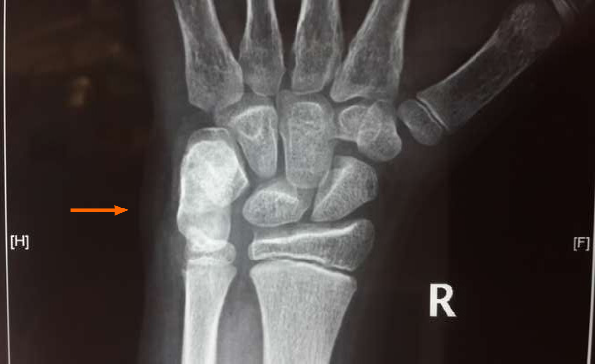
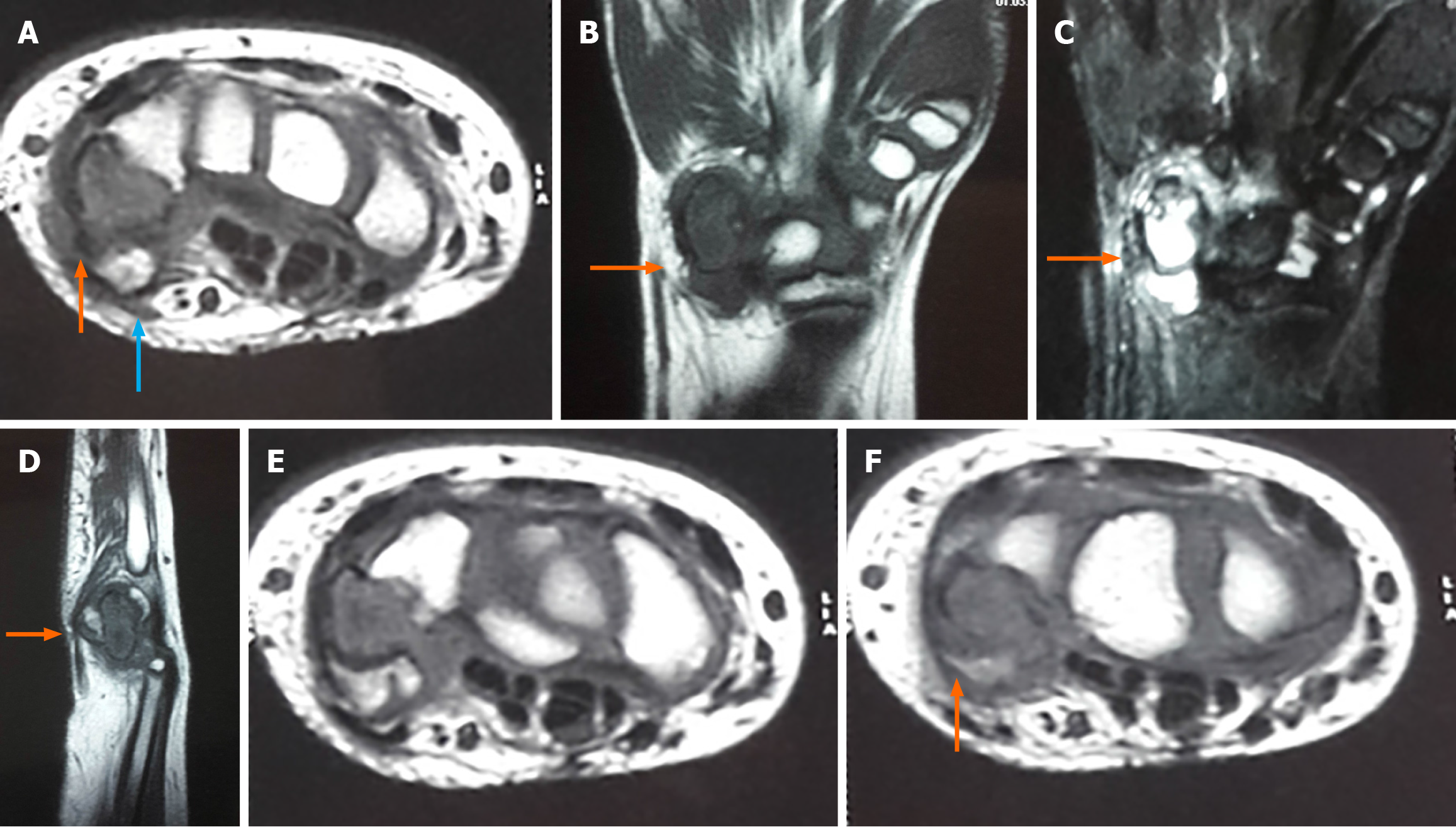
Radiographs revealed an elongated ossified mass arising from the volar aspect of the pisiform bone (Figure 1). Computed tomography scan demonstrated a well-circumscribed lesion and a small bump ventrally suggestive of pisiform (pisiform with dorsal osteochondroma) with no stalk and no periosteal reaction (Figure 2). Magnetic resonance imaging showed T1 hypointense lesion with ventral isointense bone suggestive of native pisiform (Figure 3A marked orange) with dorsal lesion appearing hypointense (Figure 3A marked white) due to cartilage cap and T2 STIR pictures were suggestive of oedema of the pisiform and triquetrum without adjacent soft tissue infiltration (Figure 3).
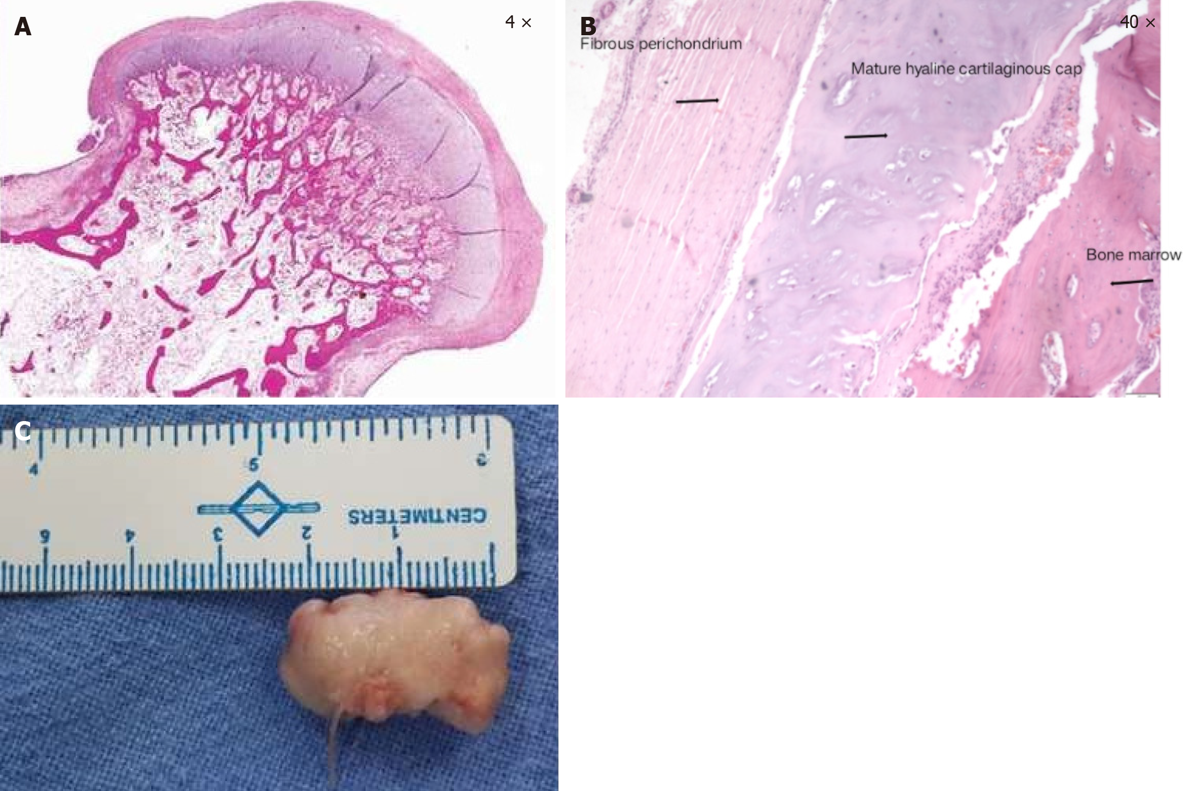
The findings were suggestive of an osteochondroma of the pisiform radiologically and clinically and confirmed by the histopathological examination (Figure 4A and B) which showed hyaline cartilage cap surrounding the bone marrow elements.
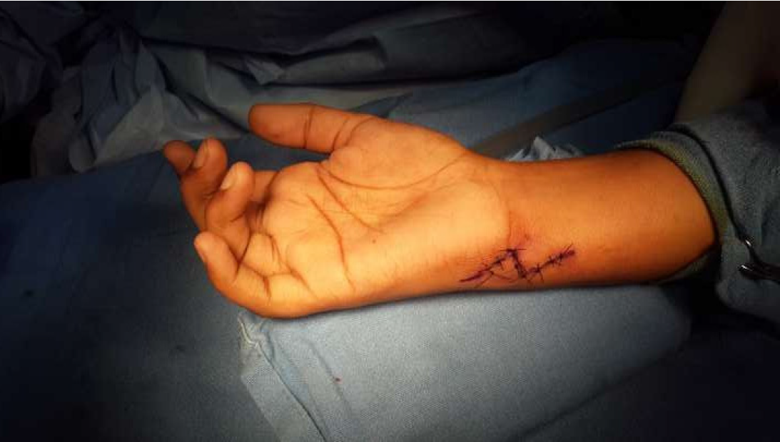
We planned for an excisional biopsy of pisiform in our hand unit. A 5 cm Bruner incision was made and centered over the flexor carpi ulnaris (FCU) (Figure 5). After superficial dissection, the FCU tendon was identified and retracted radially (Figure 6). The ulnar nerve and artery were secured, and the ulnar nerve with both branches was followed to the Guyon’s canal. Pisiform was excised with sharp subperiosteal dissection to preserve FCU insertion and sent for pathological examination (Figure 4). The pisiform ligaments were restored by suturing the FCU tendon sheath with piso-metacarpal and piso-hamate ligaments using nonabsorbable sutures. A below-elbow slab was applied.
The patient was reviewed 10 days after surgery, at which point the sutures were removed, and a below-elbow cast was applied. Plaster was discontinued after 6 weeks, and wrist physiotherapy was started. The child was relieved of wrist pain and had no distal neurological deficit. A full range of motion of the wrist was achieved in 3 months. The child had no complaints or recurrence at one year follow up.
Osteochondromas are benign bone tumours that most commonly arise from long bones. There are a handful of case reports documenting the osteochondromas of the carpal bones[2]. Scaphoid osteochondromas are more frequent when compared to other carpals[3-6]. Osteochondromas have also been documented from lunate[6], trapezium[8], capitate[9] and hamate[10]. We could find only one case report of osteochondroma from pisiform[11].
Osteochondromas elsewhere mostly cause cosmetic concerns and, in some subset of patients, complaints due to pressure effects. In carpal bones, they can cause arthritis, restriction of the range of movements, instabilities, pressure effects and cosmetic concerns. Rimmer et al[3] have described a scaphoid osteochondroma with radio carpal arthritis due to impingement with the radial styloid process. Koshi et al[8] has described a rare case report of solitary osteochondroma of a trapezium with trapezio-metacarpal arthrosis, which was managed successfully with marginal excision. In our patient, there were no features of arthritis. The child’s main complaint was swelling with pain in the terminal range of movement.
The pressure effects can cause neurological symptoms, attritional rupture of tendons, etc. Shah et al[9] have reported a case of capitate osteochondroma arising from the dorsal aspect, causing attritional rupture of the extensor digit minimi tendon, which was treated by excision and repair of the tendon. Such attritional ruptures of the tendons have also been described with osteochondromas of scaphoid and lunate[4,7]. O'Dwyer and Jefferiss[4] reported a rare case report of scaphoid exostosis causing attrition and rupture of the flexor pollicis longus tendon. Katayama et al[7] described a rare case report of lunate exostosis causing attritional rupture of extensor tendons, including extensor indicis proprium and slip of extensor digitorum communis of 2nd finger.
A few reports of scaphoid osteochondromas demonstrated pressure effects on median nerves causing features of carpal tunnel syndrome, and in a few reports, there was documentation of the compression of the superficial branch of radial nerve as well, which was treated by excision biopsy that relieved the symptoms[6]. In some case reports, there were features of scapholunate dissociation leading to dorsal intercalated segmental instability, which was treated by excision biopsy[8]. In our child there were no features of instability, neruological complaints or tendon attrition.
The gold standard treatment of choice for osteochondroma, irrespective of the presentation, is excision biopsy. Ventura-Parellada et al[11] in their patient with pisiform osteochondroma have excised the entire pisiform. The literature regarding pisiform osteochondroma is scarce. In our patient there was no stalk connecting the osteochondroma to the parent bone and it being a sessile osteochondroma, we have excised the entire bone with lesion and sent for histopathological examination. In a few patients, along with excision, it required repair of tendons or ligaments. In our patient, as pisiform is a sesamoid bone after excision, we restored the ligaments by suturing the ligaments of pisiform to the FCU tendon sheath and providing plaster immobilisation for six weeks.
The current report describes a child with wrist swelling which on evaluation was found to be an osteochondroma of the pisiform, which is seldom described in the literature, and managed by excision biopsy. Though osteochondromas of the carpal bones are rare they should be included in the differential of any wrist swelling with the symptom complex varying from pressure effects to surrounding soft tissues and nerves, attritional tendon ruptures, carpal instabilities and arthritis.
| 1. | Fabbri N, De Paolis M, Bertoni F. Benign cartilage tumours. Curr Orthop. 2001;15:7-30. [DOI] [Full Text] |
| 2. | Sze-Yan C, Fu-Keung I, Tak-Cheun W, Siu-Ho W. Exostosis in the Hand: Case Series and Literature Review. J Orthop Trauma Rehabil. 2012;16:66-69. [RCA] [DOI] [Full Text] [Cited by in Crossref: 1] [Cited by in RCA: 3] [Article Influence: 0.2] [Reference Citation Analysis (0)] |
| 3. | Rimmer SG, Bhoora IG, Hooper G. Scaphoid exostosis and radio-carpal osteoarthrosis. J Hand Surg Br. 1995;20:741-744. [RCA] [PubMed] [DOI] [Full Text] [Cited by in Crossref: 8] [Cited by in RCA: 11] [Article Influence: 0.4] [Reference Citation Analysis (0)] |
| 4. | O'Dwyer KJ, Jefferiss CD. Scaphoid exostosis causing rupture of the flexor pollicis longus. Acta Orthop Belg. 1994;60:124-126. [PubMed] |
| 5. | Stahl S, Rayek S. Scaphoid osteochondroma with scapholunate dissociation--a case report. Hand Surg. 2000;5:73-75. [RCA] [PubMed] [DOI] [Full Text] [Cited by in Crossref: 6] [Cited by in RCA: 7] [Article Influence: 0.3] [Reference Citation Analysis (0)] |
| 6. | Wong A, Watson S, Bakula A, Ashmead D. Carpal tunnel syndrome caused by a large osteochondroma. Hand (N Y). 2012;7:438-441. [RCA] [PubMed] [DOI] [Full Text] [Cited by in Crossref: 2] [Cited by in RCA: 5] [Article Influence: 0.4] [Reference Citation Analysis (0)] |
| 7. | Katayama T, Ono H, Furuta K. Osteochondroma of the lunate with extensor tendons rupture of the index finger: a case report. Hand Surg. 2011;16:181-184. [RCA] [PubMed] [DOI] [Full Text] [Cited by in Crossref: 5] [Cited by in RCA: 8] [Article Influence: 0.5] [Reference Citation Analysis (0)] |
| 8. | Koshi H, Shinozaki T, Hosokawa T, Yanagawa T, Takagishi K. Solitary osteochondroma of the trapezium: case report. J Hand Surg Am. 2011;36:428-431. [RCA] [PubMed] [DOI] [Full Text] [Cited by in Crossref: 8] [Cited by in RCA: 10] [Article Influence: 0.7] [Reference Citation Analysis (0)] |
| 9. | Shah NR, Wilczynski M, Gelberman R. Osteochondroma of the capitate causing rupture of the extensor digiti minimi: case report. J Hand Surg Am. 2009;34:46-48. [RCA] [PubMed] [DOI] [Full Text] [Cited by in Crossref: 12] [Cited by in RCA: 14] [Article Influence: 0.8] [Reference Citation Analysis (0)] |
| 10. | Cha SM, Shin HD, Kim DY. A solitary unilobed osteochondroma of the hamate: a case report. J Pediatr Orthop B. 2017;26:274-276. [RCA] [PubMed] [DOI] [Full Text] [Cited by in Crossref: 4] [Cited by in RCA: 6] [Article Influence: 0.7] [Reference Citation Analysis (0)] |
| 11. | Ventura-Parellada C, Subirà-I-Álvarez T, Martínez-Ruiz A. Solitary osteochondroma in the pisiform bone with pisotriquetal osteoarthritis. A case study. Rev Esp Cir Ortop Traumatol (Engl Ed). 2021;65:9-12. [RCA] [PubMed] [DOI] [Full Text] [Cited by in RCA: 1] [Reference Citation Analysis (0)] |
Open Access: This article is an open-access article that was selected by an in-house editor and fully peer-reviewed by external reviewers. It is distributed in accordance with the Creative Commons Attribution NonCommercial (CC BY-NC 4.0) license, which permits others to distribute, remix, adapt, build upon this work non-commercially, and license their derivative works on different terms, provided the original work is properly cited and the use is non-commercial. See: https://creativecommons.org/Licenses/by-nc/4.0/


