©The Author(s) 2025.
World J Clin Oncol. Sep 24, 2025; 16(9): 108585
Published online Sep 24, 2025. doi: 10.5306/wjco.v16.i9.108585
Published online Sep 24, 2025. doi: 10.5306/wjco.v16.i9.108585
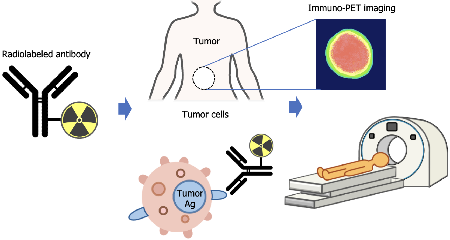
Figure 1 Concept of immuno-positron emission tomography using radiolabeled antibody for tumor detection and imaging.
Immuno-PET: Immuno-positron emission tomography.
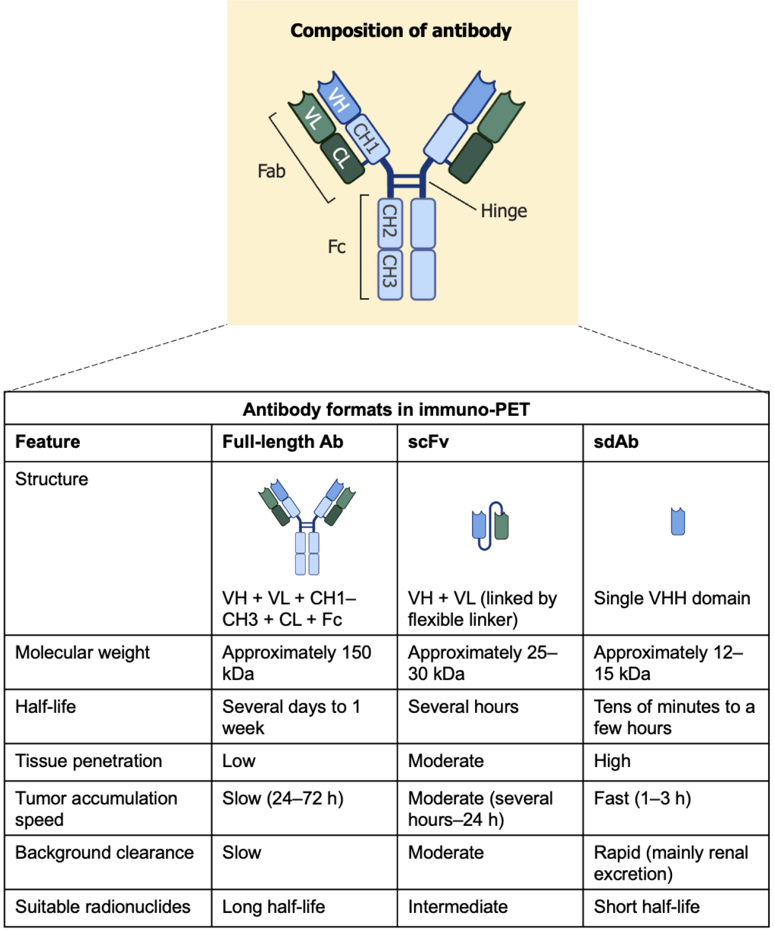
Figure 2 Comparison of antibody formats in immuno-positron emission tomography.
Figure 2 was in part created in BioRender. Luanpitpong, S. (2025) https://BioRender.com/ymggrq9. Ab: Antibody; scFv: Single-chain variable fragment; sdAb: Single-domain antibody; VH: Variable region of the heavy chain; VL: Variable region of the light chain; CH: Constant region of the heavy chain; CL: Constant region of the light chain; VHH: Variable domain of the heavy chain of the heavy-chain-only antibody; Immuno-PET: Immuno-positron emission tomography.
- Citation: Goto H, Takano M, Shiraishi Y, Luanpitpong S. Immuno-positron emission tomography as a new frontier in imaging hematologic malignancies. World J Clin Oncol 2025; 16(9): 108585
- URL: https://www.wjgnet.com/2218-4333/full/v16/i9/108585.htm
- DOI: https://dx.doi.org/10.5306/wjco.v16.i9.108585


