Copyright
©The Author(s) 2025.
World J Clin Oncol. Mar 24, 2025; 16(3): 101240
Published online Mar 24, 2025. doi: 10.5306/wjco.v16.i3.101240
Published online Mar 24, 2025. doi: 10.5306/wjco.v16.i3.101240
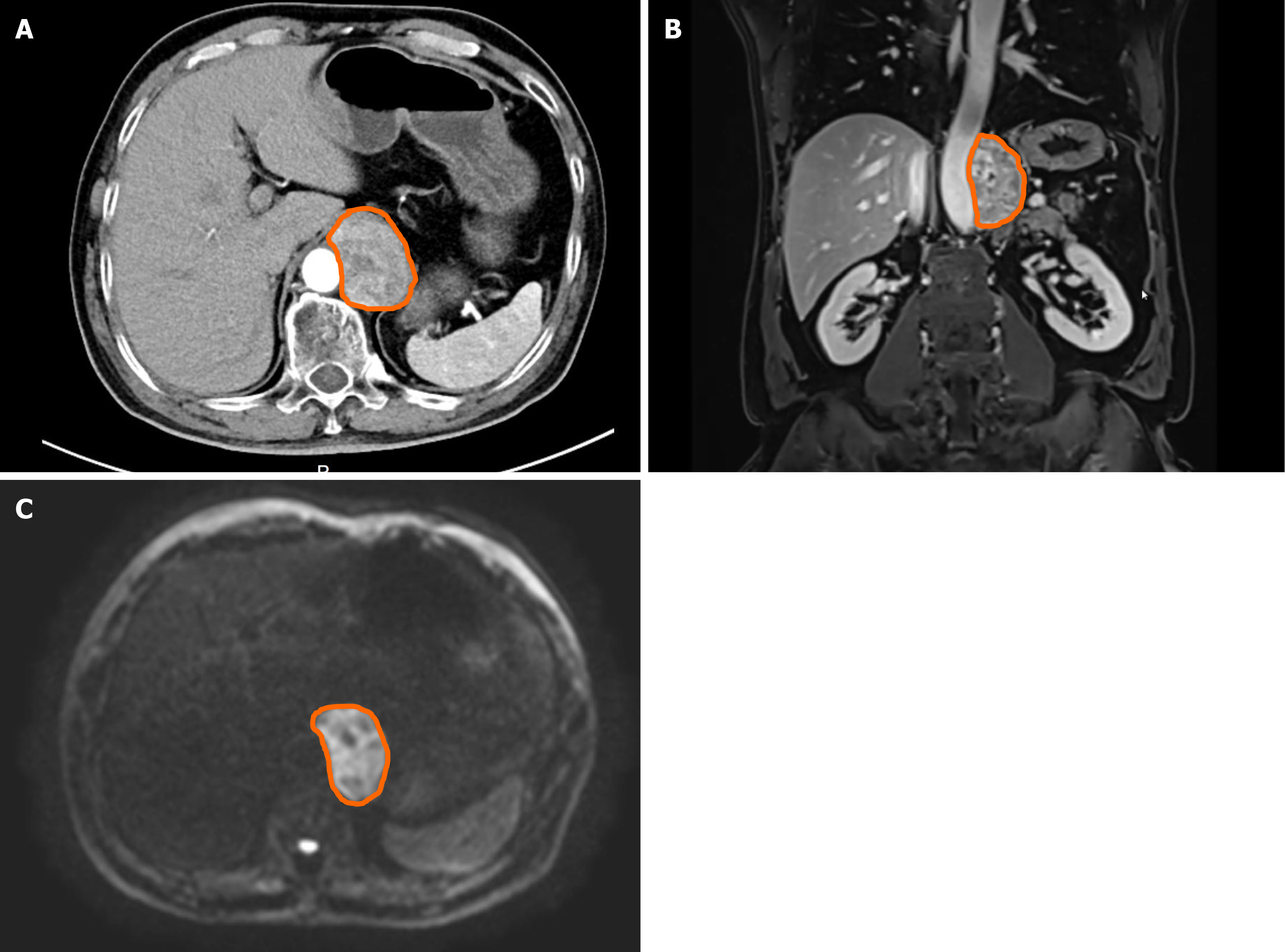
Figure 1 Imaging of the retroperitoneal mass.
A and B: Contrast-enhanced computed tomography scan of the abdomen showing a well-defined, heterogeneous retroperitoneal mass; C: Diffusion-weighted imaging reveals restricted diffusion in the area.
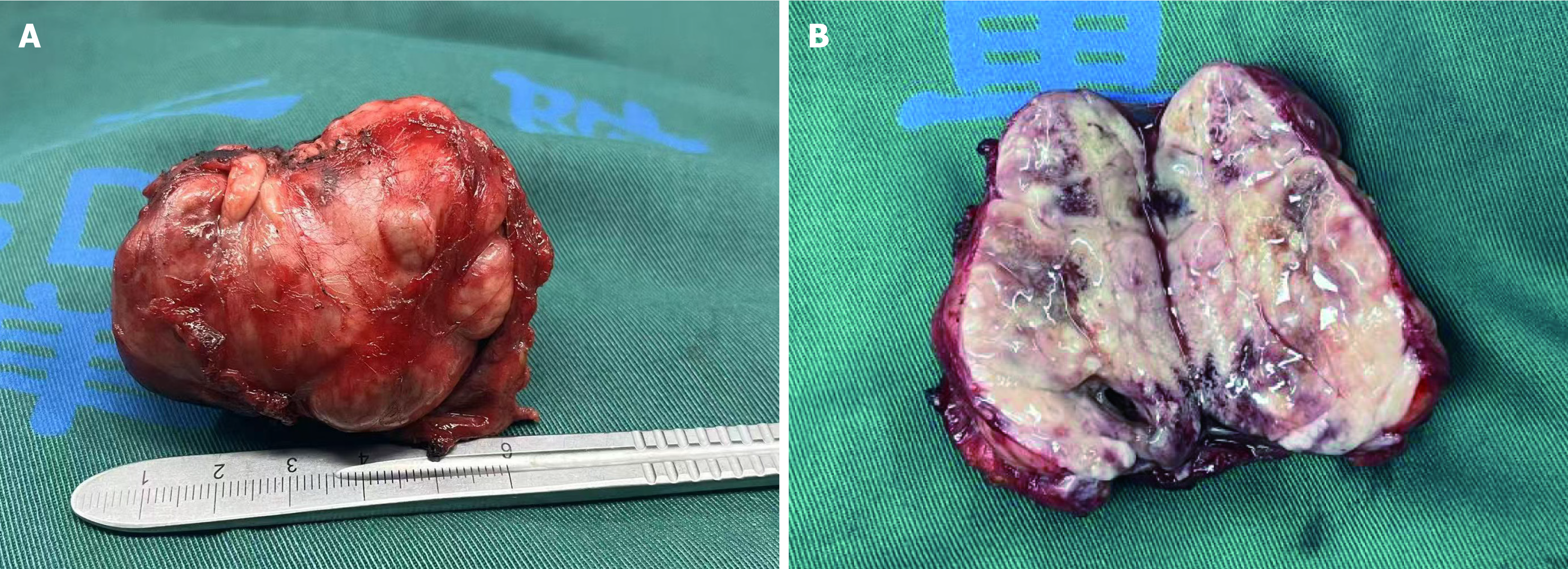
Figure 2 Intraoperative specimen photographs.
A: The resected specimen measured approximately 6 cm in diameter; B: Internal inspection of the tumor.
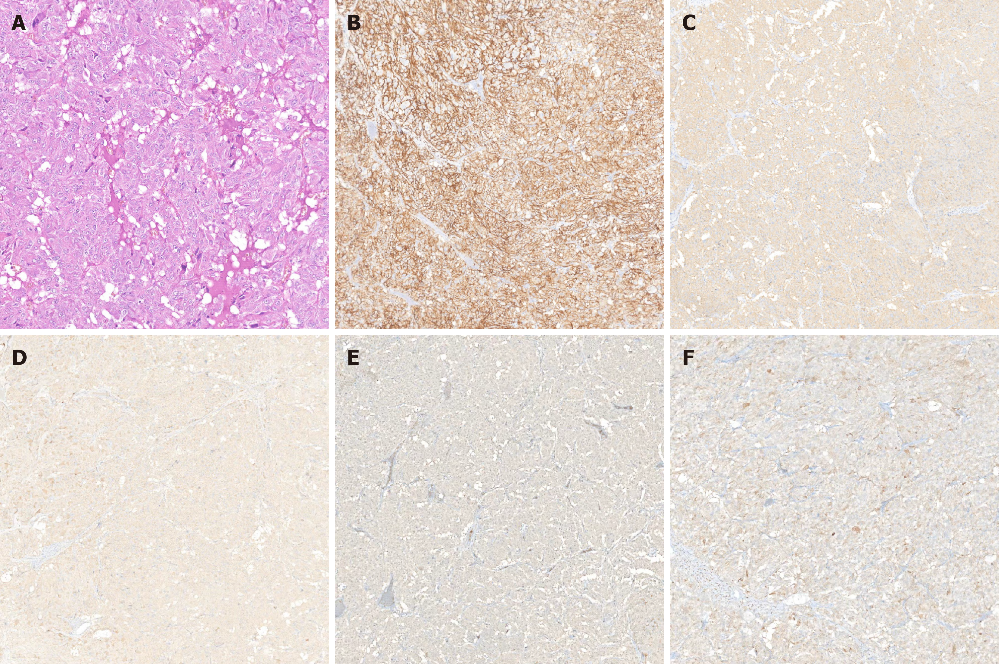
Figure 3 Histopathological and immunohistochemical examination of the resected tumor.
A: Hematoxylin and eosin staining (40 ×); B: CD56 (40 ×); C: Synaptophysin (40 ×); D: Chromogranin A (40 ×); E: S100 (40 ×); F: Neuron-specific enolase (40 ×).
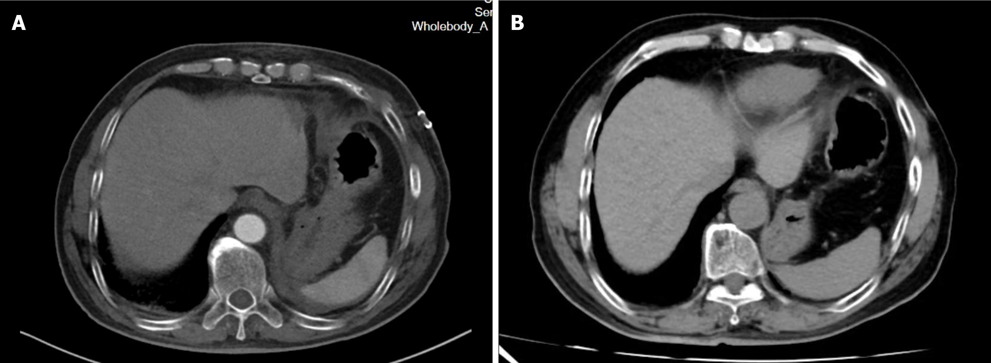
Figure 4 Postoperative computed tomography scan of the abdomen.
A: Computed tomography (CT) images of the abdomen 2 weeks after surgery indicated that the tumor was completely resected, with no significant residual tissue observed; B: CT images of the abdomen 6 months after surgery indicated a few scar strips in the surgical area.
- Citation: Feng YF, Pan YF, Zhou HL, Hu ZH, Wang JJ, Chen B. Surgical resection of a recurrent retroperitoneal paraganglioma: A case report. World J Clin Oncol 2025; 16(3): 101240
- URL: https://www.wjgnet.com/2218-4333/full/v16/i3/101240.htm
- DOI: https://dx.doi.org/10.5306/wjco.v16.i3.101240


