©The Author(s) 2024.
World J Clin Oncol. Sep 24, 2024; 15(9): 1245-1250
Published online Sep 24, 2024. doi: 10.5306/wjco.v15.i9.1245
Published online Sep 24, 2024. doi: 10.5306/wjco.v15.i9.1245
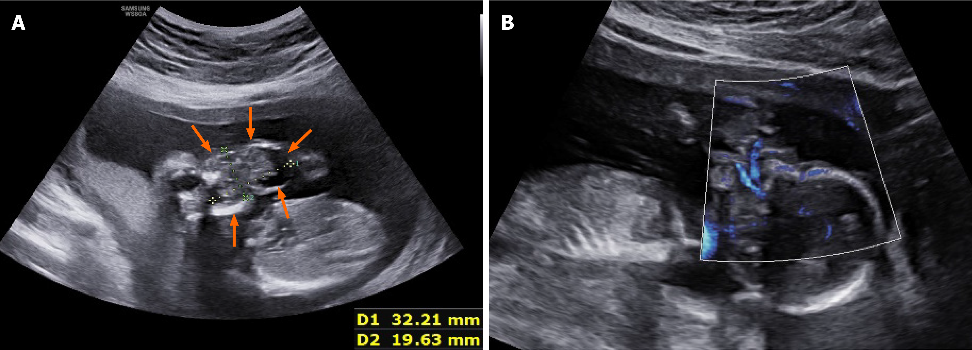
Figure 1 Prenatal ultrasound of a teratoma at 16 weeks of gestation (case 1).
A: Cystic-solid echo of the fetal orofacial exophytic process (orange arrows); B: Microvascular flow imaging showed that blood flow originates from the deep oral cavity.
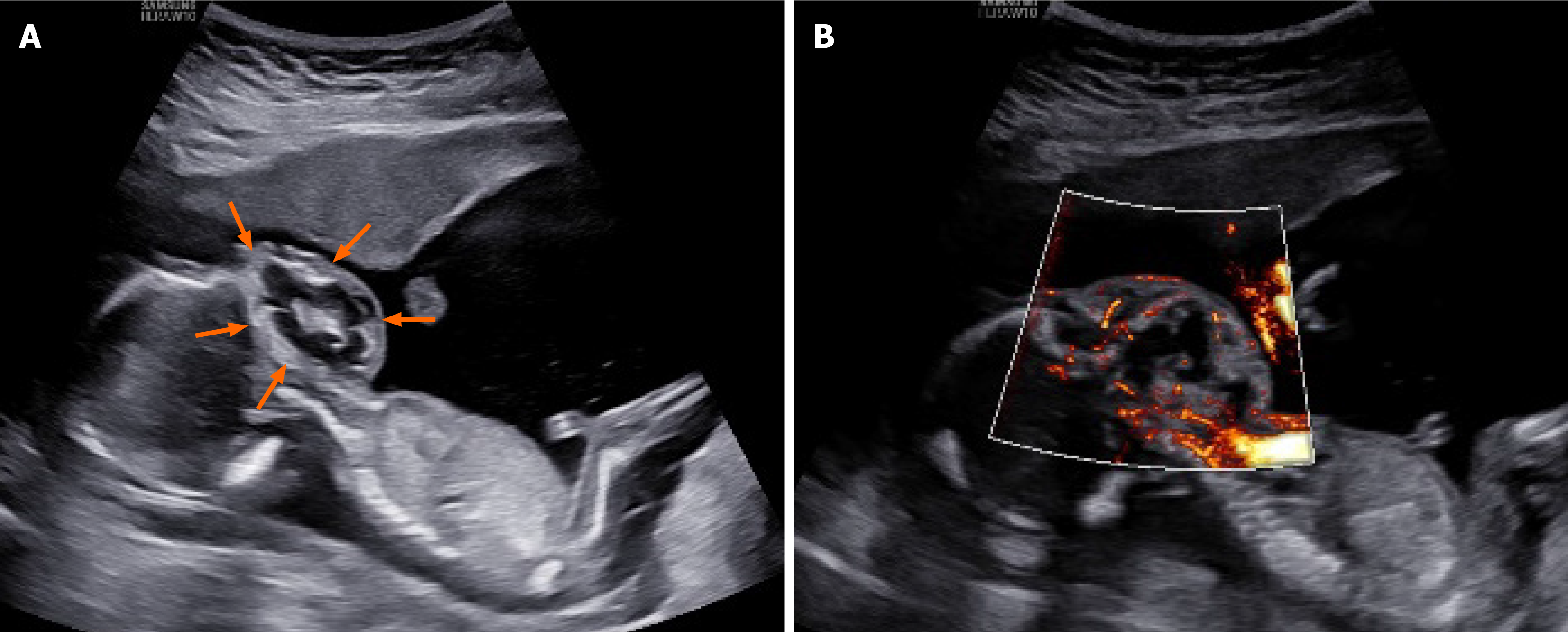
Figure 2 Prenatal ultrasound of an orofacial teratoma at 16 weeks of gestation (case 2).
A: Cystic-solid echo of the fetal face (orange arrow); B: Mic
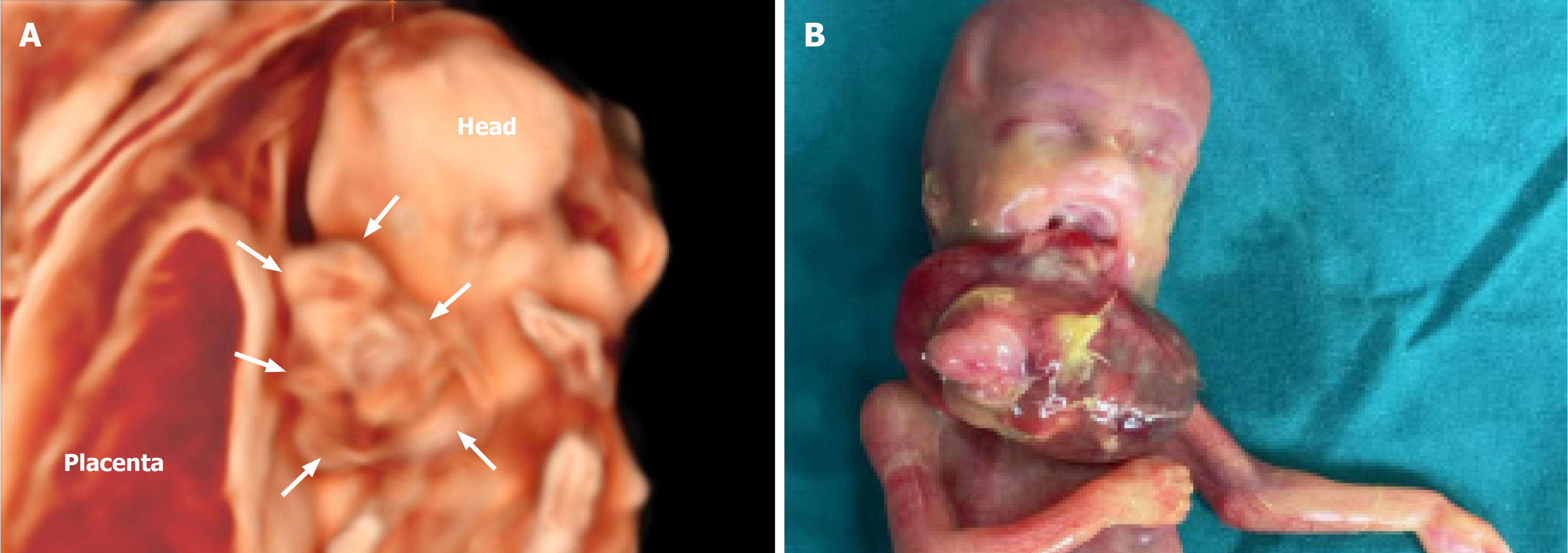
Figure 3 Gross specimen after induction of labor (case 1).
A: Three-dimensional ultrasound imaging of the fetal face shows the outline of the mass (white arrows); B: The gross specimen after induction showed an extraoral mass with a disorderly structure, originating from the palate.
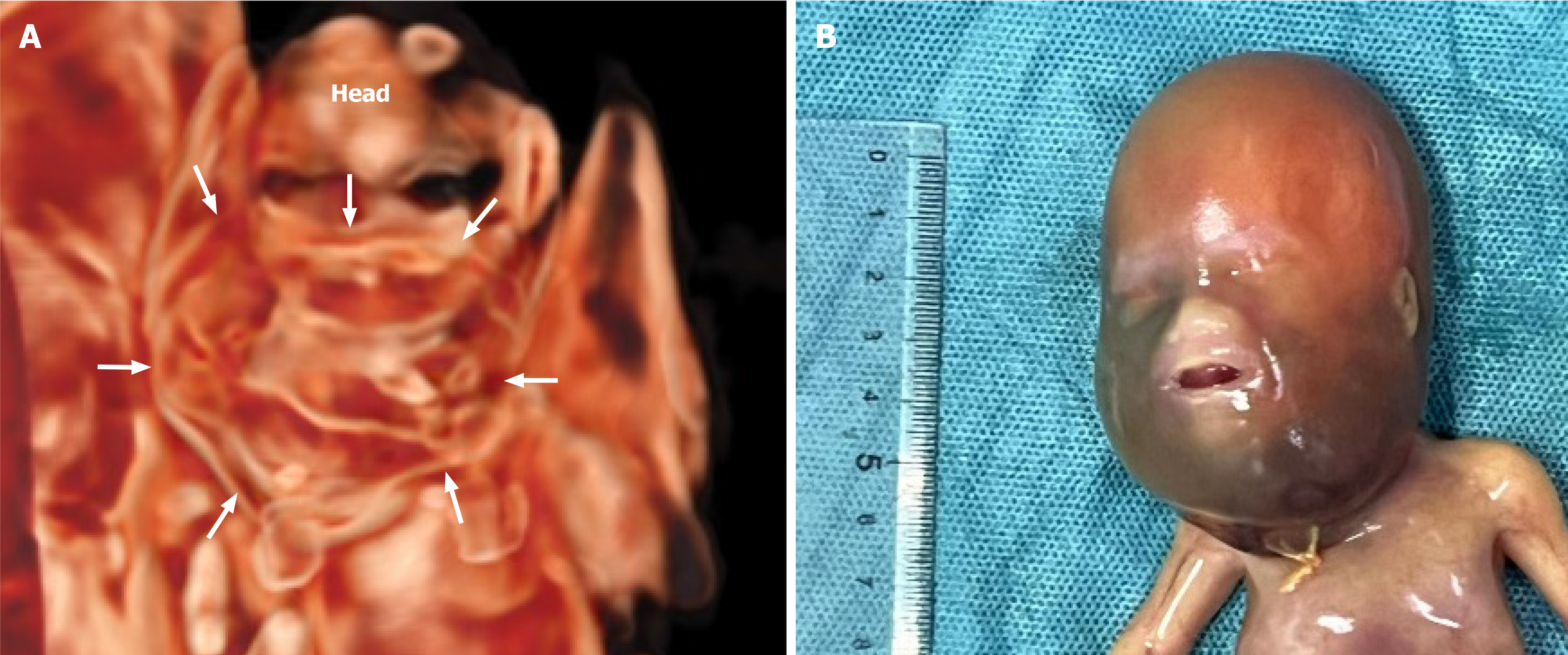
Figure 4 Gross specimen after induction of labor (case 2).
A: Three-dimensional crystal imaging of the fetal face shows the internal outline of the mass in the coronal plane (white arrows); B: After induction of labor, the gross specimen showed swelling of the neck and dark color.
- Citation: Gao CF, Zhou P, Zhang C. Prenatal ultrasound diagnosis of fetal maxillofacial teratoma: Two case reports. World J Clin Oncol 2024; 15(9): 1245-1250
- URL: https://www.wjgnet.com/2218-4333/full/v15/i9/1245.htm
- DOI: https://dx.doi.org/10.5306/wjco.v15.i9.1245


