©The Author(s) 2024.
World J Clin Oncol. Sep 24, 2024; 15(9): 1239-1244
Published online Sep 24, 2024. doi: 10.5306/wjco.v15.i9.1239
Published online Sep 24, 2024. doi: 10.5306/wjco.v15.i9.1239
Figure 1 Computed tomography scan.
A and B: Computed tomography (CT) scan of the whole abdomen, showing an irregular mass on the right anterior wall of the bladder. The mass had a size of approximately 30 mm × 29 mm and was observed to project into the lumen (orange arrow); C: Abdominal CT enhancement. The lesion exhibited multiple speckles of hyperdense shadows, which became more pronounced after enhancement (orange arrow).
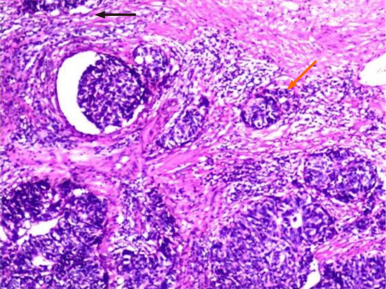
Figure 2 Histopathologic analysis of the resected specimen.
Tumor with a large nest arrangement, showing peripheral fenestrations and rosette knots (orange arrow), together with abundant bright eosinophilic cytoplasm and apoptotic vesicles (black arrow).
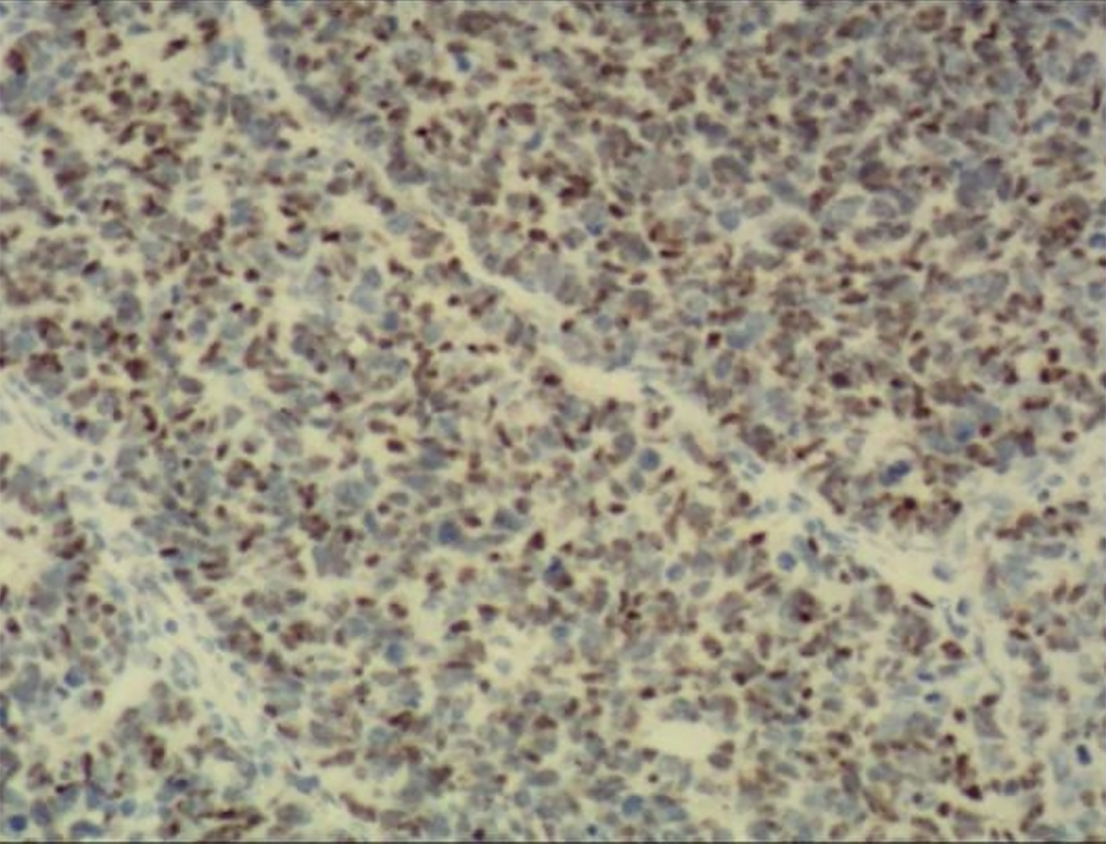
Figure 3 Immunohistochemical examination of the resected specimen.
Tumor showing a sheet-like growth pattern.
- Citation: Zhou Y, Yang L. Large-cell neuroendocrine carcinoma of the bladder: A case report. World J Clin Oncol 2024; 15(9): 1239-1244
- URL: https://www.wjgnet.com/2218-4333/full/v15/i9/1239.htm
- DOI: https://dx.doi.org/10.5306/wjco.v15.i9.1239


