©The Author(s) 2024.
World J Clin Oncol. Dec 24, 2024; 15(12): 1514-1519
Published online Dec 24, 2024. doi: 10.5306/wjco.v15.i12.1514
Published online Dec 24, 2024. doi: 10.5306/wjco.v15.i12.1514
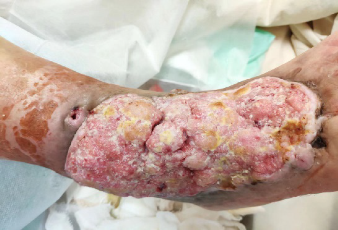
Figure 1 The foot ulcer area in 2018, showing an irregular shape resembling a rotten vegetable pattern, accompanied by soybean curd secretions as well as significant blood and liquid oozing.
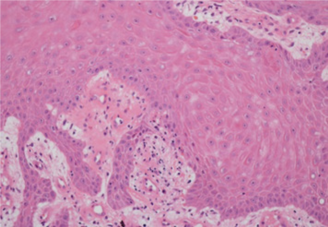
Figure 2 Pathological examination conducted on February 21st, 2019, revealed pseudo-epitheliomatous hyperplasia of the squamous epithelium (right foot) with partial atypical hyperplasia.
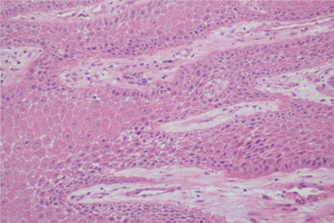
Figure 3 In July 2020, histopathological analysis confirmed well-differentiated cutaneous squamous cell carcinoma once again within the wound tissue sample.
- Citation: Luo Y, Li CY, Wang YQ, Xiang SM, Zhao C. Diabetic ulcer with cutaneous squamous cell carcinoma: A case report. World J Clin Oncol 2024; 15(12): 1514-1519
- URL: https://www.wjgnet.com/2218-4333/full/v15/i12/1514.htm
- DOI: https://dx.doi.org/10.5306/wjco.v15.i12.1514


