Copyright
©The Author(s) 2021.
World J Clin Oncol. Apr 24, 2021; 12(4): 282-289
Published online Apr 24, 2021. doi: 10.5306/wjco.v12.i4.282
Published online Apr 24, 2021. doi: 10.5306/wjco.v12.i4.282
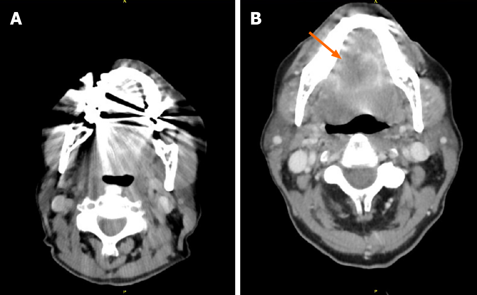
Figure 1 Computed tomography of neck.
A: Computed tomography of neck visualization significantly limited due to motion and dental artifact; B: Pulmonary sarcomatoid carcinoma metastasis to tongue. A partially visualized peripherally irregular lesion measuring 2.7 cm × 3.2 cm × 1.9 cm within the tongue (arrow), with peripherally enhancement with central hypoattenuation, only partially visualized due to streak artifact from dental amalgam.
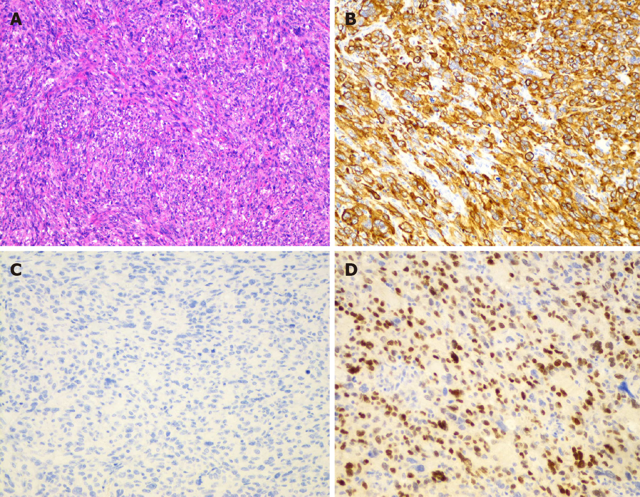
Figure 2 Histologic examination of the tongue mass.
A: Tongue mass characterized by high-grade, poorly-differentiated spindle and epithelioid malignant neoplasm (magnification, 100 ×); B and C: Positive for cytokeratin AE1/AE3 (B) and TTF-1 (C), consistent with a metastatic carcinoma of pulmonary origin (magnification, 200 ×); D: The neoplastic cells are completely negative for p40 (magnification, 200 ×).
- Citation: Guo MN, Jalil A, Liu JY, Miao RY, Tran TA, Guan J. Tongue swelling as a manifestation of tongue metastasis from pulmonary sarcomatoid carcinoma: A case report. World J Clin Oncol 2021; 12(4): 282-289
- URL: https://www.wjgnet.com/2218-4333/full/v12/i4/282.htm
- DOI: https://dx.doi.org/10.5306/wjco.v12.i4.282


