©The Author(s) 2016.
World J Radiol. Apr 28, 2016; 8(4): 419-427
Published online Apr 28, 2016. doi: 10.4329/wjr.v8.i4.419
Published online Apr 28, 2016. doi: 10.4329/wjr.v8.i4.419
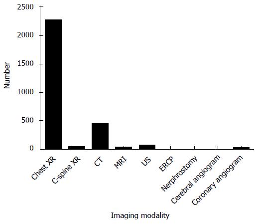
Figure 1 Modality contributions to the total number of investigations performed in intensive care unit patients over a 1-year period.
XR: X-ray; CT: Computed tomography; MRI: Magnetic resonance imaging; US: Ultrasound; ERCP: Endoscopic retrograde cholangiopancreatography.
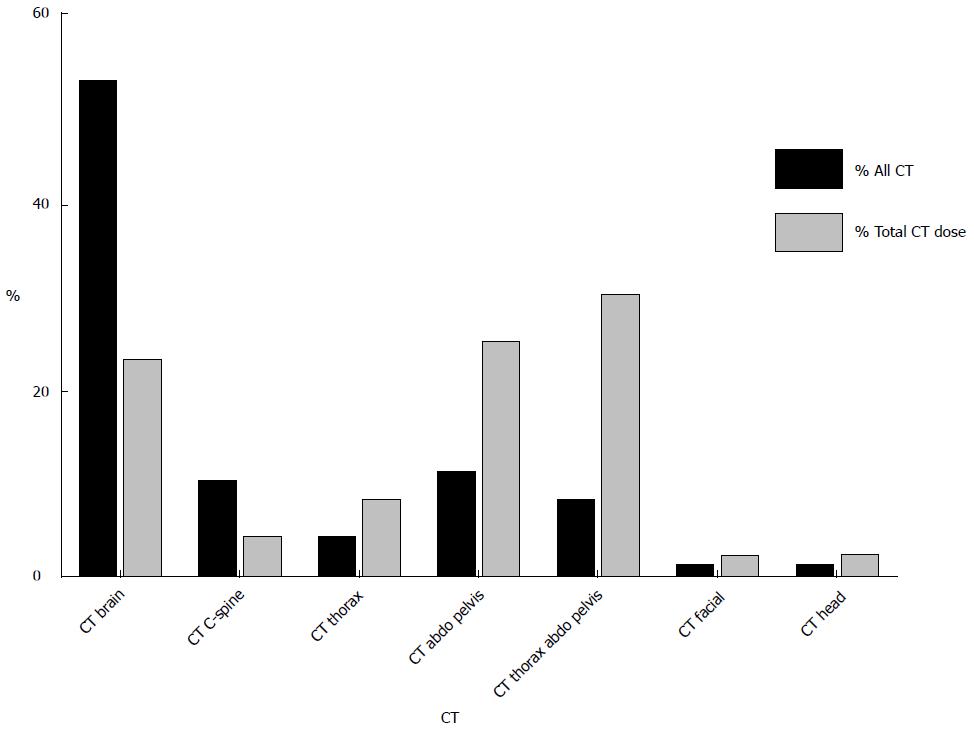
Figure 2 Contribution of individual types of computed tomography to total number of computed tomography examinations and total dose from computed tomography.
CT brain was the most commonly performed CT examination followed by CT abdomen and pelvis and CT thorax, abdomen, and pelvis. CT: Computed tomography; C-spine: Cervical spine; Abdo: Abdomen; CTA: Computed tomography angiography.
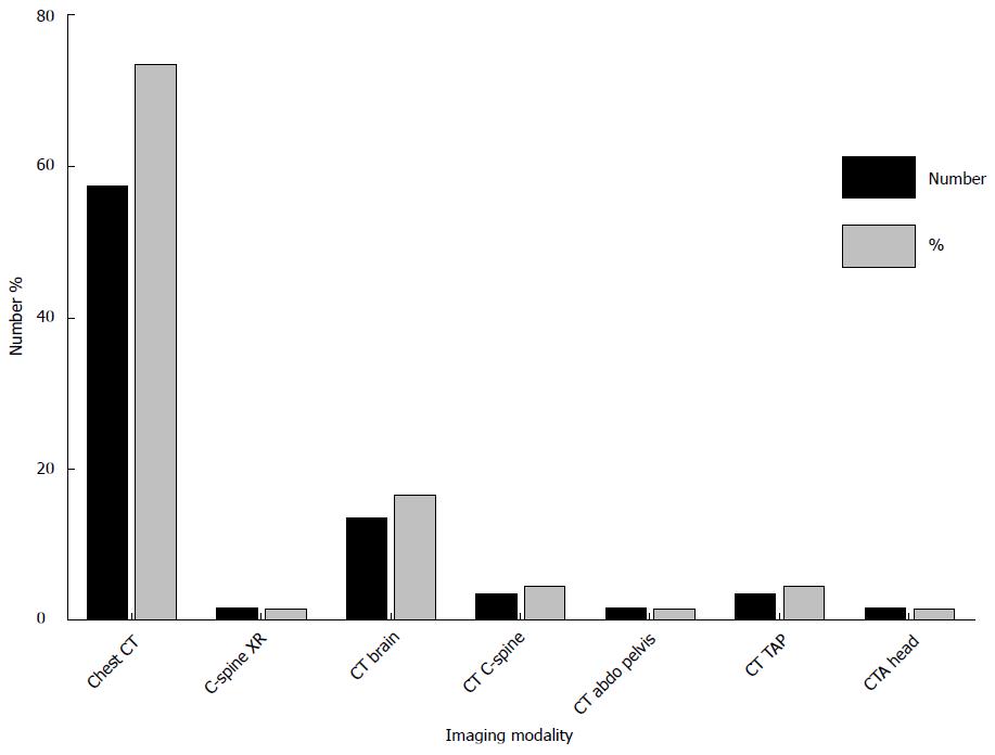
Figure 3 Modality contributions to the total number of investigations performed in a cohort of 23 pediatric intensive care unit patients over a 1-year period.
XR: X-ray; CT: Computed tomography; C-spine: Cervical spine; Abdo: Abdomen; CTA: Computed tomography angiography.
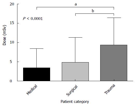
Figure 4 Comparison of cumulative effective dose in medical, surgical, and trauma patients.
Trauma patients received a statistically significant higher dose [median CED; 7.7 mSv (IQR: 3.5-13.8 mSv)] than medical [median CED: 1.4 mSv (IQR: 0.05-5.4 mSv)] and surgical [median CED: 1.6 mSv (IQR: 0.04-7.5mSv)] patients. (Dunn’s multiple comparisons test aP < 0.0001, bP < 0.0001). CED: Cumulative effective dose.
- Citation: Moloney F, Fama D, Twomey M, O’Leary R, Houlihane C, Murphy KP, O’Neill SB, O’Connor OJ, Breen D, Maher MM. Cumulative radiation exposure from diagnostic imaging in intensive care unit patients. World J Radiol 2016; 8(4): 419-427
- URL: https://www.wjgnet.com/1949-8470/full/v8/i4/419.htm
- DOI: https://dx.doi.org/10.4329/wjr.v8.i4.419


