Copyright
©The Author(s) 2025.
World J Radiol. Sep 28, 2025; 17(9): 110267
Published online Sep 28, 2025. doi: 10.4329/wjr.v17.i9.110267
Published online Sep 28, 2025. doi: 10.4329/wjr.v17.i9.110267
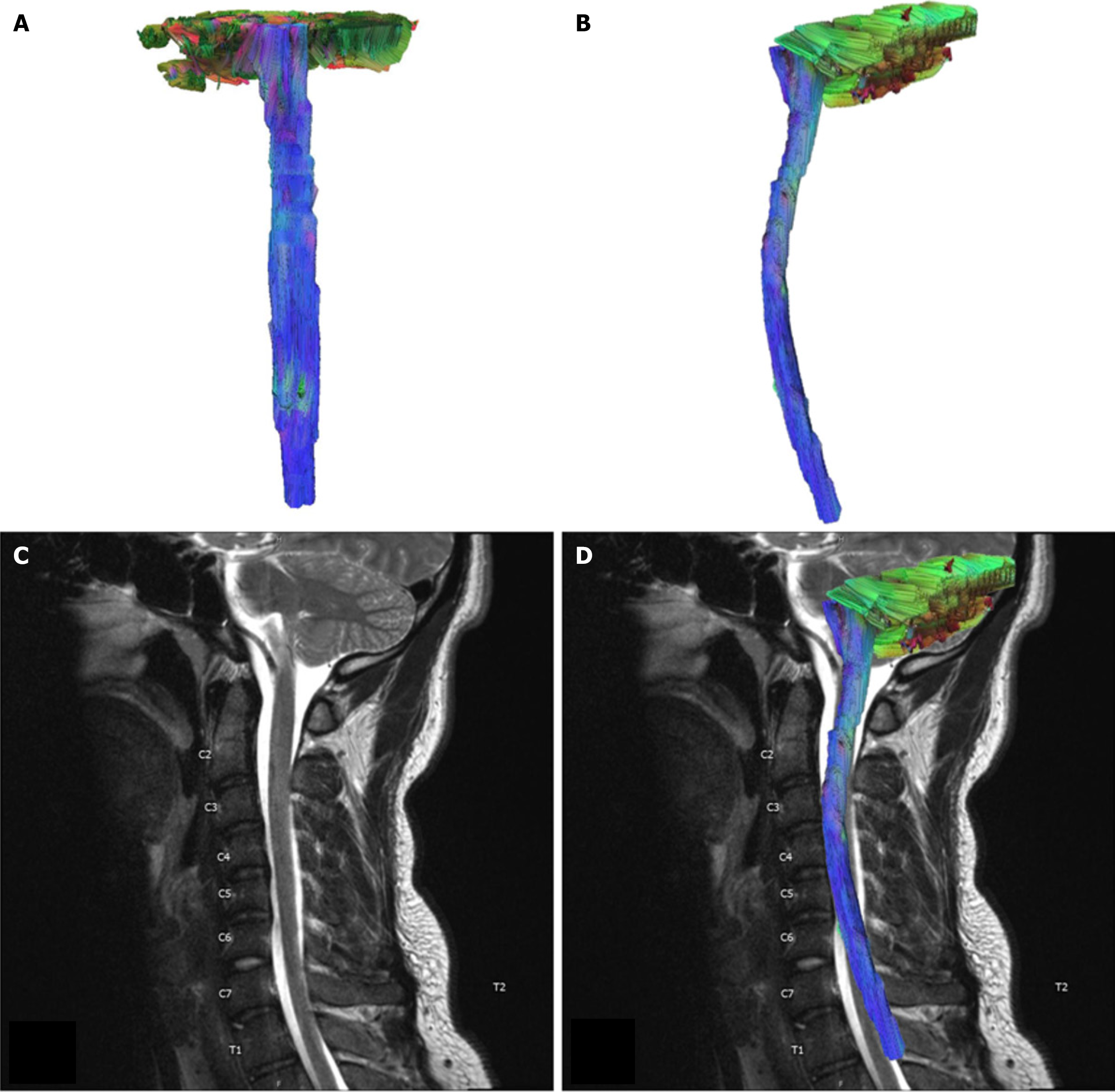
Figure 1 Control case.
A: AP view of tractography model of a 22 year old male control case; B: Sagittal view of the tractography model; C: T2-weighted Sagittal view of the cervical spinal cord. No discernable signal abnormality in the cervical spinal cord; D: Tractography of control case superimposed over T2-weighted sagittal view of the cervical spinal cord.
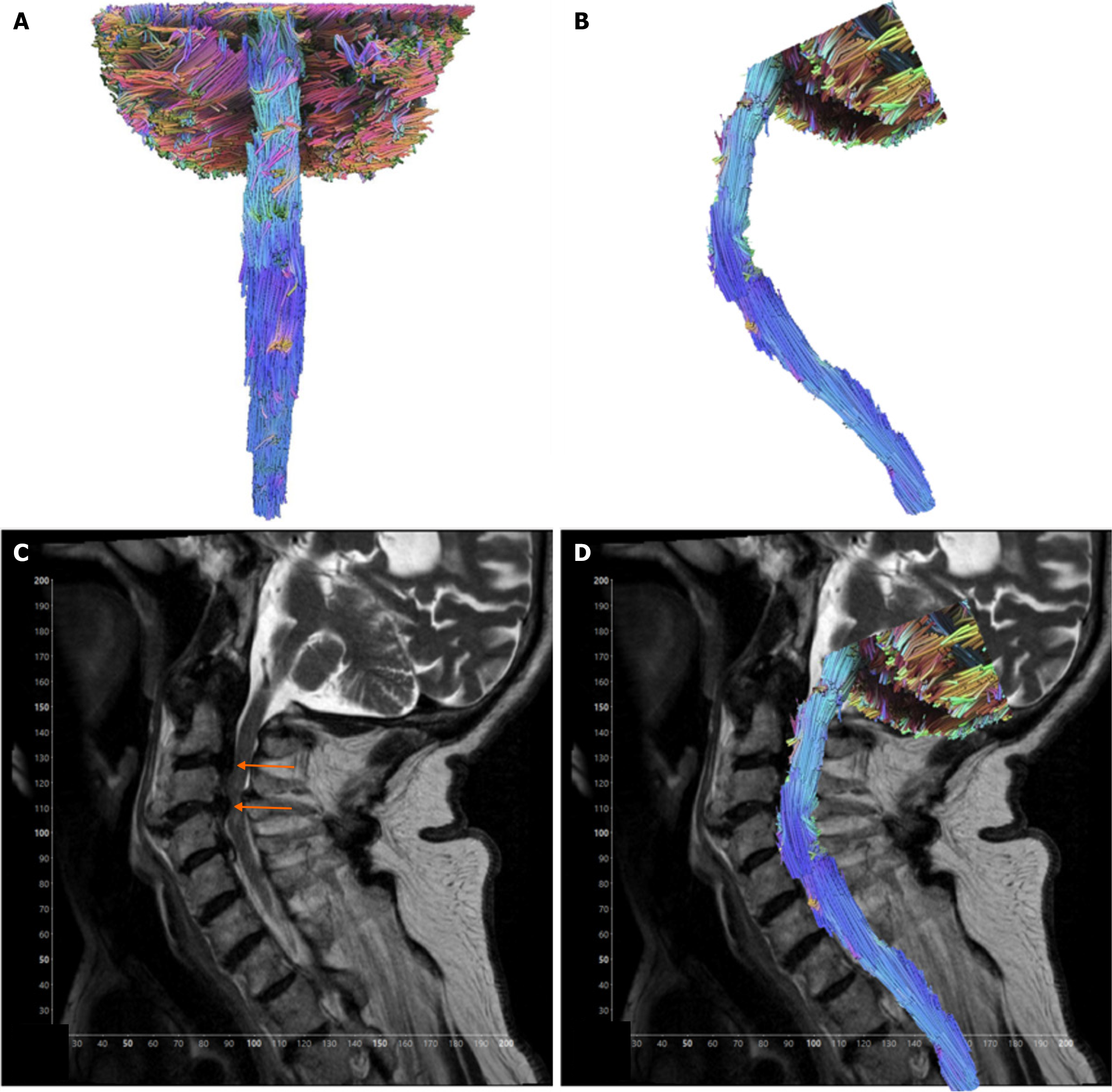
Figure 2 Contusion case.
A: AP view of tractography model. Case of an 82-year-old male presenting with spinal cord contusion; B: Sagittal view of the tractography model; C: T2-weighted sagittal view of the cervical spinal cord showing severe spinal canal stenosis from disc herniation and degenerative spondylosis at the levels of C2-C3 and C3-C4, with severe spinal cord flattening at C3-C4 (orange arrows); D: Sagittal view tractography model of contusion case superimposed over T2-weighted imaging of the cervical spinal cord.
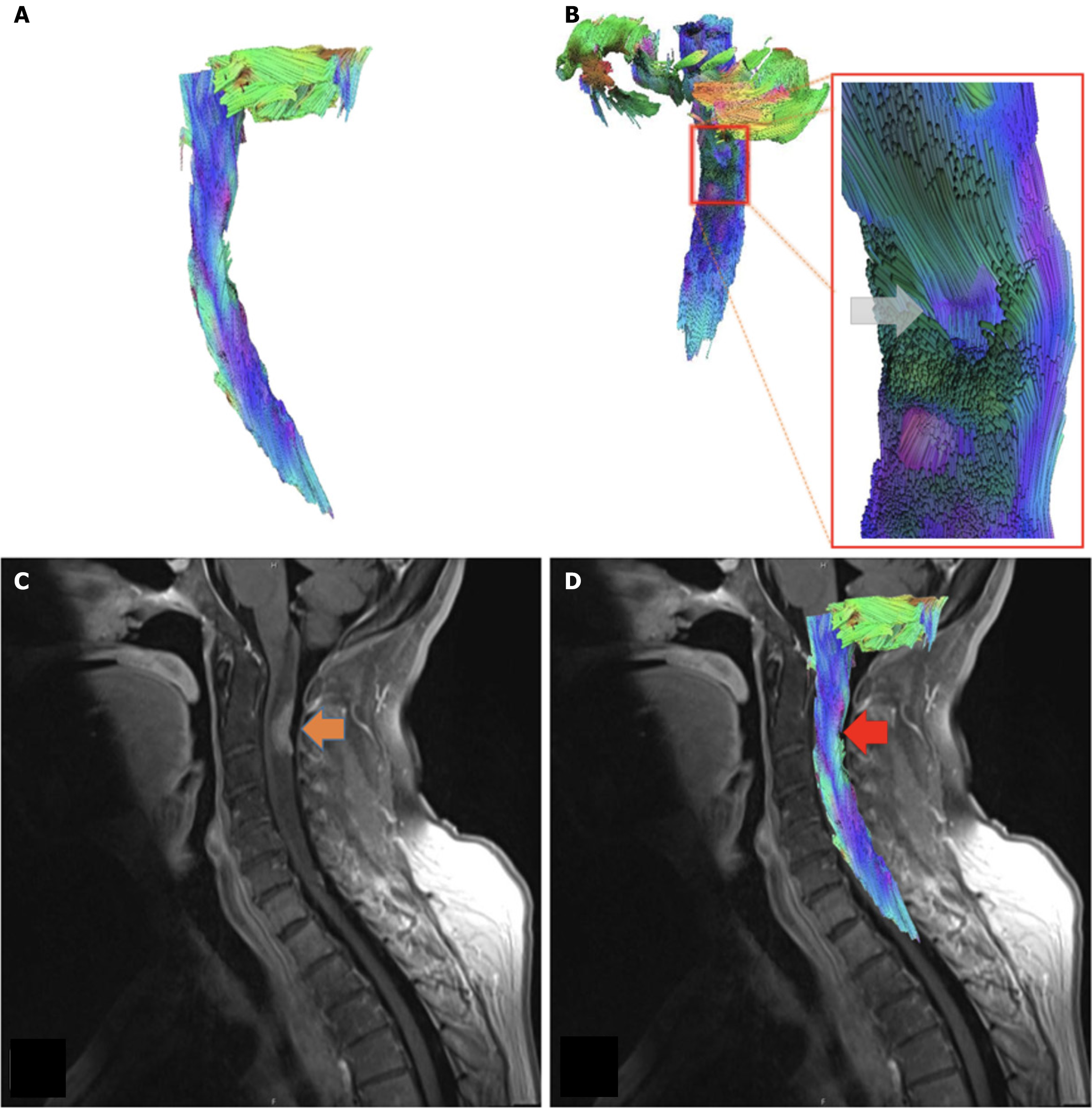
Figure 3 Metastasis case.
A: Sagittal view of tractography model. Case of 62-year-old female with primary breast cancer, displaying metastases to the lungs, spine, and brain; B: Posterior oblique view of the cervical spinal cord tractography model, with associated close up at the level of C2-C3. Note displaced fibers at the site of lesion (gray arrow); C: T1 FR STIR sagittal view of the cervical spinal cord. Metastatic lesion located at C2-C3 (orange arrow); D: Tractography model of metastasis case superimposed over T1 FR STIR sagittal view of the cervical spinal cord. The metastatic lesion can be seen at the level C2-C3 (red arrow).
Figure 4 Overall fractional anisotropy values are lower at the pathological region while mean diffusivity values are higher than in the control.
Fractional anisotropy and mean diffusivity values are similar at the inferior and superior areas of the pathology compared to the control. FA: Fractional anisotropy; MD: Mean diffusivity.
- Citation: Supsupin EP, Serrano A, Louviere C, Pearson L, Hernandez M, Sekar V, Amer A, Cikla U, Virarkar M, Gumus KZ. Magnetic resonance tractography of the cervical spine: A rapid diffusion tensor imaging protocol to serve as a clinical evaluation tool. World J Radiol 2025; 17(9): 110267
- URL: https://www.wjgnet.com/1949-8470/full/v17/i9/110267.htm
- DOI: https://dx.doi.org/10.4329/wjr.v17.i9.110267


