©The Author(s) 2025.
World J Radiol. May 28, 2025; 17(5): 106975
Published online May 28, 2025. doi: 10.4329/wjr.v17.i5.106975
Published online May 28, 2025. doi: 10.4329/wjr.v17.i5.106975
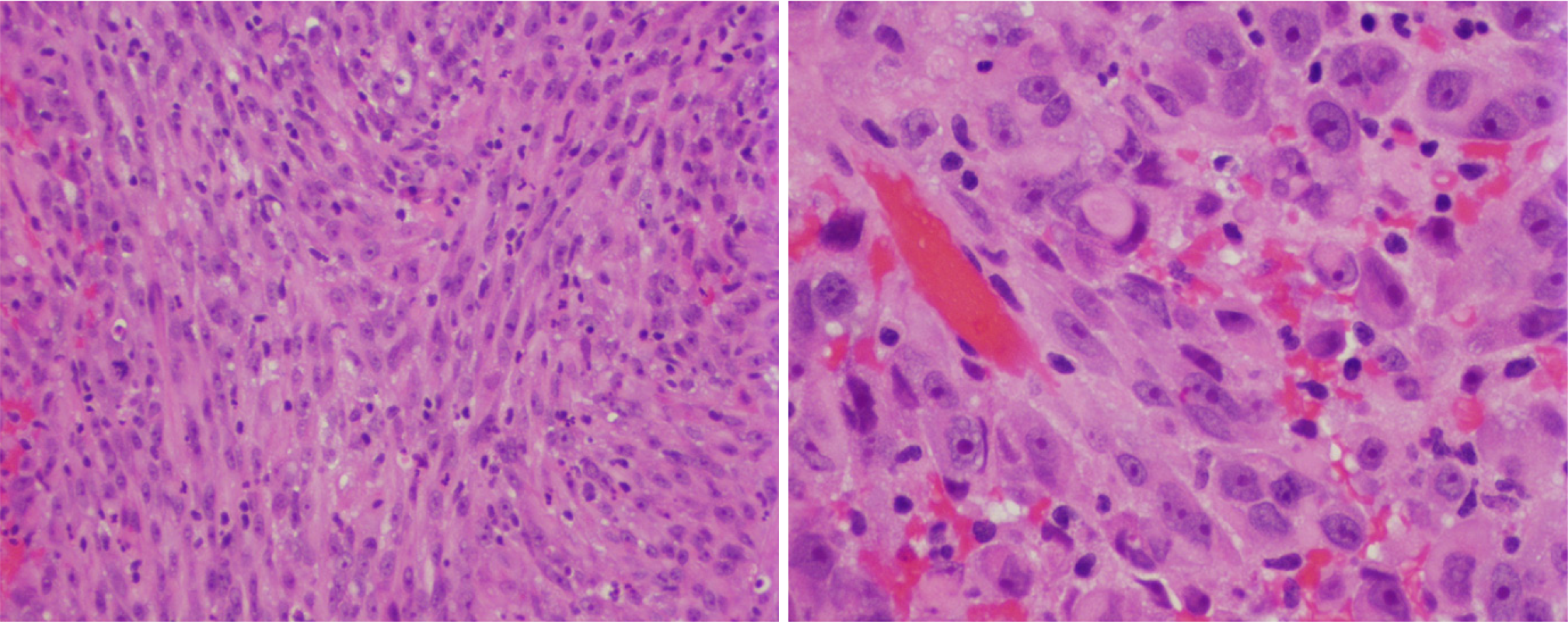
Figure 1 Histological features of sellar atypical teratoid/rhabdoid tumor.
Hematoxylin and eosin stain (Left). Epithelioid-like spindle cells with pink cytoplasm and prominent nuclei arranged in fascicular pattern, × 20 (Right). Focal tumor cells exhibiting rhabdoid features, × 40. Reproduced from Lev et al[16]. Citation: Lev I, Fan X, Yu R. Sellar Atypical Teratoid/Rhabdoid Tumor: Any Preoperative Diagnostic Clues? AACE Clin Case Rep 2015; 1: e2-e7. Copyright© 2015 Elsevier Inc. Published by Elsevier Inc. This is an open access article with User License: Creative Commons Attribution-NonCommercial-NoDerivs (CC BY-NC-ND 4.0). See: https://creativecommons.org/licenses/by-nc-nd/4.0/.
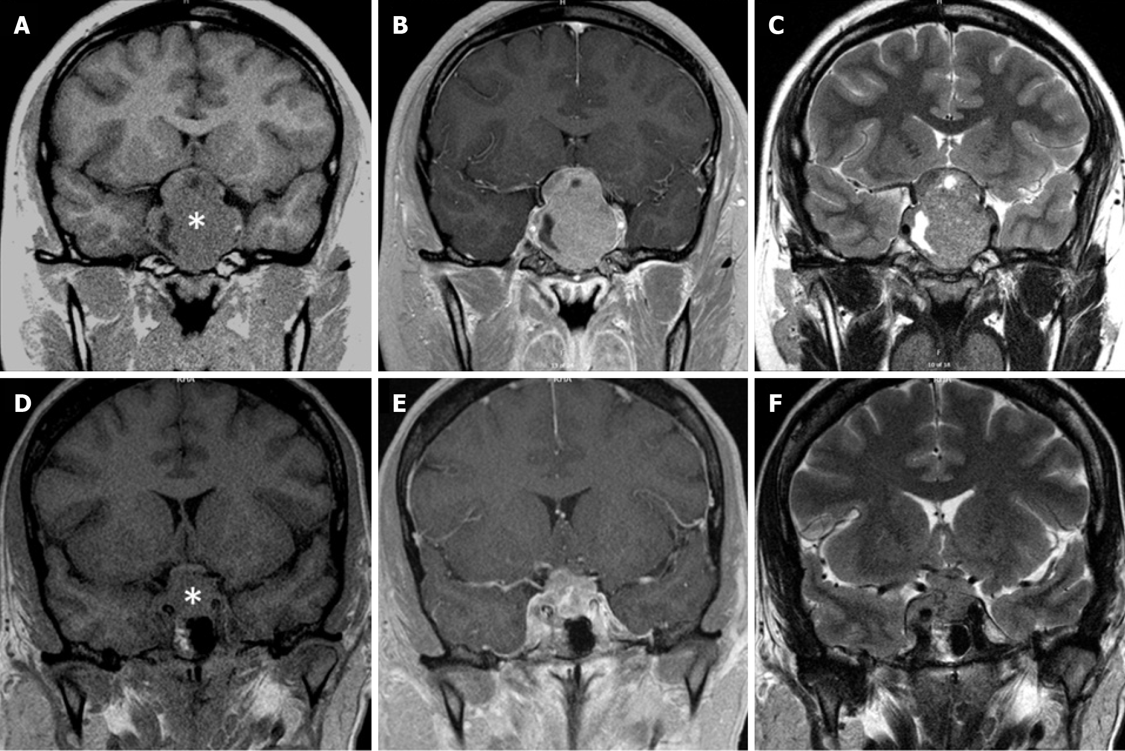
Figure 2 Magnetic resonance imaging of pituitary macroadenoma and sellar atypical teratoid/rhabdoid tumor.
A-C: A 4.1 cm × 4.4 cm macroprolactinoma of a 19-year-old male; D-F: A 2.2 cm × 1.8-cm sellar atypical teratoid/rhabdoid tumor of a 45-year-old female. T1 imaging without contrast (A and D). T1 imaging after contrast administration (B and E). T2 imaging (C and F). Asterisk, sellar mass. Note the limited cavernous sinus invasion despite large tumor size and the optic chiasm thinning in the patient with macroprolactinoma. Also note the extensive bilateral cavernous sinus invasion but intact optic chiasm in the patient with sellar atypical teratoid/rhabdoid tumor. Panel E is reproduced from Yu[17]. Citation: Yu R. Sellar Mass in 2 Patients With Acute-Onset Headache and Visual Symptoms: Not Your Usual Pituitary Adenoma. AACE Clin Case Rep 2023; 9: 197-200. Copyright © 2015 Elsevier Inc. Published by Elsevier Inc. This is an open access article with User License: Creative Commons Attribution-NonCommercial-NoDerivs (CC BY-NC-ND 4.0). See: https://creativecommons.org/licenses/by-nc-nd/4.0/.
- Citation: Yu R. Specific imaging features of sellar atypical teratoid/rhabdoid tumor or the lack of thereof. World J Radiol 2025; 17(5): 106975
- URL: https://www.wjgnet.com/1949-8470/full/v17/i5/106975.htm
- DOI: https://dx.doi.org/10.4329/wjr.v17.i5.106975


