©The Author(s) 2025.
World J Radiol. Nov 28, 2025; 17(11): 111651
Published online Nov 28, 2025. doi: 10.4329/wjr.v17.i11.111651
Published online Nov 28, 2025. doi: 10.4329/wjr.v17.i11.111651
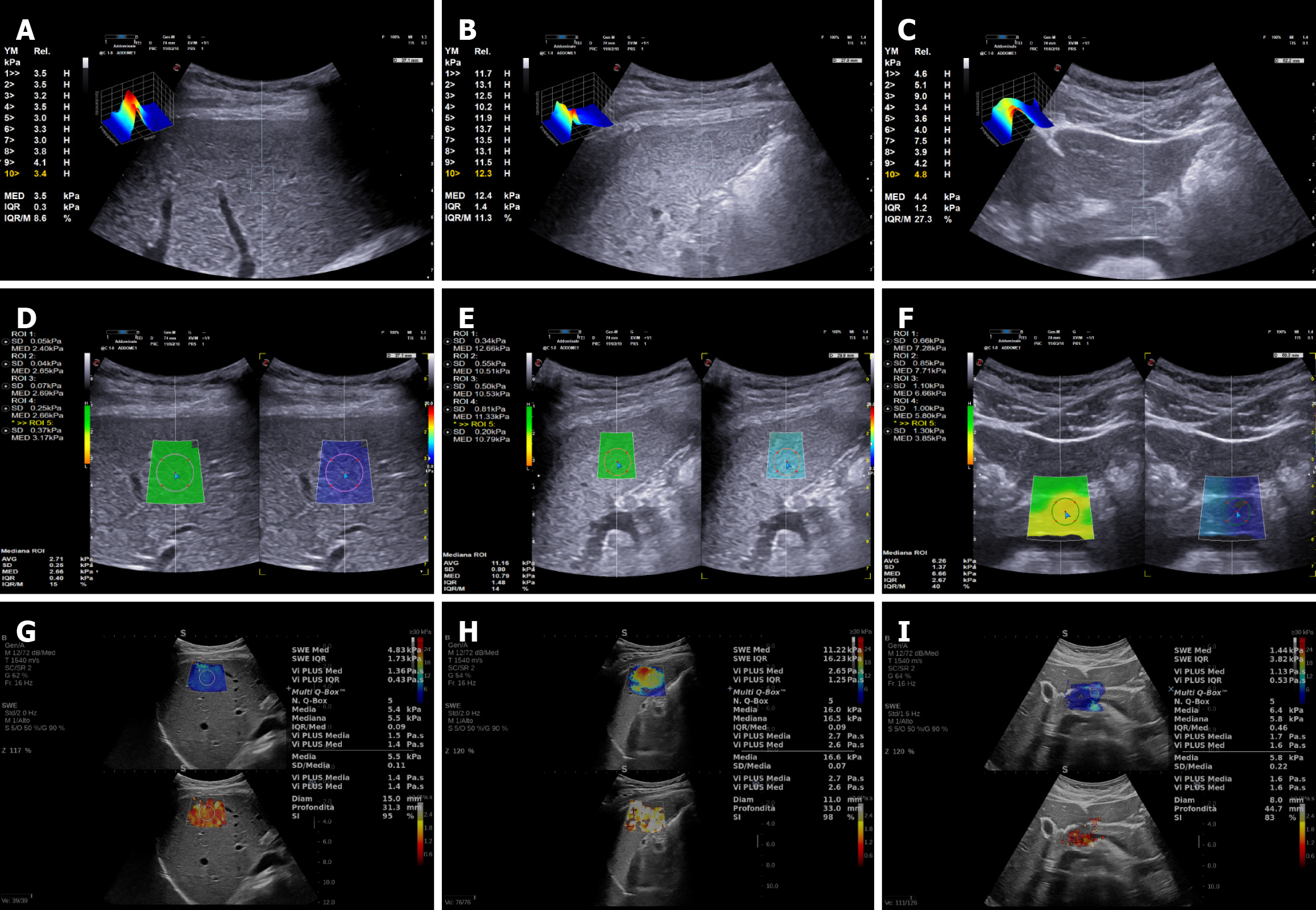
Figure 1 Shear wave elastography in healthy patients.
A-C: Point-shear wave elastography of liver (A), spleen (B), and pancreas (C); D-F: 2-dimensional-shear wave elastography of liver (D), spleen (E), and pancreas (F); G-I: 2-dimensional-SuperSonic Imagine Aixplorer of liver (G), spleen (H), and pancreas (I).
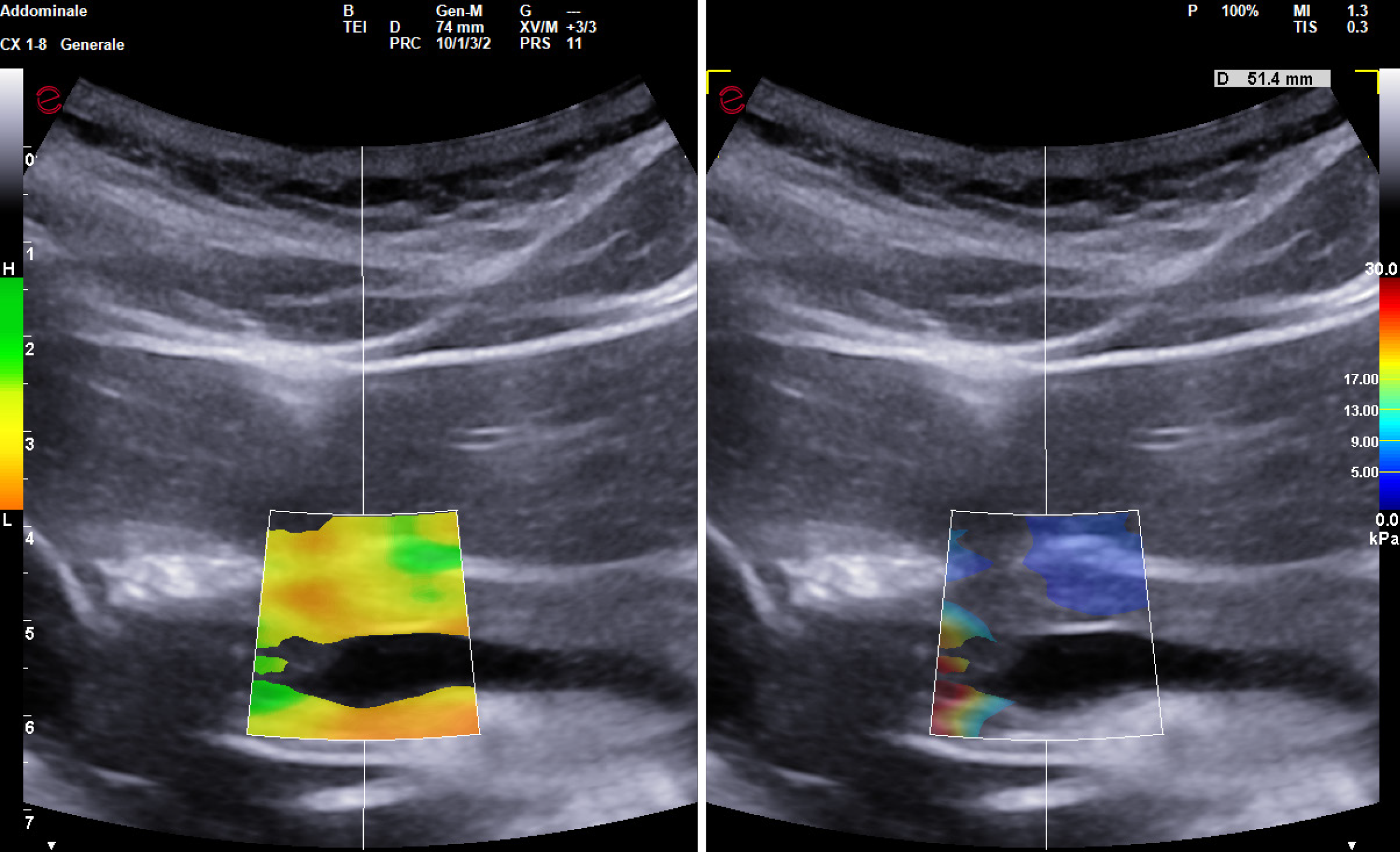
Figure 2 2-dimensional-shear wave elastography of pancreas: Inhomogeneous filling of the colorimetric map suggests an unreliable measurement.
- Citation: Viceconti N, Paratore M, Del Zompo F, Zocco MA, Ainora ME, Esposto G, Gasbarrini A, Pompili M, Riccardi L, Garcovich M. Shear wave elastography in healthy patients: Pancreatic stiffness is less reliable than liver and spleen measurements. World J Radiol 2025; 17(11): 111651
- URL: https://www.wjgnet.com/1949-8470/full/v17/i11/111651.htm
- DOI: https://dx.doi.org/10.4329/wjr.v17.i11.111651


