©The Author(s) 2024.
World J Radiol. Oct 28, 2024; 16(10): 561-568
Published online Oct 28, 2024. doi: 10.4329/wjr.v16.i10.561
Published online Oct 28, 2024. doi: 10.4329/wjr.v16.i10.561
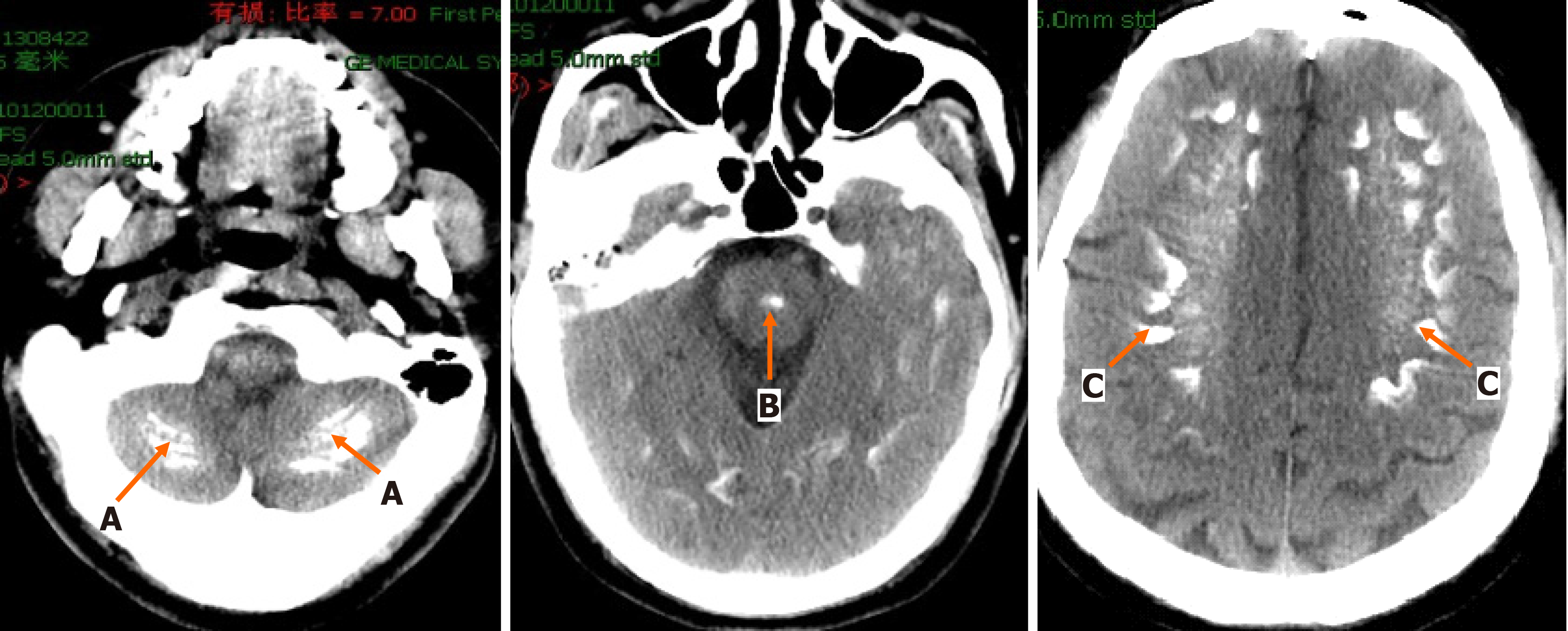
Figure 1 Cranial computed tomography scan.
A: Bilateral cerebellar calcifications; B: Brainstem; C: Bilateral cerebral calcifications.
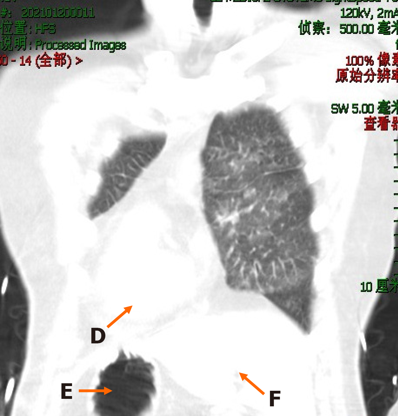
Figure 2 Chest computed tomography scan.
(Coronal view: D: Heart; E: Stomach; F: Liver)
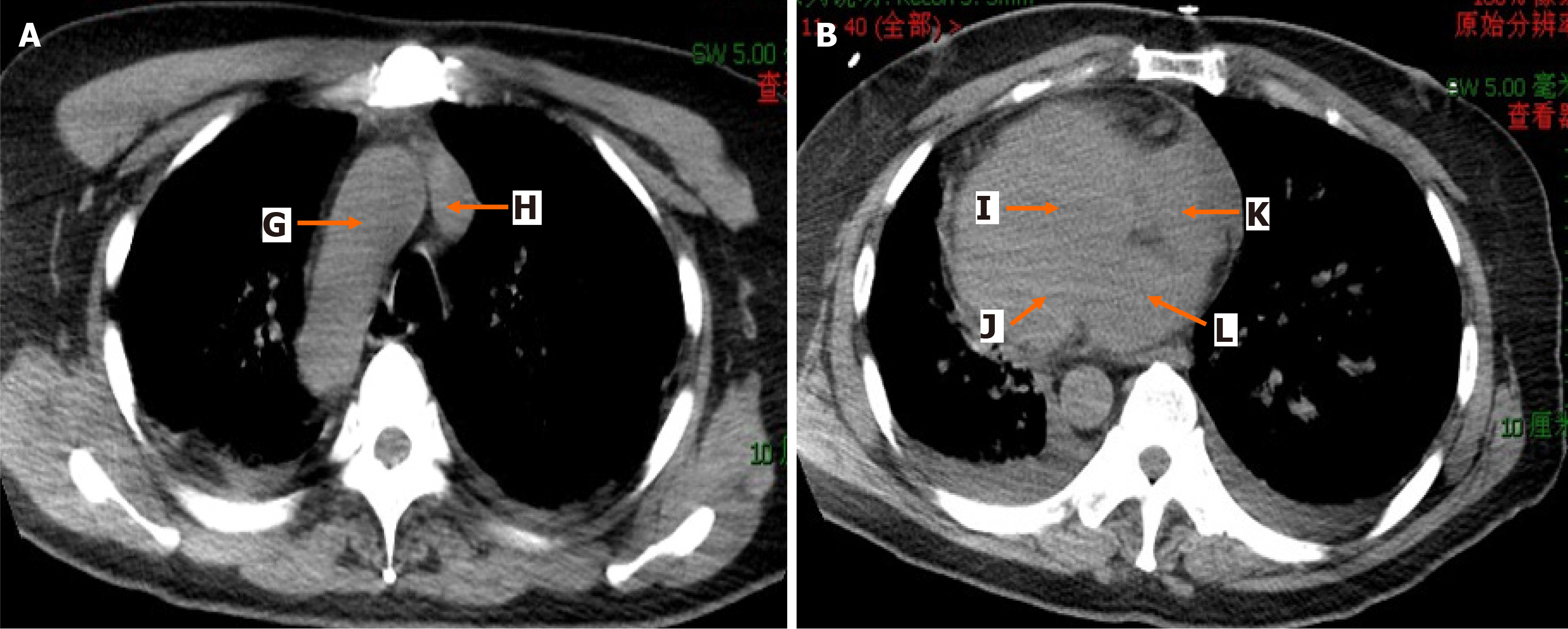
Figure 3 Chest computed tomography scan, A: Aortic arch level (G: Aortic arch; H: Superior vena cava); B: Four-chamber heart level (I: Right ventricle; J: Left ventricle; K: Right atrium; L: Left atrium)
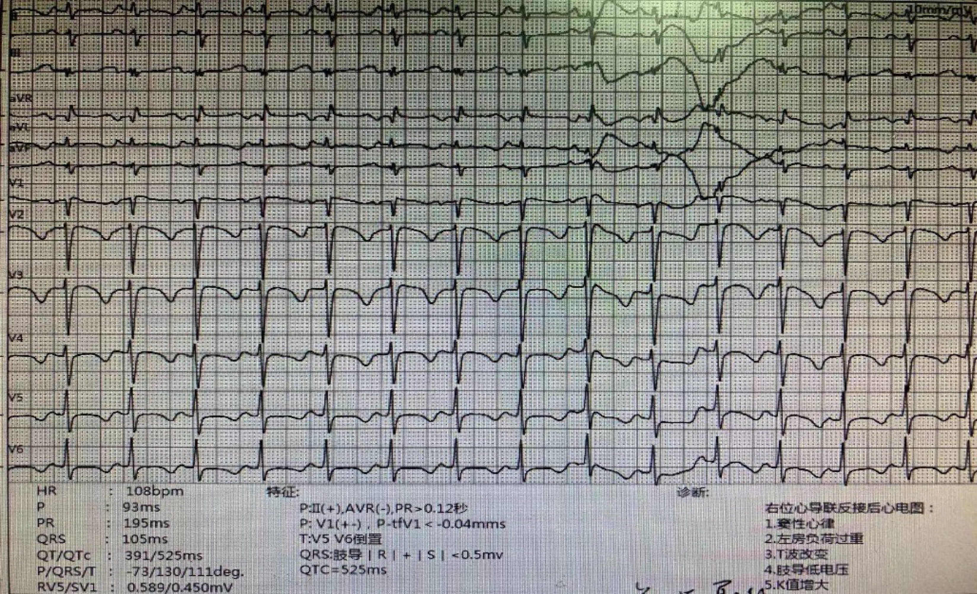
Figure 4 Electrocardiogram.
(1) Sinus tachycardia; (2) Electrocardiogram changes indicating dextrocardia.
- Citation: Yang M, Pu SL, Li L, Ma Y, Qin Q, Wang YX, Huang WL, Hu HY, Zhu MF, Li CZ. Hypoparathyroidism with situs inversus totalis: A case report. World J Radiol 2024; 16(10): 561-568
- URL: https://www.wjgnet.com/1949-8470/full/v16/i10/561.htm
- DOI: https://dx.doi.org/10.4329/wjr.v16.i10.561


