Case Report Open Access
Copyright ©The Author(s) 2024. Published by Baishideng Publishing Group Inc. All rights reserved.
World J Radiol. Oct 28, 2024; 16(10): 569-578
Published online Oct 28, 2024. doi: 10.4329/wjr.v16.i10.569
Mesenteric venous thrombosis in a young adult: A case report and review of the literature
Jiao-Jiao Yuan, Jian Zhang, Jun-Zhi Li, Department of Emergency Medicine, Ninth Hospital of Xi'an, Xi’an 710000, Shaanxi Province, China
Hai-Fu Zhang, Department of Vascular Intervention, Ninth Hospital of Xi'an, Xi’an 710000, Shaanxi Province, China
ORCID number: Jiao-Jiao Yuan (0000-0003-3867-8709); Jun-Zhi Li (0009-0001-7306-553X).
Co-first authors: Jiao-Jiao Yuan and Hai-Fu Zhang.
Co-corresponding authors: Jiao-Jiao Yuan and Jun-Zhi Li.
Author contributions: Yuan JJ and Li JZ drafted the conception and design of the study; Yuan JJ wrote the article; Zhang HF participated in data collection and organization; Zhang J handled image sorting and summarization; Yuan JJ and Zhang HF made crucial and indispensable contributions towards the completion of the study and were thus qualified as the co-first authors of the paper. Li JZ and Yuan JJ revised the article; Li JZ and Yuan JJ provided final approval for the version submitted for publication. All authors have read and approved the final manuscript. The collaboration between Yuan JJ and Li JZ as the co-corresponding authors is crucial for the publication of this manuscript. Yuan JJ searched the literature, and drafted, revised, and submitted the early version of the manuscript. Li JZ supervised the whole process of the study, was instrumental and responsible for data re-analysis and re-interpretation, and revised the article.
Informed consent statement: Informed consent was obtained from the patient for publication of this case report and any accompanying images.
Conflict-of-interest statement: All authors declare that they have no competing interests to disclose.
CARE Checklist (2016) statement: The authors have read the CARE Checklist (2016), and the manuscript was prepared and revised according to the CARE Checklist (2016).
Open-Access: This article is an open-access article that was selected by an in-house editor and fully peer-reviewed by external reviewers. It is distributed in accordance with the Creative Commons Attribution NonCommercial (CC BY-NC 4.0) license, which permits others to distribute, remix, adapt, build upon this work non-commercially, and license their derivative works on different terms, provided the original work is properly cited and the use is non-commercial. See: https://creativecommons.org/Licenses/by-nc/4.0/
Corresponding author: Jun-Zhi Li, Chief Doctor, Professor, Department of Emergency Medicine, Ninth Hospital of Xi'an, No. 151 Dongduan Nanerhuan, Beilin District, Xi’an 710000, Shaanxi Province, China. hess2009@126.com
Received: May 14, 2024
Revised: August 22, 2024
Accepted: September 6, 2024
Published online: October 28, 2024
Processing time: 166 Days and 23.8 Hours

Abstract
BACKGROUND

Acute mesenteric vein thrombosis (MVT) accounts for only 2%–10% of all cases of acute mesenteric ischaemia, with an incidence rate of ~0.1% in Europe and the United States. It represents < 10% of mesenteric infarction cases and is seen predominantly in older adults. In younger individuals, MVT is uncommon, with 36% of cases having unidentified mechanisms and causes.

CASE SUMMARY

A 27-year-old man presented to the emergency department on February 29, 2024, with a chief complaint of intermittent abdominal pain for 3 day. He was previously in good health. As the abdominal pain was not alleviated by conventional treatment, an abdominal computed tomography (CT) scan was performed, which showed increased density in the portal and mesenteric veins. Further imaging, including portal vein ultrasound, mesenteric CT angiography, and enhanced abdominal CT, revealed widespread thrombosis of the portal vein system (including the main portal vein, left and right branches, proximal mesenteric vein, and splenic vein). After 10 day of thrombectomy and anticoagulation therapy, the patient’s abdominal pain had improved significantly. Follow-up assessments indicated that portal venous blood flow had largely returned to normal. He was discharged on March 9, 2024. During a follow-up exam 2 months later, repeat abdominal enhanced CT showed that the previously detected thrombi were no longer visible.

CONCLUSION

Clinicians should remain vigilant for acute MVT in young patients presenting with abdominal pain, to prevent misdiagnosis of this fatal condition.

Key Words: Acute mesenteric ischaemia; Acute extensive portal vein system thrombosis; Portal vein system thrombosis; Mesenteric vein thrombosis; Mesenteric artery embolism; Young adults; Case report

Core Tip: Acute mesenteric venous thrombosis accounts for < 10% of mesenteric infarction cases. It is rare in younger individuals, with unidentified mechanisms and causes in 36% of the cases. This case report provides a detailed description of a 27-year-old man diagnosed with acute mesenteric vein thrombosis. The rarity of this condition in younger patients and the critical importance of early diagnosis for clinical decision-making are highlighted. Our aim is to enhance clinicians' awareness of acute mesenteric vein thrombosis in young patients presenting with abdominal pain, to prevent the potentially fatal consequences of a missed diagnosis.


INTRODUCTION

Acute extensive thrombosis of the portal vein system is a rare but severe condition characterised by the formation of blood clots in two or more blood vessels, including the portal vein, splenic vein, superior mesenteric vein (SMV), and inferior mesenteric vein. The condition manifests as a critical acute abdomen with complex clinical symptoms. The incidence rate is relatively low (0.6%-1.0%), with the disease accounting for 5%-15% of intestinal vascular diseases[1]. According to the American Association of Hepatology Guidelines for the Diagnosis and Treatment of Hepatic Vascular Disease[2], patients with persistent abdominal pain for > 24 h should be evaluated for portal vein thrombosis, irrespective of the presence of fever or intestinal obstruction. The high rates of misdiagnosis can lead to significant treatment challenges and, in part, account for the high mortality of this condition.

Mesenteric vein thrombosis (MVT) constitutes only 2%-10% of all acute mesenteric ischaemia cases, with an incidence of ~0.1% in Europe and the United States[3]. MVT is primarily seen in older adults, occurring rarely in younger individuals among which 36% have unidentified mechanisms and causes[4].

Half of the deaths from acute mesenteric infarction (AMI) are attributed to superior mesenteric artery (SMA) occlusion, divided equally between non-occlusive infarction and inferior mesenteric artery occlusion/MVT[5]. The incidence rate of AMI ranges from 4.3 to 10 per 10000 individuals[6-9]; as a percentage of all acute surgical admissions, the incidence of AMI increases with age, ranging from 0.09% to 0.2%. If left untreated, the mortality rate is between 50% and 90%. Timely diagnosis and intervention can reduce the mortality rate by half. Like MVT, AMI mainly impacts older adults, with a mortality rate estimated between 68% and 75%, which rises in parallel with age. Conversely, AMI is uncommon in individuals under 40. While the exact incidence in this age group is uncertain, the mortality rate for AMI in younger patients is between 45% and 74.8% and age < 60 is linked to better survival rates than in older age groups[9]. Early diagnosis is often challenging, but if the condition goes unrecognised, it can lead to fatal outcomes, even in younger patients. Here we present our experience in the diagnosis and treatment of a 27-year-old patient with sub-mesenteric vein embolism, aiming to encourage clinicians to consider acute mesenteric ischaemia when assessing unexplained abdominal pain in young patients.

CASE PRESENTATION
Chief complaints

Persistent abdominal discomfort for 3 d along with nausea and vomiting lasting 5 h.

History of present illness

On February 29, 2024, a 27-year-old man presented to the emergency department of our hospital with persistent abdominal discomfort for 3 d along with nausea and vomiting lasting 5 h prior to admission.

History of past illness

The patient’s overall health status was good and he had no prior surgical interventions or traumatic events.

Personal and family history

The patient denied any family history of genetic disorders.

Physical examination

The initial assessment revealed stable vital signs but localised tenderness in various abdominal regions (upper, periumbilical, and lower right abdomen), without evidence of rebound tenderness or muscular rigidity. Left kidney percussion elicited discomfort while the bowel sounds were diminished, without any other notable abnormalities.

Laboratory examinations

Subsequent laboratory investigations revealed an elevated white blood cell (WBC) count (11.39 × 10⁹/L) and an increased C-reactive protein (CRP) level (10.31 mg/L). The urinalysis findings included urinary protein (1+), urocholinogen (1+), urobilirubin (2+), and specific gravity > 1.030, as well as an elevated WBC count (11/µL), without detectable red blood cells. Amylase and lipase levels were within the normal range. These findings effectively excluded acute pancreatitis but suggested a urinary tract infection rather than the presence of calculi. Nevertheless, the patient complained of intense abdominal pain. He was administered phloroglucinol and scopolamine, which provided temporary spasm relief but the pain persisted.

Imaging examinations

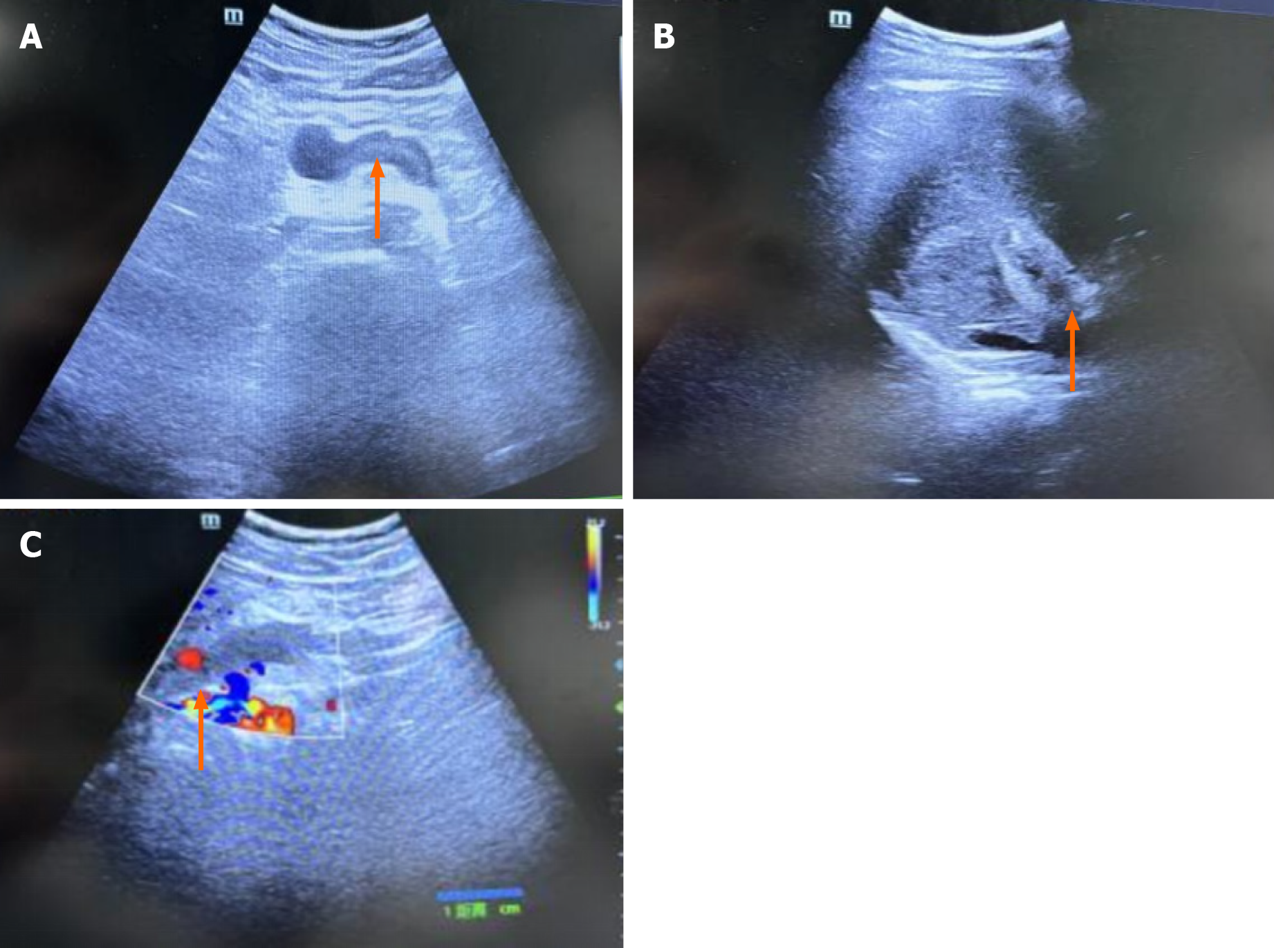
Subsequent abdominal computed tomography (CT) scanning revealed increased density in the portal and sub-mesenteric veins as well as the presence of a fecalith in the appendix (without appendicitis). Enhanced CT scans were then performed, which ruled out intestinal obstruction and appendicitis. The patient was informed about his condition and received dezocine for pain relief due to persistent abdominal pain. He underwent portal vein ultrasound (Figure 1), mesenteric CT angiography (CTA), and a whole-abdominal enhanced CT scan in the emergency department. Multiple emboli in the main portal vein as well as in the left and right branches of the portal vein, proximal SMV, and proximal splenic vein were detected. A slight decrease in the density of the sub-mesenteric vein lumen was observed, potentially attributable to an embolus. Weak enhancement in the hepatic gallbladder bed during the arterial phase and local perfusion abnormalities were also noted.

Figure 1
Figure 1 Ultrasonography of the abdominal and portal veins. A: The portal vein lumen displays hypoechoic filling with a solid echo (orange arrows), indicating thrombosis; B: Thrombosis in the lumen of the posterior pancreatic spleen vein (orange arrows) was suspected; C: The absence of a blood flow signal in the portal vein (orange arrows) suggests thrombosis.
MULTIDISCIPLINARY EXPERT CONSULTATION

The medical team informed the patient about his condition and recommended further examinations: Portal vein ultrasound, mesenteric CTA, and a comprehensive abdominal enhanced CT scan.

FINAL DIAGNOSIS

The patient received a prompt diagnosis of acute extensive portal vein system thrombosis and was admitted to the hospital for further treatment.

TREATMENT

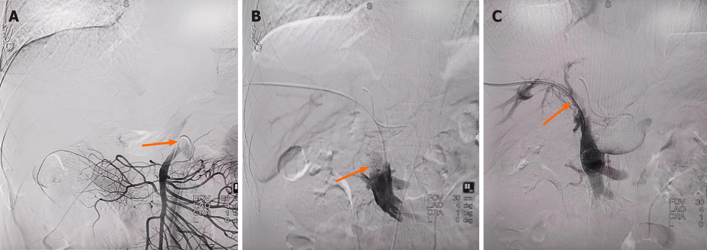
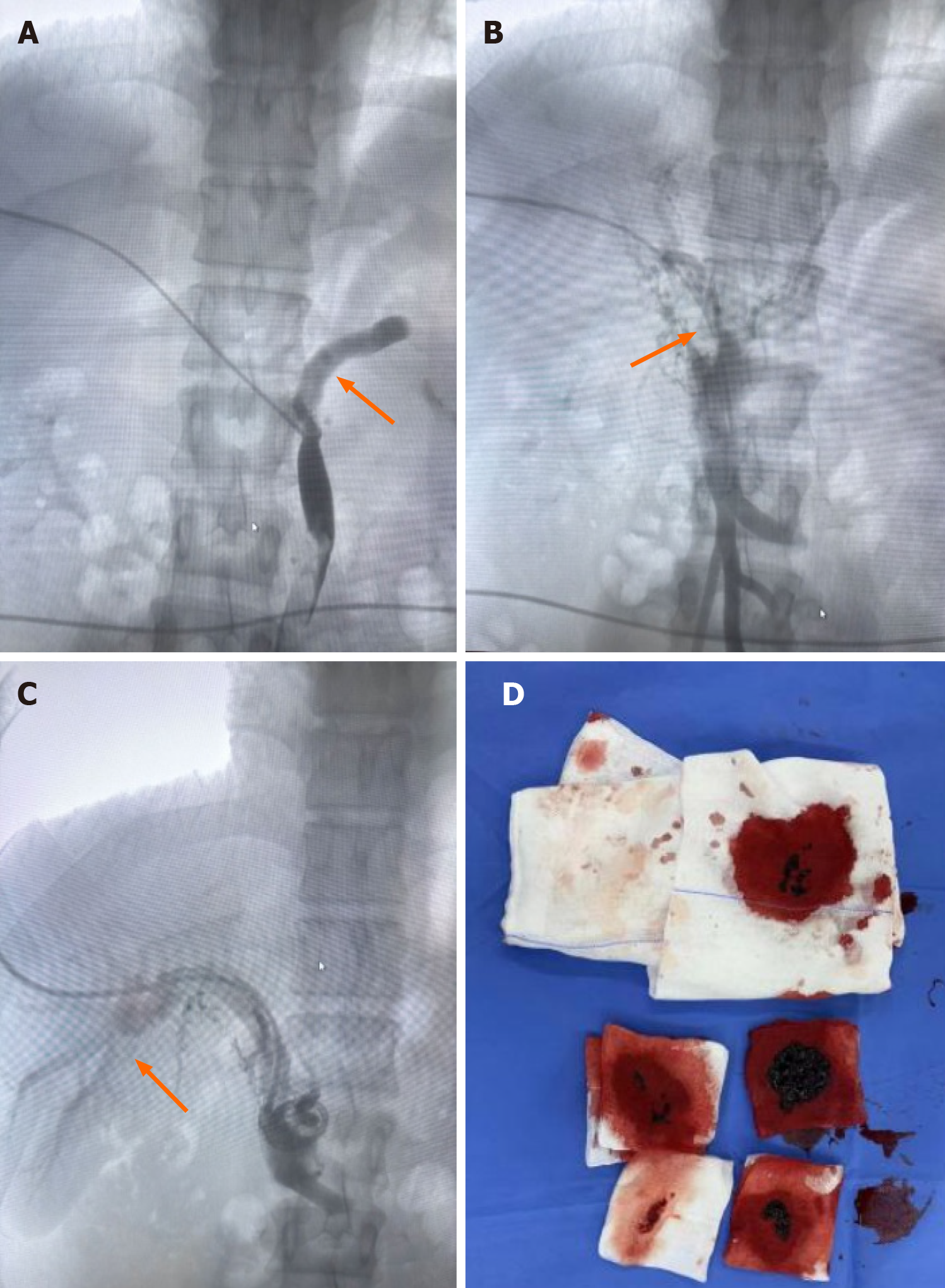
Anticoagulation treatment with 5000 IU of low molecular weight heparin was administered subcutaneously every 12 h. Emergency mesenteric venography and catheter-directed thrombolysis were performed to prevent paralytic ileus and intestinal ischaemic necrosis (Figure 2). The treatment regimen included continuous infusion of 100000 IU of urokinase along with 50 mL of 0.9% sodium chloride at a rate of 20000 IU/h. The patient underwent additional testing, including routine nasopharyngeal polymerase chain reaction (PCR) screening, which yielded a positive result for coronavirus disease 2019 (COVID-19). The results of additional laboratory investigations for protein S, protein C, antithrombin III, coagulation factor II, coagulation factor V, histidine-rich glycoprotein, thrombomodulin, and anti-cardiolipin antibodies were negative. Genetic testing revealed a heterozygous mutation in the upstream 2-kb region of plasminogen activator inhibitor-1, potentially linked to low-dose heparin usage. A homozygous missense mutation in methylenetetrahydrofolate reductase, suggesting an increased genetic predisposition to hyperhomocysteinaemia and an elevated thrombotic risk, was identified as well. Subsequently, the patient underwent mechanical thrombectomy on March 5, 2024 (Figure 3), during which 20 mL of thrombus was removed; he was then administered 15 mg of rivaroxaban twice daily.

Figure 2
Figure 2 Indirect and direct portal venography conducted on February 29, 2024. A: Indirect portal venography showed an underdeveloped portal vein (orange arrows); B: Direct portal vein angiography showed significant filling defects (orange arrows), indicating thrombosis; C: Partial visualisation of the portal vein (orange arrows) during intraoperative angiography after thrombolysis via an indwelling biliary drainage tube.
Figure 3
Figure 3 Portal venography and mechanical thrombectomy performed on March 5, 2024. A: Portal vein angiography showed persistent filling defects in the inferior mesenteric and splenic veins (orange arrows), suggesting thrombosis; B: Filling defects in the superior mesenteric vein (orange arrows), indicating thrombosis; C: Successful visualisation of the left and right branches of the portal vein (orange arrows) after mechanical thrombectomy; D: Approximately 20 mL of thrombus was aspirated following mechanical thrombectomy.

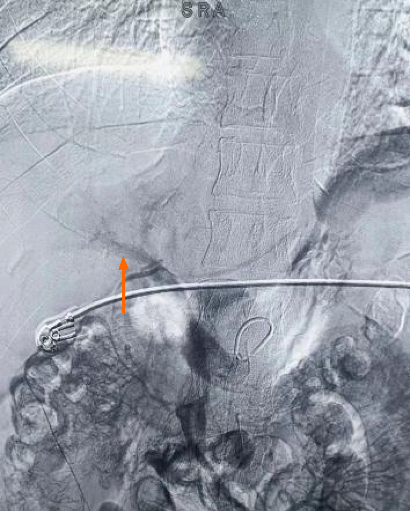
On March 7, 2024, mesenteric artery and vein venography with catheter-directed thrombolysis was performed again (Figure 4). Further treatment consisted of a continuous infusion of 200000 IU of urokinase and 50 mL of 0.9% sodium chloride at a rate of 20000 IU/h, in conjunction with 20 mg rivaroxaban taken once daily.

Figure 4
Figure 4 Indirect portal vein angiography after thrombolysis on March 7, 2024. Indirect portal vein angiography after repeated thrombolysis showed the development of the portal vein.
OUTCOME AND FOLLOW-UP

After 10 d of treatment, the patient's abdominal pain had significantly improved and indicators of infection had decreased notably. Repeat angiography demonstrated that blood flow in the portal vein had largely returned to normal. Due to the early diagnosis and prompt intervention, no complications, such as intestinal obstruction and bowel necrosis or shock, developed during hospitalisation. The patient was discharged on March 9, 2024. The 2-mo follow-up included an enhanced abdominal CT scan, which showed that the initial splenic vein thrombosis was no longer visible, the slightly high-density shadow in the gallbladder was absent, and there was a significant reduction in thrombosis in both the left and right branches of the portal vein and some of its tributaries. The presence of multiple enlarged, tortuous vascular shadows around the main trunk and branches of the portal vein suggested the development of collateral circulation (Figure 5).

Figure 5
Figure 5 Portal venography before and after treatment. A: Computed tomography angiography (CTA) performed on February 29, 2024, revealed an underdeveloped portal vein; B: Repeat CTA of the portal vein confirmed its development, and revealed that the portal vein was visible and collateral circulation has formed.
DISCUSSION
Causes of abdominal pain

There are two main types of mesenteric ischaemia: Non-occlusive and occlusive. The latter is subdivided into acute mesenteric artery embolism, acute mesenteric arterial thrombosis, and MVT. Mesenteric ischaemia is classified as acute or chronic based on disease severity. In 2017, the European Society for Vascular Surgery (ESVS)[10] defined AMI as a sudden cessation of blood flow to the small intestine, colon, or both, accompanied by symptoms that may last a few minutes (due to embolism) or a few hours (due to atherosclerosis). The European Guidelines for Chronic Mesenteric Ischemia, developed with research collaboration from seven professional societies, describe the symptoms of chronic mesenteric ischaemia as recurrent abdominal pain that intensifies after a meal, typically beginning 10-30 min after eating and lasting 1-2 h[11]. After a meal, the demand for oxygen to carry out digestion increases markedly, resulting in a rise in mesenteric blood flow of 30%-150%. MVT due to mechanical obstruction accounts for 5%-10% of ischaemia cases, while arterial ischaemia is primarily the result of obstruction (60%-70%) or low blood flow (20%-30%)[12]. A previous study[13] showed that patients with SMA embolism are at increased risk of developing an embolus in other solid organs, such as the spleen, liver, kidneys, and even brain. Chronic obstruction of the SMA is an uncommon cause of acute abdomen, and its diagnosis is often complicated by subtle symptoms, such that it may take several days for the condition to manifest clearly. However, the delayed onset of precursor symptoms and AMI are linked to survival outcomes, described as an ‘acute-chronic’ syndrome[14].

Given the potentially gradual progression of intestinal damage, diagnosing and treating AMI can present significant challenges, due to its diverse clinical presentations and non-specific initial symptoms, particularly in patients with arterial blockage. However, the incidence of AMI is low, although it increases exponentially with age and is similar for both men and women[15]. In older adults, AMI occurs more frequently than ruptured abdominal aortic aneurysm, appendicitis, and other causes of acute abdominal pain. Our patient presented with recurrent abdominal pain for a 3-d duration. Considering his medical history and current auxiliary examinations, his abdominal pain was likely to be due to obstructive mesenteric ischaemia caused by MVT, with clinical manifestations resembling an ‘acute-chronic’ syndrome (duration of symptoms lasting up to 3 d). Abdominal pain caused by MVT accounts for only a small fraction of ischaemia cases and is extremely rare, particularly in the younger population. A primary cause of mesenteric ischaemia in young adults, besides susceptibility due to hypercoagulable states or underlying vasculitis, is cocaine abuse. Heavy smoking may also increase the risk of mesenteric arterial occlusive disease and mesenteric ischaemia in young patients[9].

Diagnosis of the cause of abdominal pain (MVT)

Early enhanced CT scans are critical in patients with suspected acute mesenteric ischaemia, to determine treatment strategies. Dynamic enhanced CT, with a sensitivity of 90%, is the preferred diagnostic method for suspected cases of MVT. CT is more sensitive in detecting venous thrombosis than other forms of acute mesenteric ischaemia, and can specifically reveal venous filling defects[16]. Case reports suggest that, in the diagnosis of AMI, repeated echocardiographic assessments of the presence of the ‘aquarium sign’, caused by the entry of hepatic portal venous gas into the right atrium and ventricle, can be valuable. Point-of-care ultrasound may also be a useful diagnostic tool for AMI in patients in whom there is active suspicion of AMI based on detection of the aquarium sign[17]. At present, no individual test or combination of tests has demonstrated consistently reliable sensitivity or specificity for diagnosing AMI[4]. Leukocyte count, serum amylase, lactate dehydrogenase, D-dimer, and lactate levels may be informative in the differential diagnosis of AMI. However, since CRP and lactate levels may be normal in the early stages of AMI, normal values of these indicators do not rule out the diagnosis. About half of the patients with AMI have elevated levels of troponin T, and D-dimer levels may increase during SMA embolism[18]. The ESVS guidelines recommend D-dimer as an exclusion test for AMI but stress its ineffectiveness in cases of acute to chronic mesenteric ischaemia[15]. In our patient, CRP and WBC levels were elevated, while troponin T, D-dimer, and lactate levels remained normal. Abdominal CT indicated increased density in the portal and inferior mesenteric veins, leading to a consideration of embolism. The diagnosis was confirmed using colour Doppler ultrasound and enhanced CT.

Factors contributing to MVT susceptibility

The patient had no personal or family history of thromboembolic events. After admission, diagnostic tests were performed to determine the potential causes of MVT, including acquired (e.g., tumours) or hereditary (e.g. genetic mutations or deficiencies) thrombophilia. His laboratory tests yielded negative results for protein S, protein C, antithrombin III, coagulation factor II, coagulation factor V, histidine-rich glycoprotein, thrombomodulin, and anti-cardiolipin antibodies. Genetic testing identified a homozygous missense mutation in methylenetetrahydrofolate reductase, indicating an increased genetic predisposition to hyperhomocysteinaemia and a heightened thrombotic risk. Additionally, routine nasopharyngeal PCR screening yielded a positive result for COVID-19. Interactions between hereditary thrombophilia factors and severe acute respiratory syndrome coronavirus 2 (SARS-CoV-2) infection may thus have played a role in triggering MVT. Viral-infection-induced thrombosis can be attributed to inflammation triggered by the infection, leading to platelet activation and clot formation as platelets adhere to damaged cells and activate clotting factors[19]. The development of MVT is associated with a broad range of factors[17], including hypercoagulable states (deficiencies in protein C, protein S, or antithrombin III, factor V Leiden mutation), anticardiolipin antibodies, malignancy, oestrogen influences, pregnancy, peripheral deep vein thrombosis, and conditions such as portal hypertension, intra-abdominal inflammation, sepsis, postoperative status, and trauma. Prothrombin mutation, protein C deficiency, protein S deficiency, antithrombin deficiency, impaired fibrinolysis (reduced levels of anti-tissue plasminogen activator), hyperviscosity syndrome, pancreatitis, various blood disorders, and the use of oral contraceptives can lead to secondary intestinal oedema due to venous thrombosis and increased vascular resistance; the resulting reduction in arterial blood flow leads to intestinal ischaemia[10]. Antithrombin-III deficiency is a significant factor in ‘primary’ MVT[20,21]. In individuals with inherited antithrombin-III deficiency, the activity of this protein usually decreases by around 50%, despite normal results in standard coagulation or fibrinolysis tests. The higher risk of venous thrombosis often manifests in adulthood.

COVID-19 not only affects the respiratory system but also induces thrombotic ischaemia, including in the gastrointestinal tract. Emerging evidence[22] links SARS-CoV-2 with endothelins, which can cause endothelial damage, inflammatory cell infiltration, and a systemic prothrombotic state triggered by excessive inflammation and elevated cytokine levels. COVID-19 is also associated with an elevated risk of diverse thromboembolic complications[23-25], including AMI, deep vein thrombosis, and pulmonary embolism. A hypercoagulable state may be induced by systemic inflammation, endothelial activation, and hypoxia. The virus's affinity for vascular endothelial cells can be attributed to their expression of ACE2, the receptor that facilitates viral entry. The resulting endothelial injury triggers the release of substantial amounts of von Willebrand factor, further increasing the risk of thrombus formation, which in turn can induce hemodynamic changes and direct injury to intestinal cells, ultimately resulting in ischaemia and necrosis of the small intestine. However, the pathogenesis of COVID-19-associated AMI has not been extensively studied, although it may involve direct viral mucosal damage, intestinal ischaemia, and the atrophy of lymphoid follicles accompanied by increased mucosal permeability[26].

MVT treatment

Non-randomised trials of patients with acute thrombotic occlusion have demonstrated[27-31] a reduced need for bowel resections and lower short- and long-term mortality with endovascular interventions rather than open surgery. The ESVS guidelines[19] state that the 30-d mortality rate after endovascular intervention is 17.2% (367/2131), while for open surgery it is 38.5%. A recent meta-analysis[32] determined a 94% technical success rate for endovascular intervention, with a 40% re-operation rate post-treatment. Various endovascular procedures for the management of portal vein thrombosis have been developed[33,34], such as transjugular intrahepatic portosystemic shunt combined with mechanical thrombectomy[35] and direct thrombolysis[36,37], percutaneous transhepatic mechanical thrombectomy[38], percutaneous transhepatic thrombolysis[39,40], SMA thrombolysis[41], and percutaneous SMV catheter placement for thrombolysis[42]. A study by Kärkkäinen et al[15] found that the mortality rate from acute mesenteric ischaemia can reach 80% without prompt surgery to restore blood flow, but early intervention can reduce mortality by 50%. In rare cases, partial intestinal resection with anticoagulant therapy may be the most effective. In unavoidable laparotomy situations, open thrombectomy, surgical bypass, or hybrid stent placement can quickly restore intestinal blood flow. Medical treatment includes the use of antibiotics, vasodilators, thrombolytic agents, and anticoagulants. Experimental drugs for primary ischaemia and reperfusion injury are also under investigation. Anticoagulation is the primary treatment for MVT, and the early administration of heparin can increase survival[30]. However, heparin cannot be the sole treatment for acute thrombosis in patients with antithrombin III deficiency, as it can lead to varying degrees of heparin resistance, potentially worsening the thrombotic condition, due to heparin's diverse effects[24].

CONCLUSION

Clinicians should remain vigilant for acute MVT in young patients presenting with abdominal pain, to prevent misdiagnosis of this potentially fatal condition.

ACKNOWLEDGEMENTS

The authors express their sincere gratitude to all individuals who contributed to this research in any capacity.

Footnotes

Provenance and peer review: Unsolicited article; Externally peer reviewed.

Peer-review model: Single blind

Specialty type: Emergency medicine

Country of origin: China

Peer-review report’s classification

Scientific Quality: Grade C, Grade C, Grade D

Novelty: Grade B, Grade C, Grade C

Creativity or Innovation: Grade B, Grade C, Grade D

Scientific Significance: Grade B, Grade C, Grade C

P-Reviewer: Shukla A; Singh SP; Zou J S-Editor: Liu JH L-Editor: Wang TQ P-Editor: Guo X

References
1.  Alzubaidi S, Patel I, Saini A, Knuttinen G, Naidu S, Kriegshuaser S, Albadawi H, Oklu R. Current concepts in portal vein thrombosis: etiology, clinical presentation and management. Abdom Radiol (NY). 2019;44:3453-3462.  [PubMed]  [DOI]  [Cited in This Article: ]  [Cited by in Crossref: 6]  [Cited by in F6Publishing: 11]  [Article Influence: 1.8]  [Reference Citation Analysis (0)]
2.  Yang F, Guo XC, Rao XL, Sun L, Xu L. Acute appendicitis complicated by mesenteric vein thrombosis: A case report. World J Clin Cases. 2021;9:11400-11405.  [PubMed]  [DOI]  [Cited in This Article: ]  [Reference Citation Analysis (0)]
3.  Furlan A, Marin D, Cabassa P, Taibbi A, Brunelli E, Agnello F, Lagalla R, Brancatelli G. Enhancement pattern of small hepatocellular carcinoma (HCC) at contrast-enhanced US (CEUS), MDCT, and MRI: intermodality agreement and comparison of diagnostic sensitivity between 2005 and 2010 American Association for the Study of Liver Diseases (AASLD) guidelines. Eur J Radiol. 2012;81:2099-2105.  [PubMed]  [DOI]  [Cited in This Article: ]  [Cited by in Crossref: 35]  [Cited by in F6Publishing: 34]  [Article Influence: 2.6]  [Reference Citation Analysis (0)]
4.  Ozturk G, Aydinli B, Atamanalp SS, Yildirgan MI, Ozoğul B, Kısaoğlu A. Acute mesenteric ischemia in young adults. Wien Med Wochenschr. 2012;162:349-353.  [PubMed]  [DOI]  [Cited in This Article: ]  [Cited by in Crossref: 15]  [Cited by in F6Publishing: 15]  [Article Influence: 1.2]  [Reference Citation Analysis (0)]
5.  Ottinger LW. Mesenteric ischemia. N Engl J Med. 1982;307:535-537.  [PubMed]  [DOI]  [Cited in This Article: ]  [Cited by in Crossref: 69]  [Cited by in F6Publishing: 69]  [Article Influence: 1.6]  [Reference Citation Analysis (0)]
6.  Acosta S, Wadman M, Syk I, Elmståhl S, Ekberg O. Epidemiology and prognostic factors in acute superior mesenteric artery occlusion. J Gastrointest Surg. 2010;14:628-635.  [PubMed]  [DOI]  [Cited in This Article: ]  [Cited by in Crossref: 40]  [Cited by in F6Publishing: 32]  [Article Influence: 2.1]  [Reference Citation Analysis (0)]
7.  Acosta S, Björck M. Acute thrombo-embolic occlusion of the superior mesenteric artery: a prospective study in a well defined population. Eur J Vasc Endovasc Surg. 2003;26:179-183.  [PubMed]  [DOI]  [Cited in This Article: ]  [Cited by in Crossref: 94]  [Cited by in F6Publishing: 85]  [Article Influence: 3.9]  [Reference Citation Analysis (0)]
8.  Kärkkäinen JM, Lehtimäki TT, Manninen H, Paajanen H. Acute Mesenteric Ischemia Is a More Common Cause than Expected of Acute Abdomen in the Elderly. J Gastrointest Surg. 2015;19:1407-1414.  [PubMed]  [DOI]  [Cited in This Article: ]  [Cited by in Crossref: 79]  [Cited by in F6Publishing: 83]  [Article Influence: 8.3]  [Reference Citation Analysis (0)]
9.  Bala M, Catena F, Kashuk J, De Simone B, Gomes CA, Weber D, Sartelli M, Coccolini F, Kluger Y, Abu-Zidan FM, Picetti E, Ansaloni L, Augustin G, Biffl WL, Ceresoli M, Chiara O, Chiarugi M, Coimbra R, Cui Y, Damaskos D, Di Saverio S, Galante JM, Khokha V, Kirkpatrick AW, Inaba K, Leppäniemi A, Litvin A, Peitzman AB, Shelat VG, Sugrue M, Tolonen M, Rizoli S, Sall I, Beka SG, Di Carlo I, Ten Broek R, Mircea C, Tebala G, Pisano M, van Goor H, Maier RV, Jeekel H, Civil I, Hecker A, Tan E, Soreide K, Lee MJ, Wani I, Bonavina L, Malangoni MA, Koike K, Velmahos GC, Fraga GP, Fette A, de'Angelis N, Balogh ZJ, Scalea TM, Sganga G, Kelly MD, Khan J, Stahel PF, Moore EE. Acute mesenteric ischemia: updated guidelines of the World Society of Emergency Surgery. World J Emerg Surg. 2022;17:54.  [PubMed]  [DOI]  [Cited in This Article: ]  [Cited by in Crossref: 10]  [Cited by in F6Publishing: 97]  [Article Influence: 32.3]  [Reference Citation Analysis (0)]
10.  Becquemin JP. Management of the Diseases of Mesenteric Arteries and Veins: Clinical Practice Guidelines of the European Society for Vascular Surgery (ESVS) [corrected]. Eur J Vasc Endovasc Surg. 2017;53:455-457.  [PubMed]  [DOI]  [Cited in This Article: ]  [Cited by in Crossref: 14]  [Cited by in F6Publishing: 17]  [Article Influence: 2.8]  [Reference Citation Analysis (0)]
11.  Terlouw LG, Moelker A, Abrahamsen J, Acosta S, Bakker OJ, Baumgartner I, Boyer L, Corcos O, van Dijk LJ, Duran M, Geelkerken RH, Illuminati G, Jackson RW, Kärkkäinen JM, Kolkman JJ, Lönn L, Mazzei MA, Nuzzo A, Pecoraro F, Raupach J, Verhagen HJ, Zech CJ, van Noord D, Bruno MJ. European guidelines on chronic mesenteric ischaemia - joint United European Gastroenterology, European Association for Gastroenterology, Endoscopy and Nutrition, European Society of Gastrointestinal and Abdominal Radiology, Netherlands Association of Hepatogastroenterologists, Hellenic Society of Gastroenterology, Cardiovascular and Interventional Radiological Society of Europe, and Dutch Mesenteric Ischemia Study group clinical guidelines on the diagnosis and treatment of patients with chronic mesenteric ischaemia. United European Gastroenterol J. 2020;8:371-395.  [PubMed]  [DOI]  [Cited in This Article: ]  [Cited by in Crossref: 104]  [Cited by in F6Publishing: 85]  [Article Influence: 17.0]  [Reference Citation Analysis (0)]
12.  Niang FG, Faye I, Ndong A, Diedhiou M, Niang I, Diop AD, Diop AN. Acute mesenteric ischemia: A case report. Radiol Case Rep. 2024;19:150-152.  [PubMed]  [DOI]  [Cited in This Article: ]  [Reference Citation Analysis (0)]
13.  Kärkkäinen JM, Lehtimäki TT, Saari P, Hartikainen J, Rantanen T, Paajanen H, Manninen H. Endovascular Therapy as a Primary Revascularization Modality in Acute Mesenteric Ischemia. Cardiovasc Intervent Radiol. 2015;38:1119-1129.  [PubMed]  [DOI]  [Cited in This Article: ]  [Cited by in Crossref: 41]  [Cited by in F6Publishing: 46]  [Article Influence: 4.6]  [Reference Citation Analysis (0)]
14.  Björnsson S, Resch T, Acosta S. Symptomatic mesenteric atherosclerotic disease-lessons learned from the diagnostic workup. J Gastrointest Surg. 2013;17:973-980.  [PubMed]  [DOI]  [Cited in This Article: ]  [Cited by in Crossref: 44]  [Cited by in F6Publishing: 45]  [Article Influence: 3.8]  [Reference Citation Analysis (0)]
15.  Kärkkäinen JM. Acute Mesenteric Ischemia: A Challenge for the Acute Care Surgeon. Scand J Surg. 2021;110:150-158.  [PubMed]  [DOI]  [Cited in This Article: ]  [Cited by in Crossref: 13]  [Cited by in F6Publishing: 15]  [Article Influence: 3.8]  [Reference Citation Analysis (0)]
16.  Bradbury MS, Kavanagh PV, Bechtold RE, Chen MY, Ott DJ, Regan JD, Weber TM. Mesenteric venous thrombosis: diagnosis and noninvasive imaging. Radiographics. 2002;22:527-541.  [PubMed]  [DOI]  [Cited in This Article: ]  [Cited by in Crossref: 144]  [Cited by in F6Publishing: 151]  [Article Influence: 6.6]  [Reference Citation Analysis (0)]
17.  Higashijima U, Sekino M, Iwasaki N, Araki H, Motokawa T, Inoue Y, Taniguchi Y, Sato S, Miyazaki Y, Hara T. Acute mesenteric ischemia diagnosed using the aquarium sign: A case report. Medicine (Baltimore). 2023;102:e33735.  [PubMed]  [DOI]  [Cited in This Article: ]  [Reference Citation Analysis (0)]
18.  Björck M, Koelemay M, Acosta S, Bastos Goncalves F, Kölbel T, Kolkman JJ, Lees T, Lefevre JH, Menyhei G, Oderich G;  Esvs Guidelines Committee; Kolh P, de Borst GJ, Chakfe N, Debus S, Hinchliffe R, Kakkos S, Koncar I, Sanddal Lindholt J, Vega de Ceniga M, Vermassen F, Verzini F, Document Reviewers, Geelkerken B, Gloviczki P, Huber T, Naylor R. Editor's Choice - Management of the Diseases of Mesenteric Arteries and Veins: Clinical Practice Guidelines of the European Society of Vascular Surgery (ESVS). Eur J Vasc Endovasc Surg. 2017;53:460-510.  [PubMed]  [DOI]  [Cited in This Article: ]  [Cited by in Crossref: 255]  [Cited by in F6Publishing: 381]  [Article Influence: 54.4]  [Reference Citation Analysis (1)]
19.  Oh GM, Jung K, Kim JH, Kim SE, Moon W, Park MI, Park SJ. Superior mesenteric vein thrombosis induced by influenza infection: A case report. World J Clin Cases. 2020;8:4193-4199.  [PubMed]  [DOI]  [Cited in This Article: ]  [Cited by in CrossRef: 2]  [Cited by in F6Publishing: 2]  [Article Influence: 0.4]  [Reference Citation Analysis (0)]
20.  Rosendaal FR, Heijboer H, Briët E, Büller HR, Brandjes DP, de Bruin K, Hommes DW, Vandenbroucke JP. Mortality in hereditary antithrombin-III deficiency--1830 to 1989. Lancet. 1991;337:260-262.  [PubMed]  [DOI]  [Cited in This Article: ]  [Cited by in Crossref: 51]  [Cited by in F6Publishing: 54]  [Article Influence: 1.6]  [Reference Citation Analysis (0)]
21.  Gruenberg JC, Smallridge RC, Rosenberg RD. Inherited antithrombin-III deficiency causing mesenteric venous infarction: a new clinical entity. Ann Surg. 1975;181:791-794.  [PubMed]  [DOI]  [Cited in This Article: ]  [Cited by in Crossref: 48]  [Cited by in F6Publishing: 54]  [Article Influence: 1.1]  [Reference Citation Analysis (0)]
22.  Sehhat S, Talebzadeh H, Hakamifard A, Melali H, Shabib S, Rahmati A, Larki-Harchegani A. Acute Mesenteric Ischemia in a Patient with COVID-19: A Case Report. Arch Iran Med. 2020;23:639-643.  [PubMed]  [DOI]  [Cited in This Article: ]  [Cited by in Crossref: 6]  [Cited by in F6Publishing: 6]  [Article Influence: 1.2]  [Reference Citation Analysis (0)]
23.  Lodigiani C, Iapichino G, Carenzo L, Cecconi M, Ferrazzi P, Sebastian T, Kucher N, Studt JD, Sacco C, Bertuzzi A, Sandri MT, Barco S; Humanitas COVID-19 Task Force. Venous and arterial thromboembolic complications in COVID-19 patients admitted to an academic hospital in Milan, Italy. Thromb Res. 2020;191:9-14.  [PubMed]  [DOI]  [Cited in This Article: ]  [Cited by in Crossref: 1298]  [Cited by in F6Publishing: 1497]  [Article Influence: 299.4]  [Reference Citation Analysis (0)]
24.  Parry AH, Wani AH, Yaseen M. Acute Mesenteric Ischemia in Severe Coronavirus-19 (COVID-19): Possible Mechanisms and Diagnostic Pathway. Acad Radiol. 2020;27:1190.  [PubMed]  [DOI]  [Cited in This Article: ]  [Cited by in Crossref: 60]  [Cited by in F6Publishing: 62]  [Article Influence: 12.4]  [Reference Citation Analysis (0)]
25.  Pirola L, Palermo A, Mulinacci G, Ratti L, Fichera M, Invernizzi P, Viganò C, Massironi S. Acute mesenteric ischemia and small bowel imaging findings in COVID-19: A comprehensive review of the literature. World J Gastrointest Surg. 2021;13:702-716.  [PubMed]  [DOI]  [Cited in This Article: ]  [Cited by in CrossRef: 11]  [Cited by in F6Publishing: 7]  [Article Influence: 1.8]  [Reference Citation Analysis (0)]
26.  Devgun P, Hassan H. Pneumatosis cystoides intestinalis: a rare benign cause of pneumoperitoneum. Case Rep Radiol. 2013;2013:353245.  [PubMed]  [DOI]  [Cited in This Article: ]  [Cited by in Crossref: 8]  [Cited by in F6Publishing: 15]  [Article Influence: 1.3]  [Reference Citation Analysis (1)]
27.  Block TA, Acosta S, Björck M. Endovascular and open surgery for acute occlusion of the superior mesenteric artery. J Vasc Surg. 2010;52:959-966.  [PubMed]  [DOI]  [Cited in This Article: ]  [Cited by in Crossref: 88]  [Cited by in F6Publishing: 86]  [Article Influence: 5.7]  [Reference Citation Analysis (0)]
28.  Ryer EJ, Kalra M, Oderich GS, Duncan AA, Gloviczki P, Cha S, Bower TC. Revascularization for acute mesenteric ischemia. J Vasc Surg. 2012;55:1682-1689.  [PubMed]  [DOI]  [Cited in This Article: ]  [Cited by in Crossref: 119]  [Cited by in F6Publishing: 127]  [Article Influence: 9.8]  [Reference Citation Analysis (0)]
29.  Arthurs ZM, Titus J, Bannazadeh M, Eagleton MJ, Srivastava S, Sarac TP, Clair DG. A comparison of endovascular revascularization with traditional therapy for the treatment of acute mesenteric ischemia. J Vasc Surg. 2011;53:698-704; discussion 704.  [PubMed]  [DOI]  [Cited in This Article: ]  [Cited by in Crossref: 174]  [Cited by in F6Publishing: 189]  [Article Influence: 13.5]  [Reference Citation Analysis (0)]
30.  Schermerhorn ML, Giles KA, Hamdan AD, Wyers MC, Pomposelli FB. Mesenteric revascularization: management and outcomes in the United States, 1988-2006. J Vasc Surg. 2009;50:341-348.e1.  [PubMed]  [DOI]  [Cited in This Article: ]  [Cited by in Crossref: 214]  [Cited by in F6Publishing: 172]  [Article Influence: 10.8]  [Reference Citation Analysis (0)]
31.  Acosta S. Surgical management of peritonitis secondary to acute superior mesenteric artery occlusion. World J Gastroenterol. 2014;20:9936-9941.  [PubMed]  [DOI]  [Cited in This Article: ]  [Cited by in CrossRef: 48]  [Cited by in F6Publishing: 37]  [Article Influence: 3.4]  [Reference Citation Analysis (0)]
32.  Heiss P, Loewenhardt B, Manke C, Hellinger A, Dietl KH, Schlitt HJ, Scheibl K, Feuerbach S, Paetzel C. Primary percutaneous aspiration and thrombolysis for the treatment of acute embolic superior mesenteric artery occlusion. Eur Radiol. 2010;20:2948-2958.  [PubMed]  [DOI]  [Cited in This Article: ]  [Cited by in Crossref: 39]  [Cited by in F6Publishing: 42]  [Article Influence: 2.8]  [Reference Citation Analysis (0)]
33.  Hawthorn BR, Ratnam LA. Acute mesenteric ischaemia: imaging and intervention. Clin Radiol. 2020;75:398.e19-398.e28.  [PubMed]  [DOI]  [Cited in This Article: ]  [Cited by in Crossref: 2]  [Cited by in F6Publishing: 4]  [Article Influence: 0.7]  [Reference Citation Analysis (0)]
34.  Acosta S, Björck M. Modern treatment of acute mesenteric ischaemia. Br J Surg. 2014;101:e100-e108.  [PubMed]  [DOI]  [Cited in This Article: ]  [Cited by in Crossref: 118]  [Cited by in F6Publishing: 97]  [Article Influence: 8.1]  [Reference Citation Analysis (0)]
35.  Alnahhal K, Toskich BB, Nussbaum S, Li Z, Erben Y, Hakaim AG, Farres H. Superior mesenteric venous thrombosis: Endovascular management and outcomes. World J Clin Cases. 2022;10:217-226.  [PubMed]  [DOI]  [Cited in This Article: ]  [Cited by in CrossRef: 3]  [Cited by in F6Publishing: 2]  [Article Influence: 0.7]  [Reference Citation Analysis (3)]
36.  Ferro C, Rossi UG, Bovio G, Dahamane M, Centanaro M. Transjugular intrahepatic portosystemic shunt, mechanical aspiration thrombectomy, and direct thrombolysis in the treatment of acute portal and superior mesenteric vein thrombosis. Cardiovasc Intervent Radiol. 2007;30:1070-1074.  [PubMed]  [DOI]  [Cited in This Article: ]  [Cited by in Crossref: 53]  [Cited by in F6Publishing: 54]  [Article Influence: 3.0]  [Reference Citation Analysis (0)]
37.  Wang MQ, Lin HY, Guo LP, Liu FY, Duan F, Wang ZJ. Acute extensive portal and mesenteric venous thrombosis after splenectomy: treated by interventional thrombolysis with transjugular approach. World J Gastroenterol. 2009;15:3038-3045.  [PubMed]  [DOI]  [Cited in This Article: ]  [Cited by in CrossRef: 17]  [Cited by in F6Publishing: 15]  [Article Influence: 0.9]  [Reference Citation Analysis (0)]
38.  Takahashi N, Kuroki K, Yanaga K. Percutaneous transhepatic mechanical thrombectomy for acute mesenteric venous thrombosis. J Endovasc Ther. 2005;12:508-511.  [PubMed]  [DOI]  [Cited in This Article: ]  [Cited by in Crossref: 23]  [Cited by in F6Publishing: 18]  [Article Influence: 0.9]  [Reference Citation Analysis (0)]
39.  Zhou W, Choi L, Lin PH, Dardik A, Eraso A, Lumsden AB. Percutaneous transhepatic thrombectomy and pharmacologic thrombolysis of mesenteric venous thrombosis. Vascular. 2007;15:41-45.  [PubMed]  [DOI]  [Cited in This Article: ]  [Cited by in Crossref: 25]  [Cited by in F6Publishing: 26]  [Article Influence: 2.9]  [Reference Citation Analysis (0)]
40.  Di Minno MN, Milone F, Milone M, Iaccarino V, Venetucci P, Lupoli R, Sosa Fernandez LM, Di Minno G. Endovascular Thrombolysis in Acute Mesenteric Vein Thrombosis: a 3-year follow-up with the rate of short and long-term sequaelae in 32 patients. Thromb Res. 2010;126:295-298.  [PubMed]  [DOI]  [Cited in This Article: ]  [Cited by in Crossref: 30]  [Cited by in F6Publishing: 24]  [Article Influence: 1.6]  [Reference Citation Analysis (0)]
41.  Hollingshead M, Burke CT, Mauro MA, Weeks SM, Dixon RG, Jaques PF. Transcatheter thrombolytic therapy for acute mesenteric and portal vein thrombosis. J Vasc Interv Radiol. 2005;16:651-661.  [PubMed]  [DOI]  [Cited in This Article: ]  [Cited by in Crossref: 208]  [Cited by in F6Publishing: 187]  [Article Influence: 9.4]  [Reference Citation Analysis (0)]
42.  Ozdogan M, Gurer A, Gokakin AK, Kulacoglu H, Aydin R. Thrombolysis via an operatively placed mesenteric catheter for portal and superior mesenteric vein thrombosis: report of a case. Surg Today. 2006;36:846-848.  [PubMed]  [DOI]  [Cited in This Article: ]  [Cited by in Crossref: 20]  [Cited by in F6Publishing: 21]  [Article Influence: 1.1]  [Reference Citation Analysis (0)]