©The Author(s) 2024.
World J Radiol. Oct 28, 2024; 16(10): 545-551
Published online Oct 28, 2024. doi: 10.4329/wjr.v16.i10.545
Published online Oct 28, 2024. doi: 10.4329/wjr.v16.i10.545
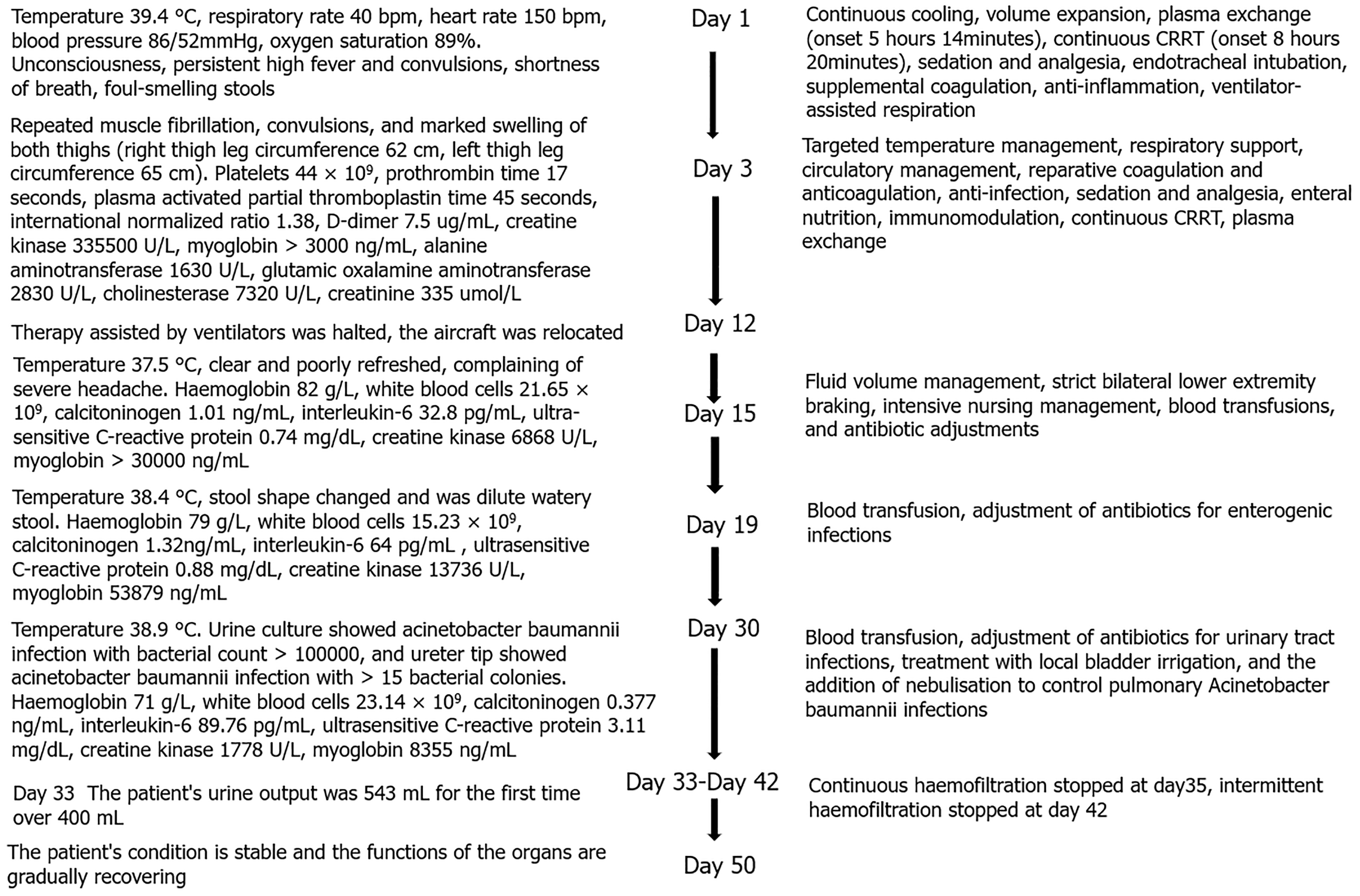
Figure 1 Significant clinical data and treatment duration axis of the patient during hospitalization.
CRRT: Continuous renal replacement therapy.
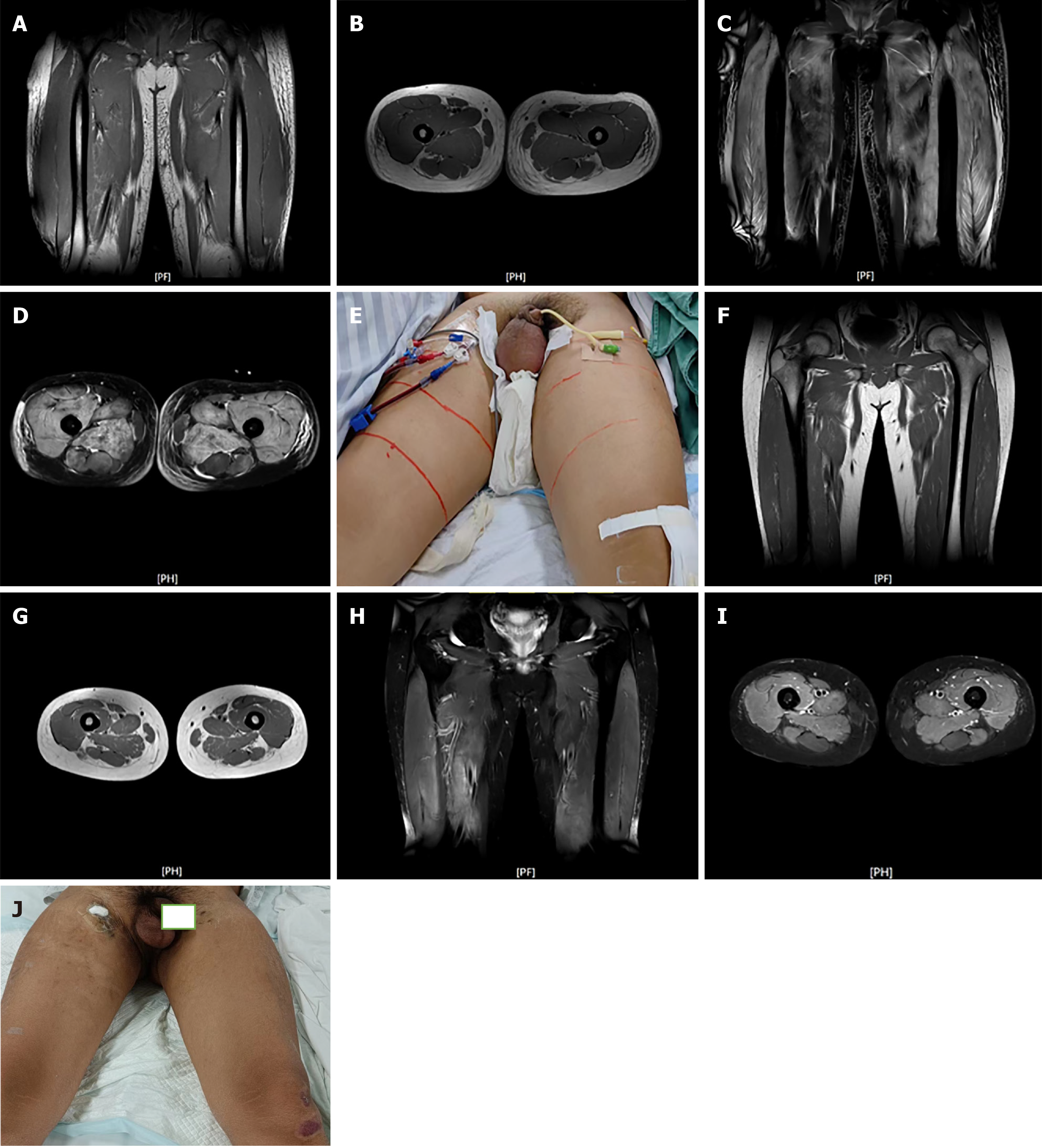
Figure 2 Upper femoral segment magnetic resonance imaging and appearance of thigh muscles.
A and B: On day 18 of disease onset, coronal and transverse positions show symmetrical swelling, edema, and abnormal patches in bilateral thigh muscle groups, and unclear inter-muscular spaces; C and D: Coronal and transverse positions show T2-weighted imaging (T2WI) fat-suppressed images revealing patches and flocculate hyperintense signals; E: Signal swelling of the thigh and scrotum; F and G: On day 64 of disease onset, T1-weighted images show coronal and transverse positions revealing significant alleviation of swelling in bilateral thigh muscle groups; H and I: No significantly abnormal signals seen in the T2WI fat-suppressed images shown in coronal and transverse positions; J: Resolution of thigh and scrotal swelling.
- Citation: Xiang CH, Zhang XM, Liu J, Xiang J, Li L, Song Q. Exertional heat stroke with pronounced presentation of microangiopathic hemolytic anemia: A case report. World J Radiol 2024; 16(10): 545-551
- URL: https://www.wjgnet.com/1949-8470/full/v16/i10/545.htm
- DOI: https://dx.doi.org/10.4329/wjr.v16.i10.545


