©The Author(s) 2023.
World J Radiol. Aug 28, 2023; 15(8): 250-255
Published online Aug 28, 2023. doi: 10.4329/wjr.v15.i8.250
Published online Aug 28, 2023. doi: 10.4329/wjr.v15.i8.250
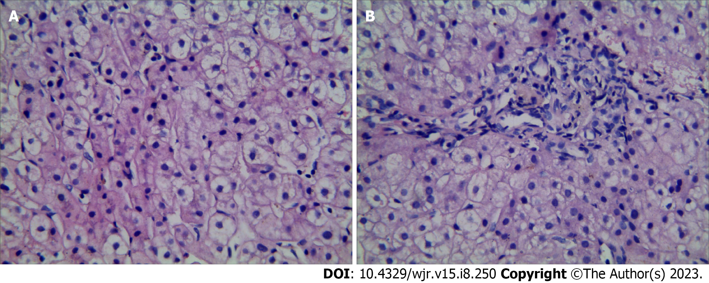
Figure 1 The number and shape of small bile ducts in the portal area were normal, and the stroma was infiltrated by a small number of chronic inflammatory cells.
A and B: Punctured hepatic tissue with hematoxylin and eosin staining (× 400).
Figure 2 After entering the splenic hilum, the splenic vein formed collateral circulation along the lower left side of the vertebral body, resulting in a venous tumor thrombus, and then flowed into the renal vein and returned to the inferior vena cava.
A: The main portal vein was thin, and no portal vein branch was found in the liver (arrow); B: The vessels were disordered at the proximal main portal vein, and esophageal varices were caused by collateral circulation upward at the portal vein origin. The superior mesenteric vein and the splenic vein formed a common trunk (arrow); C and D: After entering the splenic hilum, the splenic vein established collateral circulation (forming a venous tumour), flowed into the renal vein and returned to the inferior vena cava (arrow).
- Citation: Yao X, Liu Y, Yu LD, Qin JP. Rare portal hypertension caused by Abernethy malformation (Type IIC): A case report. World J Radiol 2023; 15(8): 250-255
- URL: https://www.wjgnet.com/1949-8470/full/v15/i8/250.htm
- DOI: https://dx.doi.org/10.4329/wjr.v15.i8.250


