Copyright
©The Author(s) 2025.
World J Gastrointest Surg. Sep 27, 2025; 17(9): 109320
Published online Sep 27, 2025. doi: 10.4240/wjgs.v17.i9.109320
Published online Sep 27, 2025. doi: 10.4240/wjgs.v17.i9.109320
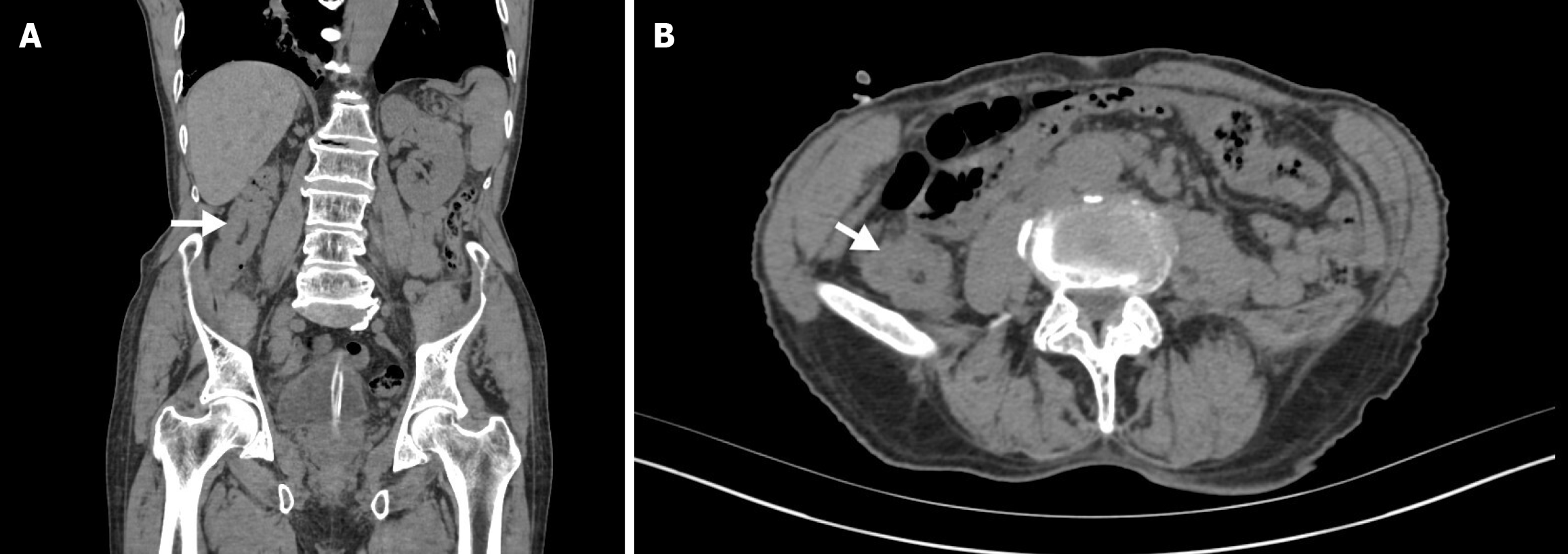
Figure 1 Abdominal computed tomography.
A: It shows abdominal computed tomography (CT) in the coronal plane; the white arrow indicates the appendix invaginated into the ascending colon, which could easily be misdiagnosed as an ascending colon tumor; B: It shows abdominal CT in the axial plane, the white arrow indicates thickening of the ascending colon wall, which could be misdiagnosed as an ascending colon tumor.
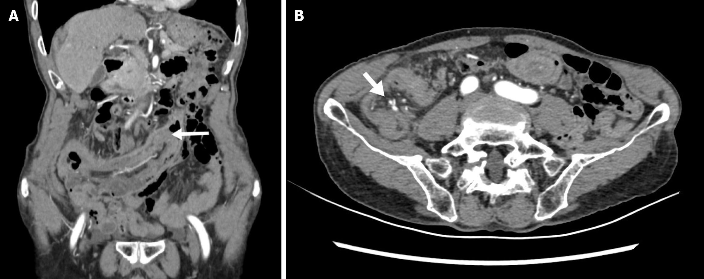
Figure 2 Enhanced abdominal computed tomography.
A: It shows enhanced abdominal computed tomography (CT) in the coronal plane; the white arrow indicates the intussuscepted distal appendix reaching the transverse colon; B: It shows enhanced abdominal CT in the axial plane, the white arrow indicates the ideal blood vessels.
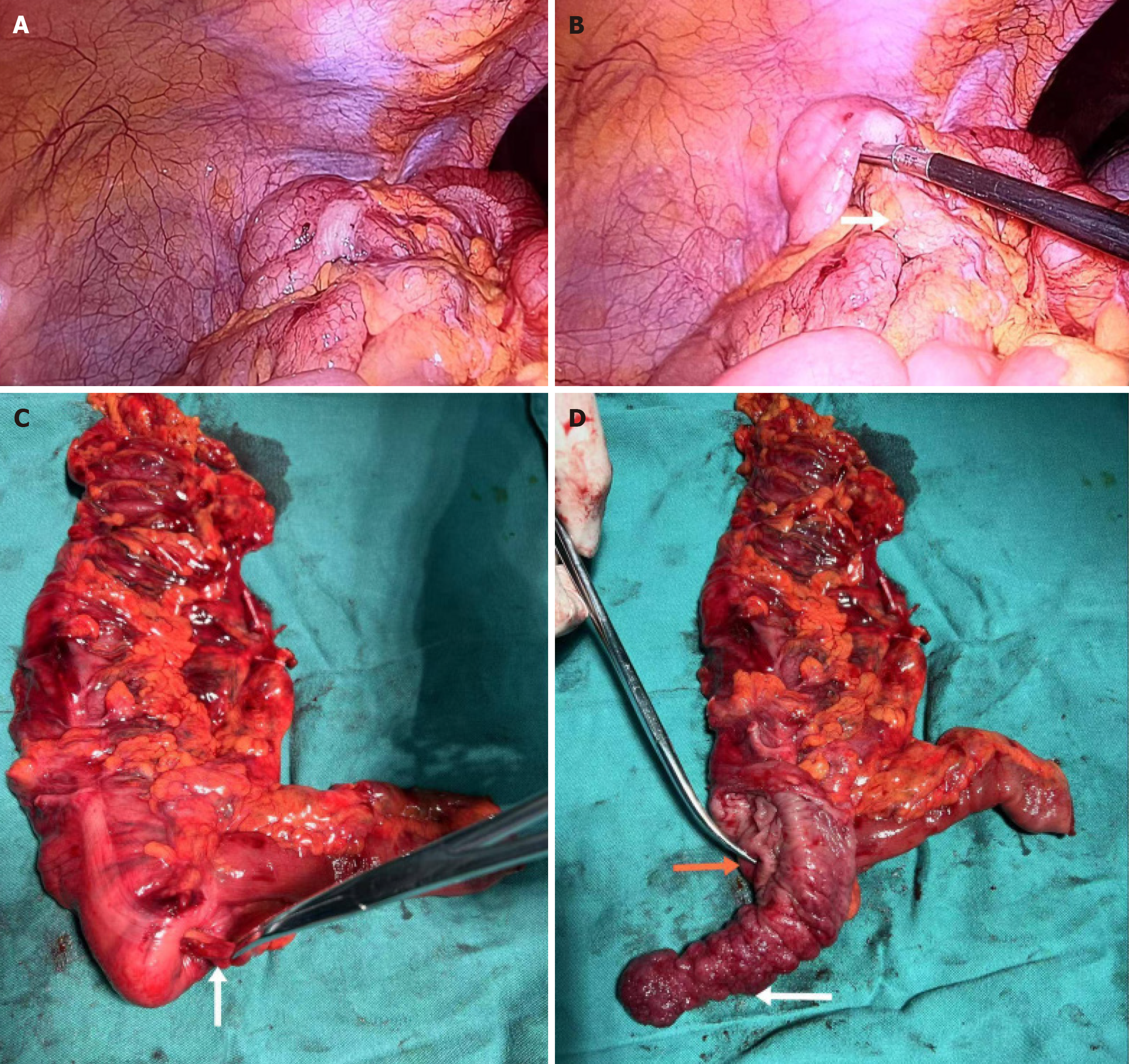
Figure 3 Laparoscopic right hemicolectomy with regional lymphadenectomy under general anaesthesia.
A: It shows laparoscopic exploration of the right abdomen; B: It shows the terminal ileum and mesentery intussuscepted into the ascending colon, with the white arrow indicating the terminal ileum; C: It shows the intussuscepted terminal ileum and mesentery after reduction, where the appendix and its mesentery could not be reduced, with the white arrow pointing to the appendix mesentery; D: It shows the postoperative specimen with the cecal wall incised laterally, with the appendix inverted outward, the orange arrow indicating the ileocecal valve, and the white arrow pointing to the inverted appendix and tumor.
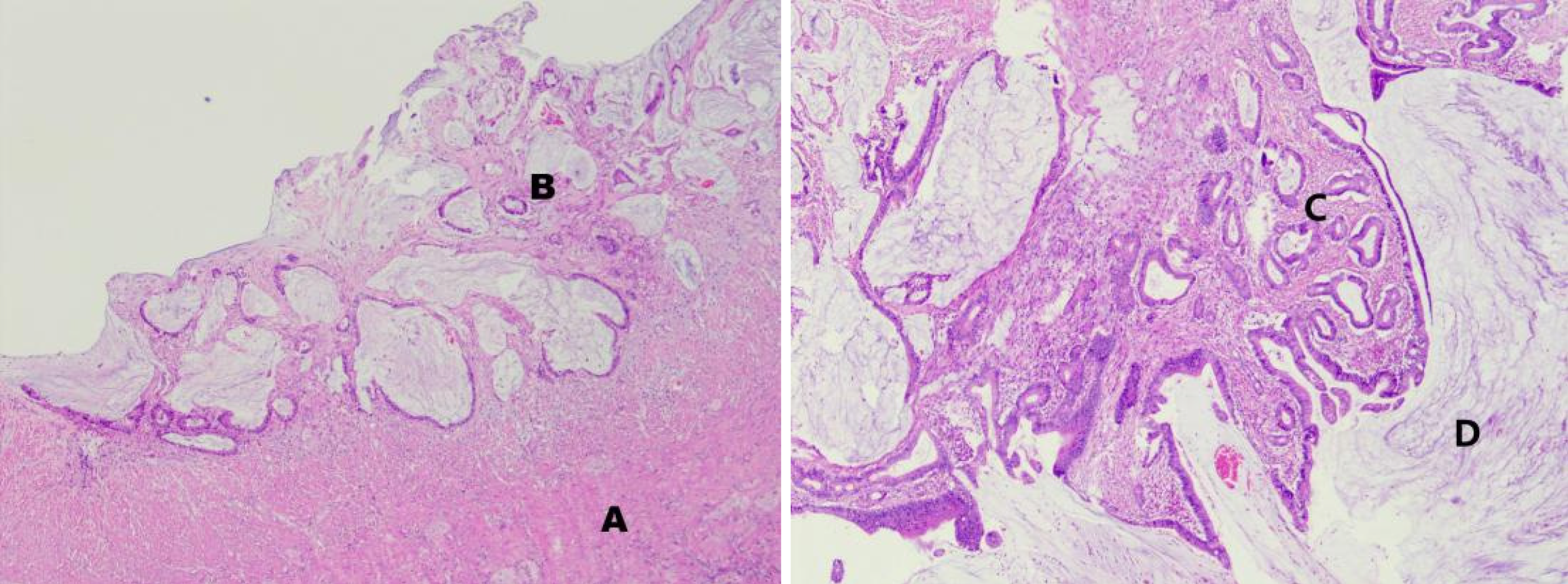
Figure 4 Histological morphology of the appendiceal lesion (hematoxylin-eosin staining).
A: The muscularis propria; B: The submucosal layer, with cancer tissue infiltrating the submucosal layer and muscularis propria, and a significant fibrotic response around the tumor; C: Disorganized structure of cancerous glands, with prominent cellular atypia and abundant mucus secretion; D: Formation of mucin lakes.
- Citation: Guo Q, Lu HY, Lyu H, Tian H, Zhao Q, Zheng YC. Complete appendiceal intussusception and appendiceal mucinous tumor: A case report and review of literature. World J Gastrointest Surg 2025; 17(9): 109320
- URL: https://www.wjgnet.com/1948-9366/full/v17/i9/109320.htm
- DOI: https://dx.doi.org/10.4240/wjgs.v17.i9.109320


