
















Copyright: ©Author(s) 2026.
World J Gastrointest Oncol. Jul 15, 2026; 18(7): 118212
Published online Jul 15, 2026. doi: 10.4251/wjgo.118212
Published online Jul 15, 2026. doi: 10.4251/wjgo.118212
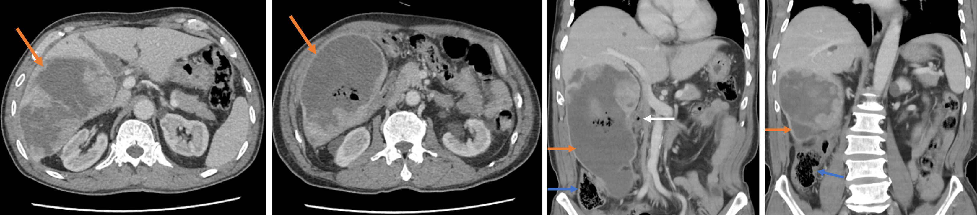
Figure 1 Abdominal contrast-enhanced computed tomography showed a large heterogeneous right hepatic lobe mass with internal high-density material, gas shadows, heterogeneous enhancement, and blurred adjacent fat planes.
Orange arrow: Liver mass; White arrow: Duodenum; Blue arrow: Colon.
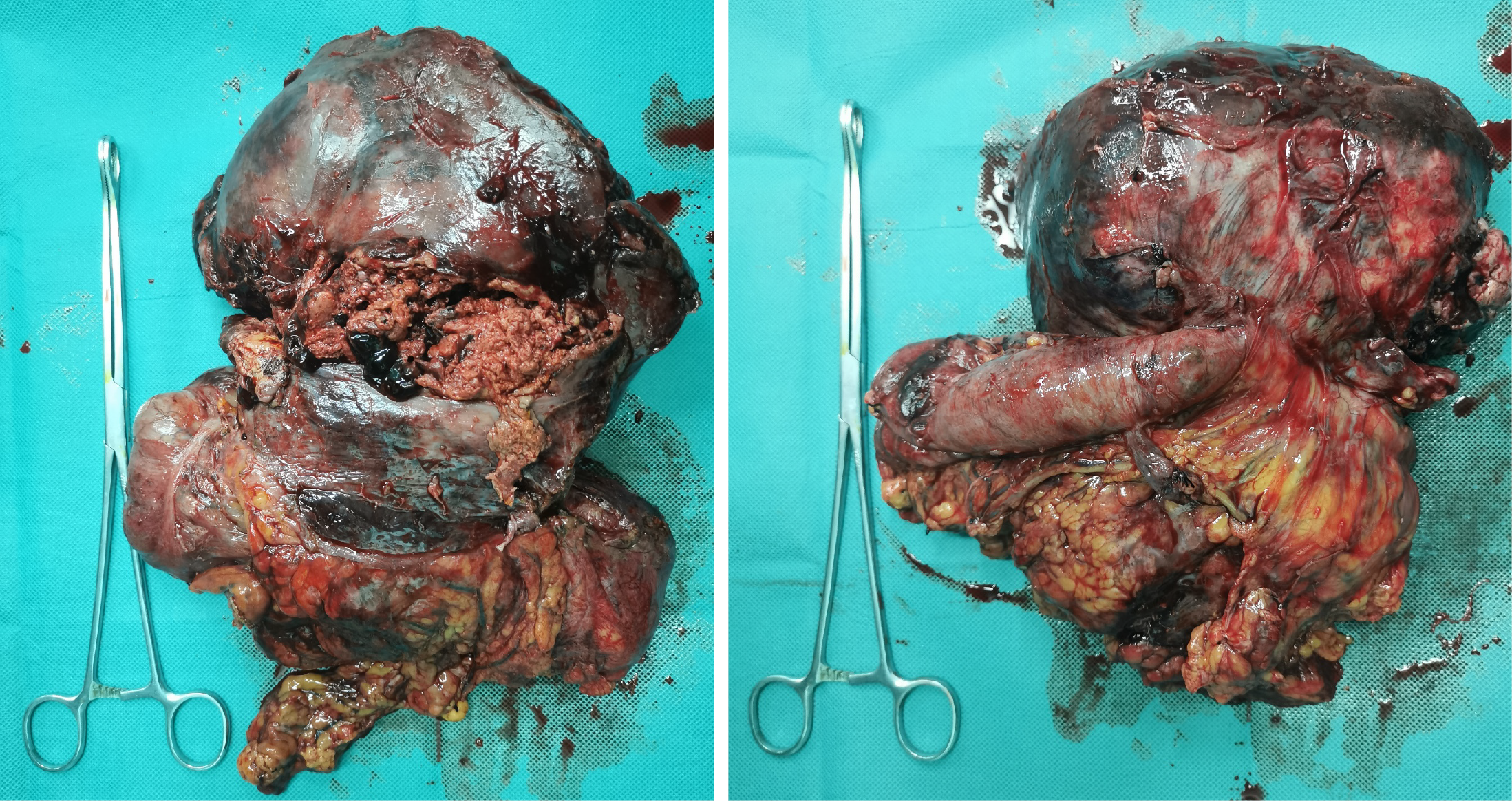
Figure 2 Gross specimen revealed the tumor adherent to the colon, with surface ulceration and an indistinct border.
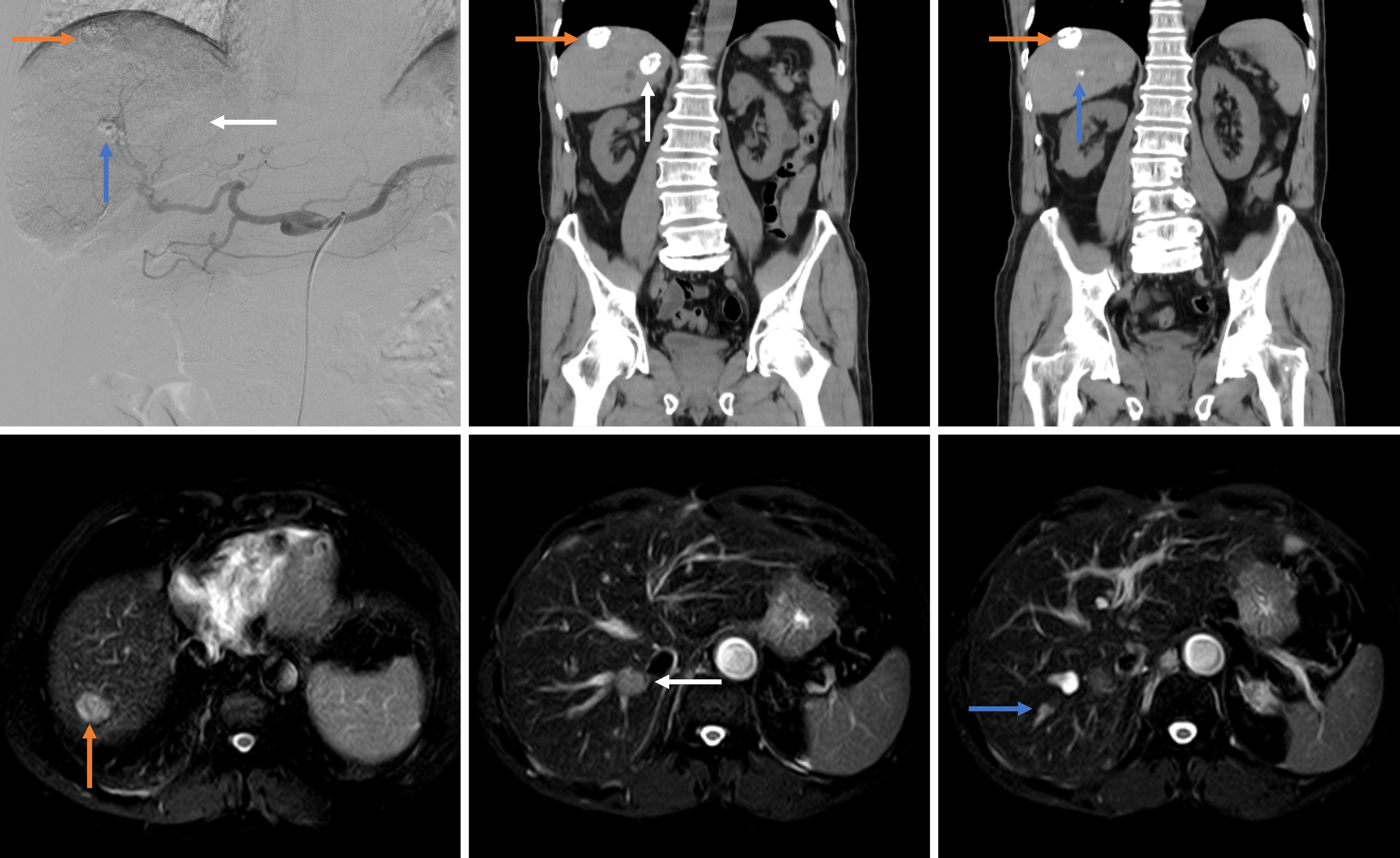
Figure 3 Transarterial chemoembolization detected three suspicious lesions.
Imaging performed 3 months after surgery showed satisfactory lipiodol deposition. Orange arrow: Dorsal portion of hepatic segment 8; White arrow: Paracaval portion of caudate lobe; Blue arrow: Segment 7.
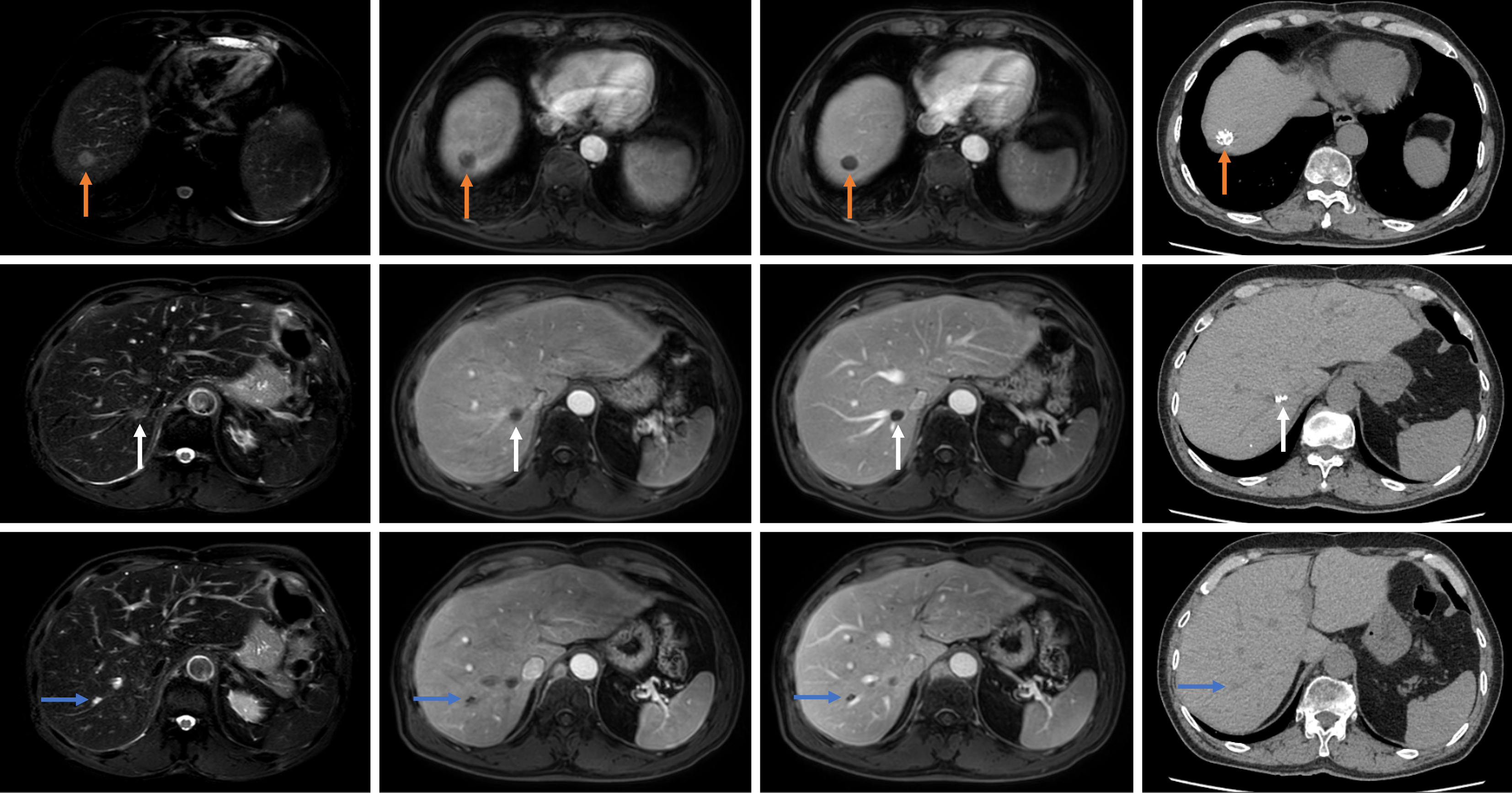
Figure 4 At the 42-month postoperative follow-up, the three occult micrometastases showed no significant blood flow or enhancement on magnetic resonance imaging during both arterial and portal venous phases, with gradual shrinkage.
Computed tomography confirmed satisfactory lipiodol deposition within some tumors. Orange arrow: Dorsal portion of hepatic segment 8; White arrow: Paracaval portion lesion; Blue arrow: Segment 7 lesion.
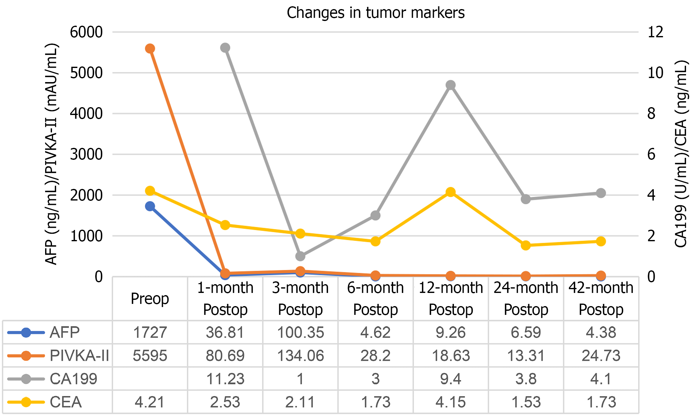
Figure 5 Changes in tumor markers.
Preoperative carbohydrate antigen 19-9 was not tested. AFP: Alpha-fetoprotein; PIVKA-II: Protein induced by vitamin K absence-II; CEA: Carcinoembryonic antigen; CA199: Carbohydrate antigen 19-9.
- Citation: Yang CW, Zhang RX, Zheng XL, Zhu L, Zheng Z, Wu XH. Long-term survival after combined hepatectomy and colectomy for ruptured combined hepatocellular-cholangiocarcinoma with direct colonic invasion: A case report. World J Gastrointest Oncol 2026; 18(7): 118212
- URL: https://www.wjgnet.com/1948-5204/full/v18/i7/118212.htm
- DOI: https://dx.doi.org/10.4251/wjgo.118212