©The Author(s) 2025.
World J Gastrointest Oncol. Sep 15, 2025; 17(9): 108960
Published online Sep 15, 2025. doi: 10.4251/wjgo.v17.i9.108960
Published online Sep 15, 2025. doi: 10.4251/wjgo.v17.i9.108960
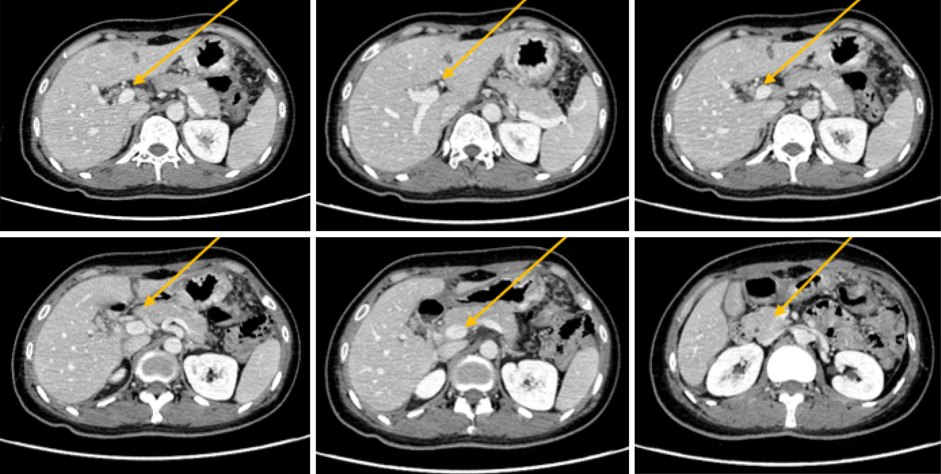
Figure 1
Computed tomography showed common bile duct wall thickening.
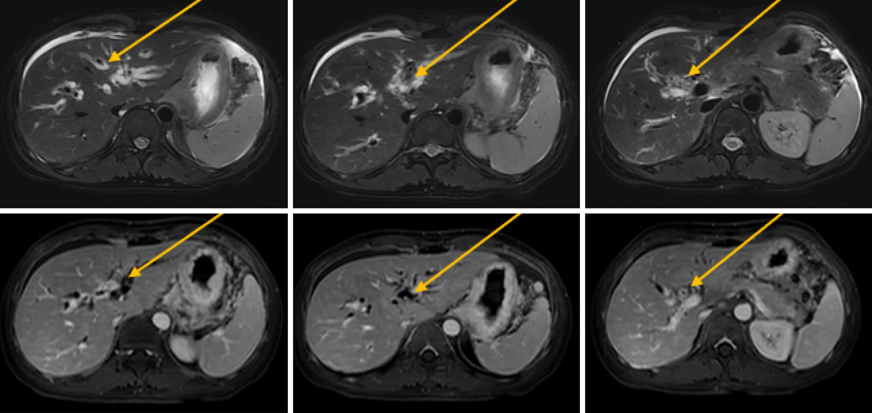
Figure 2
Magnetic resonance imaging demonstrating common bile duct stricture with upstream dilatation.
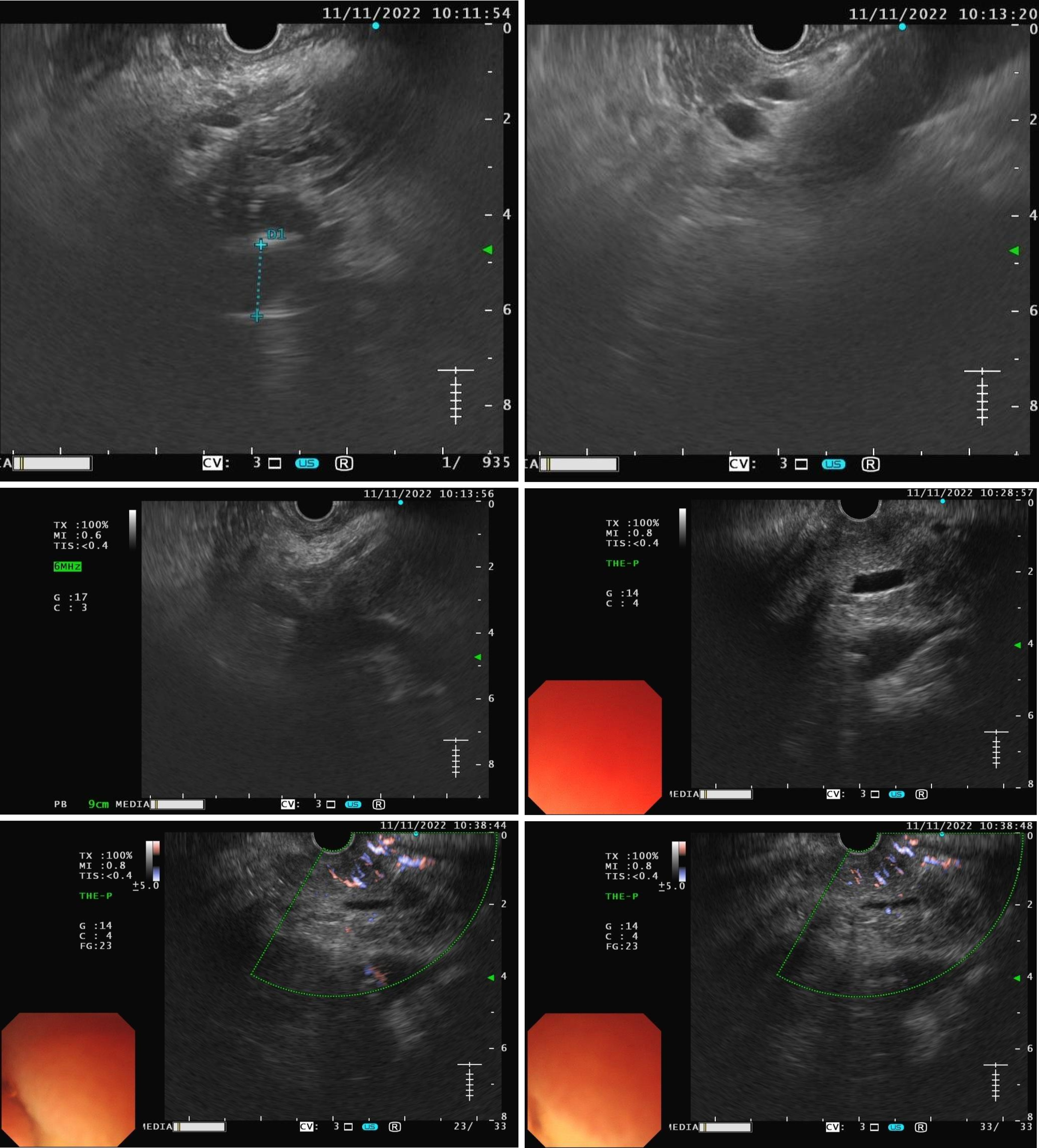
Figure 3
Endoscopic ultrasound found that the middle section of the lumen was interrupted, and the wall structure was unclear.
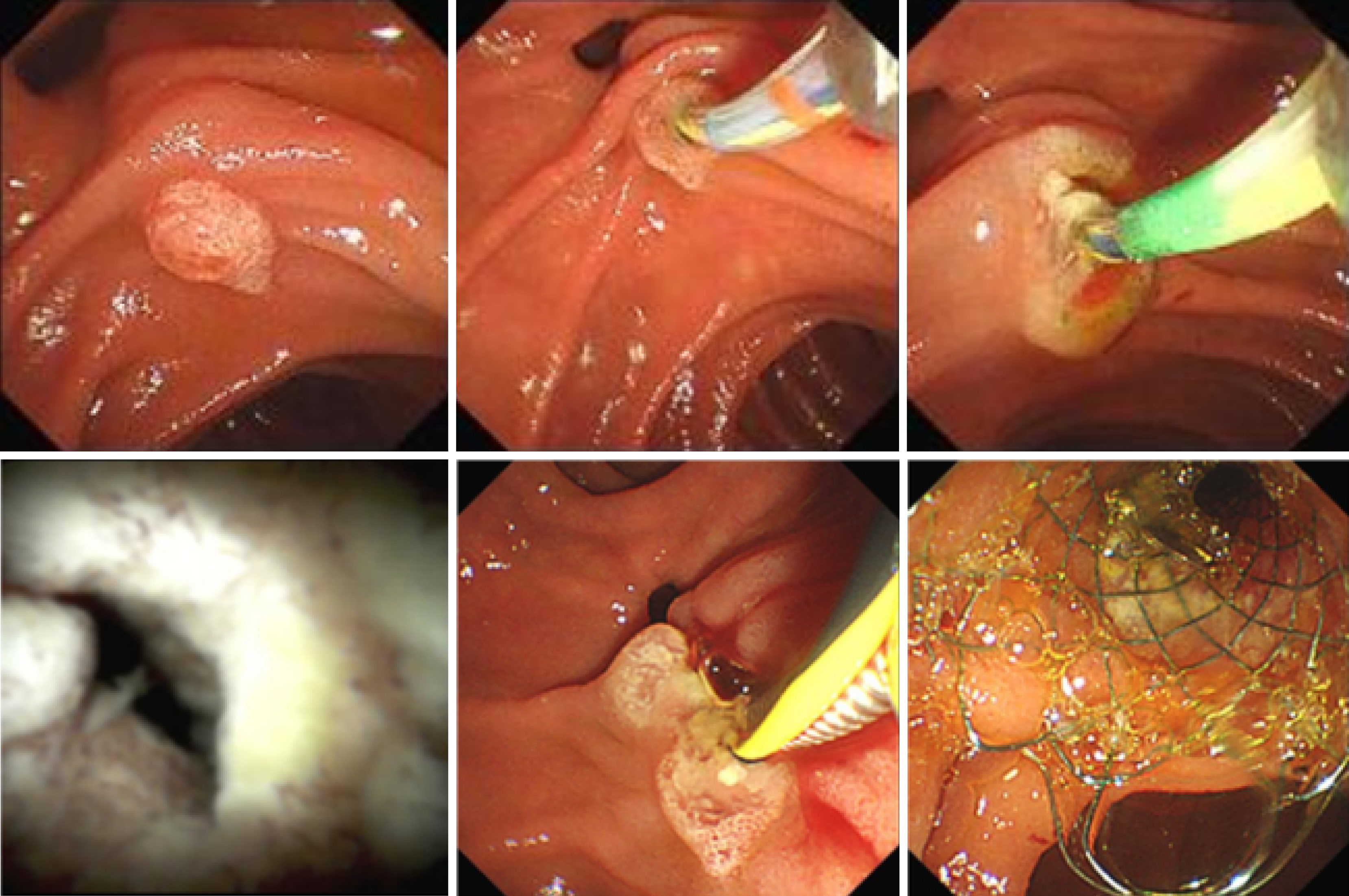
Figure 4
Endoscopic retrograde cholangiopancreatography revealed an irregular contrast defect in the middle and lower part of the common bile duct and multifocal narrowing and dilatation in the upper part of the bile duct.
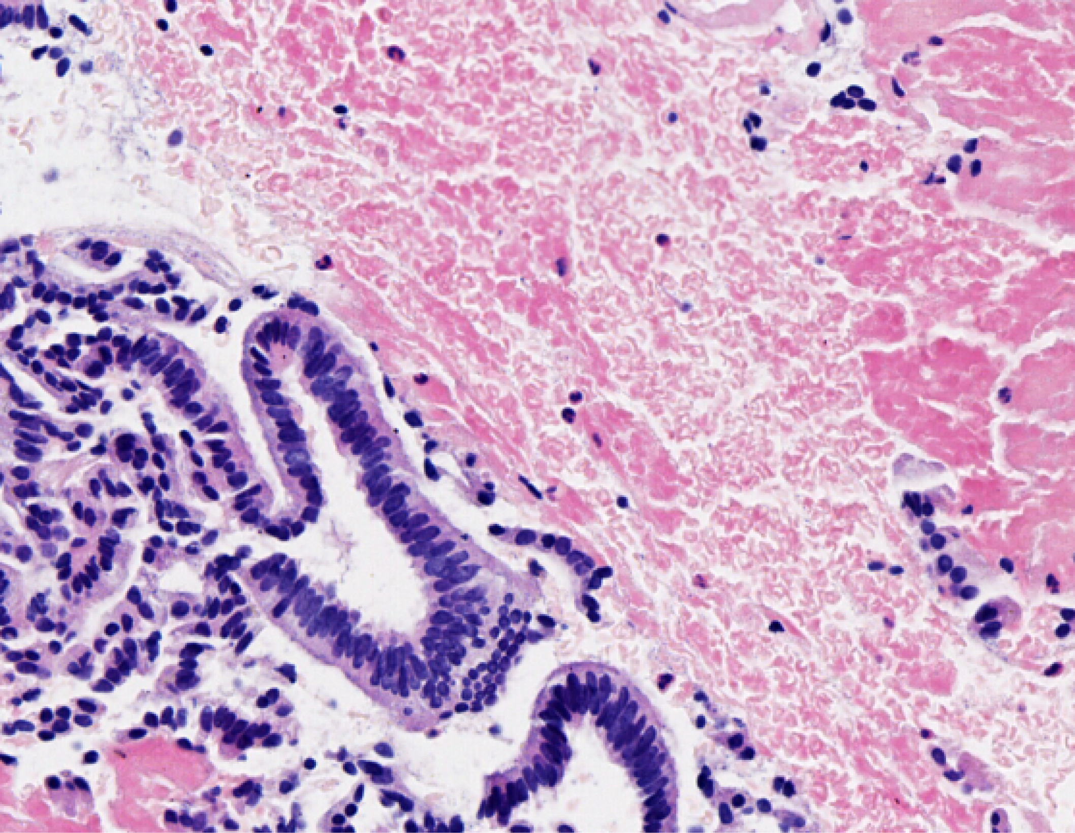
Figure 5
Histological findings of the liver (20 ×).
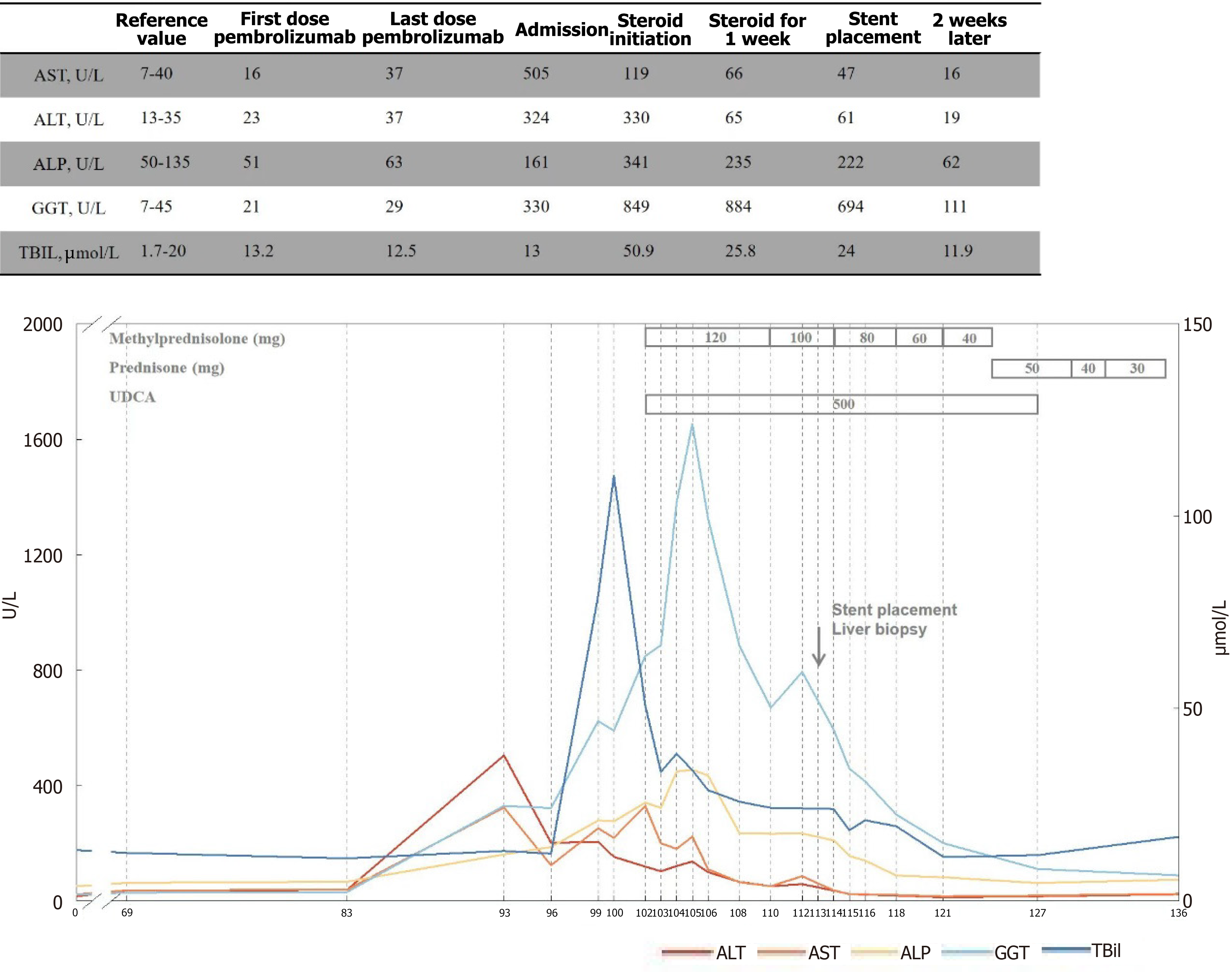
Figure 6 Longitudinal levels of serum alanine aminotransferase, aspartate aminotransferase, gamma-glutamyl transferase/γGT, Alkaline phosphatase and total bilirubin in a patient with immune-related cholangitis and the immunosuppressive agents and local stent implantation were used to control this induced immune-related cholangitis.
ALP: Alkaline phosphatase; ALT: Alanine aminotransferase; AST: Aspartate aminotransferase; GGT: Gamma-glutamyl transferase; TBil: Total bilirubin; UDCA: Ursodeoxycholic acid.
- Citation: Gao Z, Zhang JX, Tian XD, Wu SK, Jin X. Rapid cholestasis improvement as key strategy for steroid-refractory immune-related cholangitis: A case report. World J Gastrointest Oncol 2025; 17(9): 108960
- URL: https://www.wjgnet.com/1948-5204/full/v17/i9/108960.htm
- DOI: https://dx.doi.org/10.4251/wjgo.v17.i9.108960


