©The Author(s) 2025.
World J Gastrointest Oncol. Nov 15, 2025; 17(11): 112089
Published online Nov 15, 2025. doi: 10.4251/wjgo.v17.i11.112089
Published online Nov 15, 2025. doi: 10.4251/wjgo.v17.i11.112089
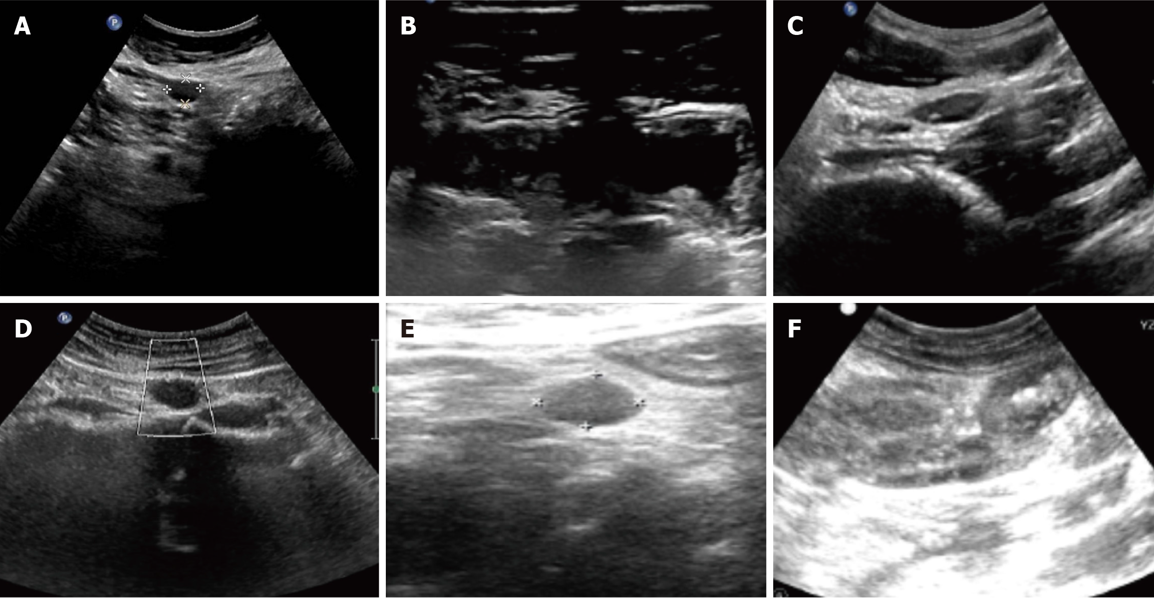
Figure 1 Ultrasonographic findings.
A: Multiple hypoechoic lesions are visualized in the peritoneal cavity. The largest, located in the perigastric region, measures 1.5 cm × 1.1 cm, demonstrates well-defined borders and a regular morphology, and exhibits homogeneous, markedly hypoechoic internal echogenicity; B: Localized thickening of the intestinal wall was observed in the terminal ileum, with a maximum thickness of approximately 0.5 cm; the layered structure of the wall remained relatively clear; C: Multiple lymph node echoes were detected in the retroperitoneum, with the largest measuring approximately 2.3 cm × 1.0 cm. These lymph nodes had clear boundaries and a regular shape; D: Lymph node echo was observed in the retroperitoneum, with the larger one located at the umbilical level in the right mid-abdomen, measuring 15 cm × 2.2 cm. The lymph node had clear boundaries and a regular shape, but the hilum structure was unclear; E: Multiple hypoechoic areas were found in the intestinal interspace, with the largest measuring 0.7 cm × 1.0 cm; F: Multiple enlarged lymph node echoes were detected in the mesentery, with the largest measuring approximately 0.8 cm × 1.8 cm. These lymph nodes exhibited relatively clear boundaries, an irregular shape, and an unclear hilum structure.
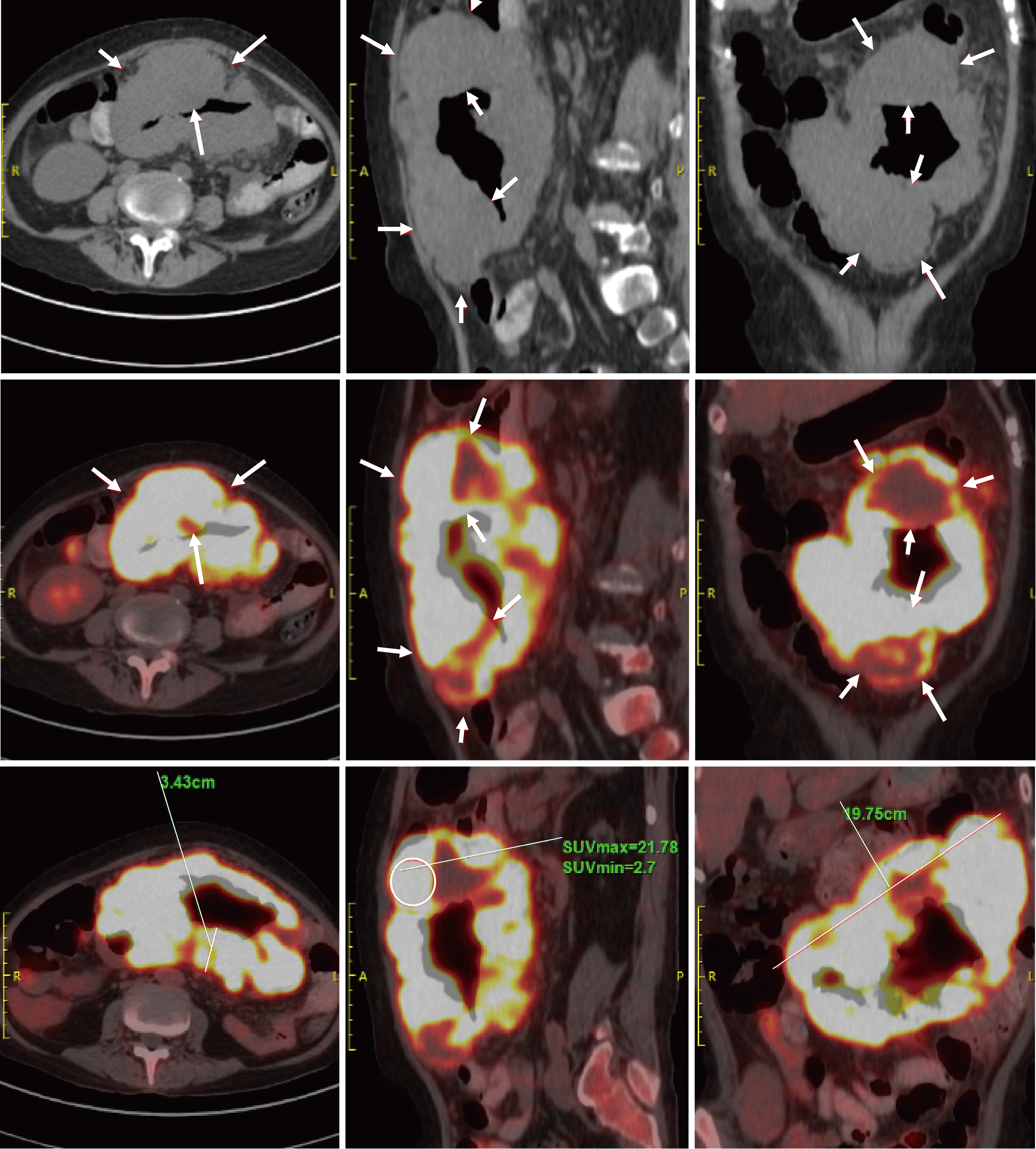
Figure 2 Positron emission tomography/computed tomography findings.
Segmental circumferential thickening and mass formation were noted in the second segment of the small intestine, extending from the left upper to mid-abdomen, with significantly increased fluorodeoxyglucose uptake. Multiple enlarged lymph nodes were observed in the abdominal cavity surrounding the small intestinal lesion, also demonstrating markedly elevated fluorodeoxyglucose uptake. Arrow: Segmental small bowel wall thickening and a mass, respectively. The circled areas highlight regions with increased fluorodeoxyglucose uptake. The solid lines mark the measured lengths.
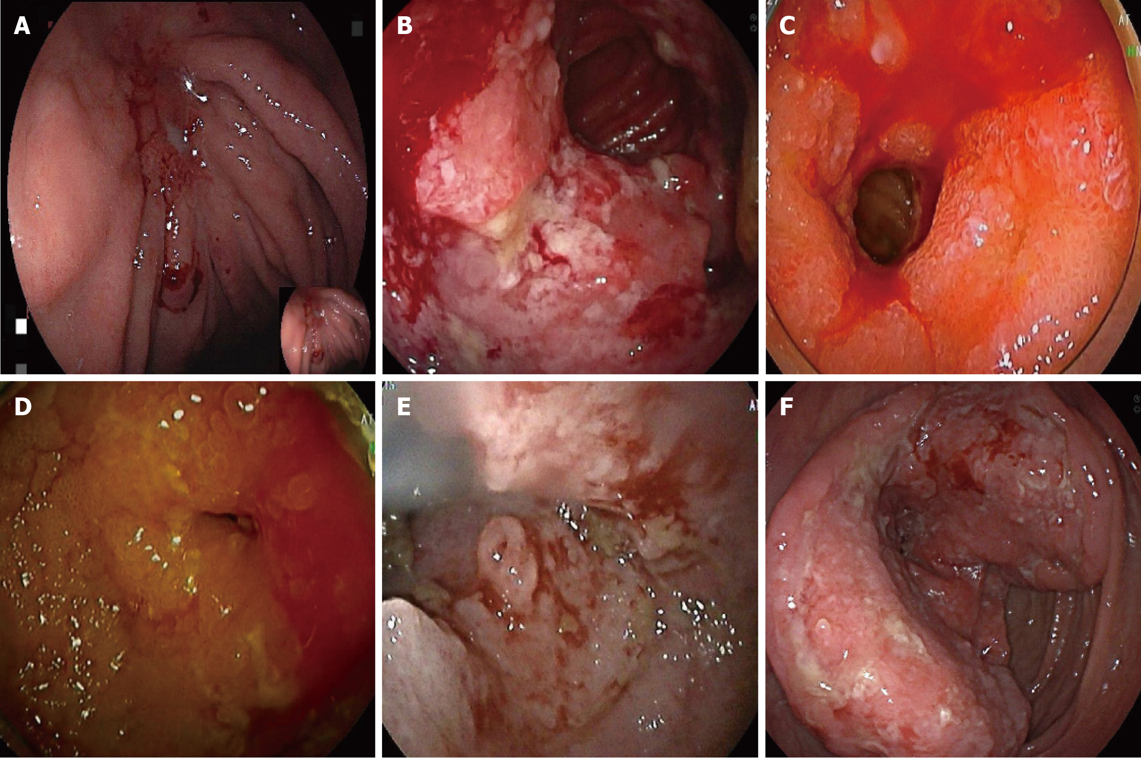
Figure 3 Endoscopic features of lymphoma.
A: An ulcer was observed at the junction of the gastric fundus and body, with a white coating in the center. The surrounding mucosa exhibited congestion, edema, and convergence, with a rigid sensation upon insufflation, poor peristalsis, and slight oozing; B: A circumferential ulcerative and proliferative lesion was found at approximately 300 cm of the small intestine, with an uneven surface covered by dirty coating, leading to intestinal stenosis; C: A circumferential ulcerative mass causing luminal stenosis was detected 160 cm from the pylorus in the small intestine. The surrounding mucosa was congested and reddened, with local mucosal rigidity and friability, making it prone to bleeding; D: A circumferential ulcerative and proliferative lesion was observed at approximately 300 cm of the small intestine via anal approach, with an uneven surface covered by dirty coating, leading to intestinal stenosis; E: A mucosal mass was found 50 cm from the ileocecal junction in the small intestine, with surface erosion and fresh blood stains. Mild luminal stenosis was noted at the lesion site; F: A circumferential space-occupying lesion was detected in the ascending colon during endoscopy, showing surface ulceration and a hard texture.
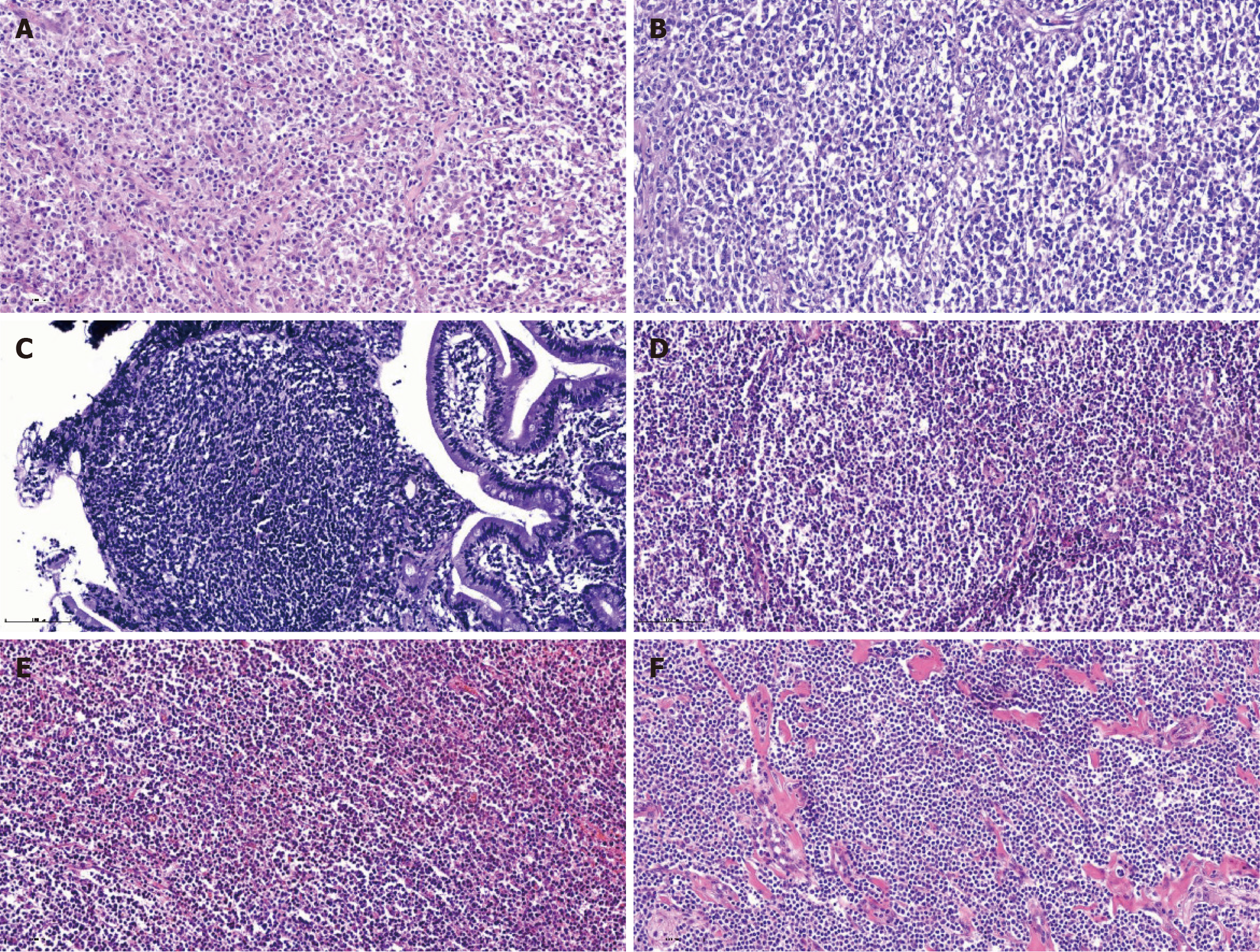
Figure 4 Histopathological features of lymphoma (hematoxylin and eosin staining).
A: Gastric diffuse large B-cell lymphoma; B: Jejunal diffuse large B-cell lymphoma; C: Duodenal follicular lymphoma (low-grade, grade 1-2); D: Axillary follicular lymphoma (grade II); E: Intestinal monomorphic T-cell lymphoma of the small intestine; F: Small intestinal extranodal marginal zone lymphoma of mucosa-associated lymphoid tissue.
- Citation: Yang CX, Xu LX, Liu J, Qiao HL, Dong ZW, Jiang D, Gu GL. Clinicopathological characteristics and surgical value of primary gastrointestinal lymphoma. World J Gastrointest Oncol 2025; 17(11): 112089
- URL: https://www.wjgnet.com/1948-5204/full/v17/i11/112089.htm
- DOI: https://dx.doi.org/10.4251/wjgo.v17.i11.112089


