©The Author(s) 2025.
World J Hepatol. Sep 27, 2025; 17(9): 109179
Published online Sep 27, 2025. doi: 10.4254/wjh.v17.i9.109179
Published online Sep 27, 2025. doi: 10.4254/wjh.v17.i9.109179
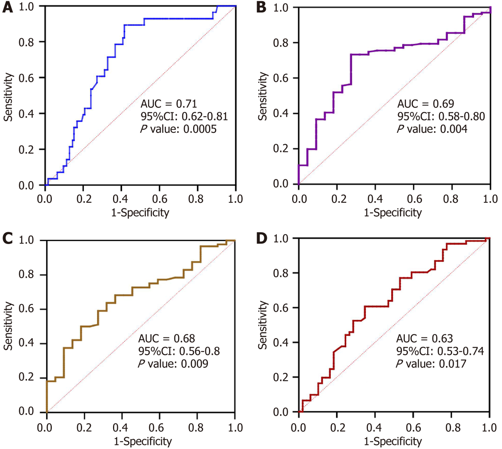
Figure 1 Area under the receiver operating characteristic curves of mac-2 binding protein glycosylation isomer in predicting hepa
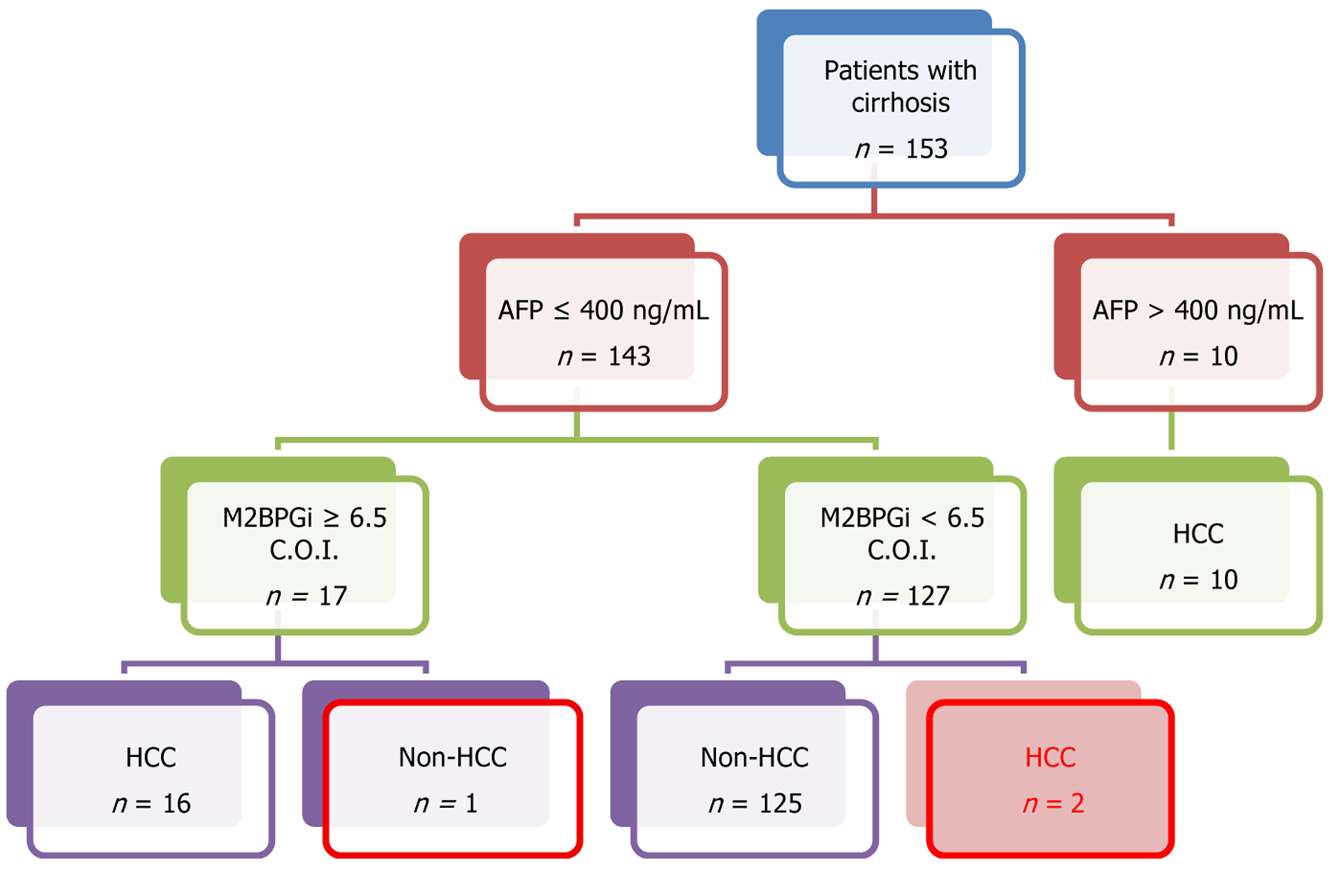
Figure 2 Algorithm combining alpha-fetoprotein and mac-2 binding protein glycosylation isomer to detect hepatocellular carcinoma in patients with cirrhosis.
Hepatocellular carcinoma was confirmed by computed tomography or magnetic resonance imaging. AFP: Alpha-fetoprotein; C.O.I.: Cutoff index; M2BPGi: Mac-2 binding protein glycosylation isomer; HCC: Hepatocellular carcinoma.
- Citation: Doan TH, Nguyen KM, Nguyen XV, Pham ATN, Le ND. Evaluating thresholds of Mac-2 binding protein glycosylation isomer in association with clinical outcomes in patients with cirrhosis. World J Hepatol 2025; 17(9): 109179
- URL: https://www.wjgnet.com/1948-5182/full/v17/i9/109179.htm
- DOI: https://dx.doi.org/10.4254/wjh.v17.i9.109179


