Copyright
©The Author(s) 2024.
World J Hepatol. Oct 27, 2024; 16(10): 1177-1187
Published online Oct 27, 2024. doi: 10.4254/wjh.v16.i10.1177
Published online Oct 27, 2024. doi: 10.4254/wjh.v16.i10.1177
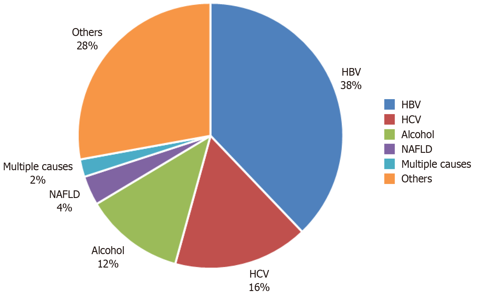
Figure 1 Etiologies of cirrhosis in patients at Tikur Anbessa Specialized Hospital and Adera Medical Center.
HBV: Hepatitis B virus; HCV: Hepatitis C virus; NAFLD: Non-alcoholic fatty liver disease.
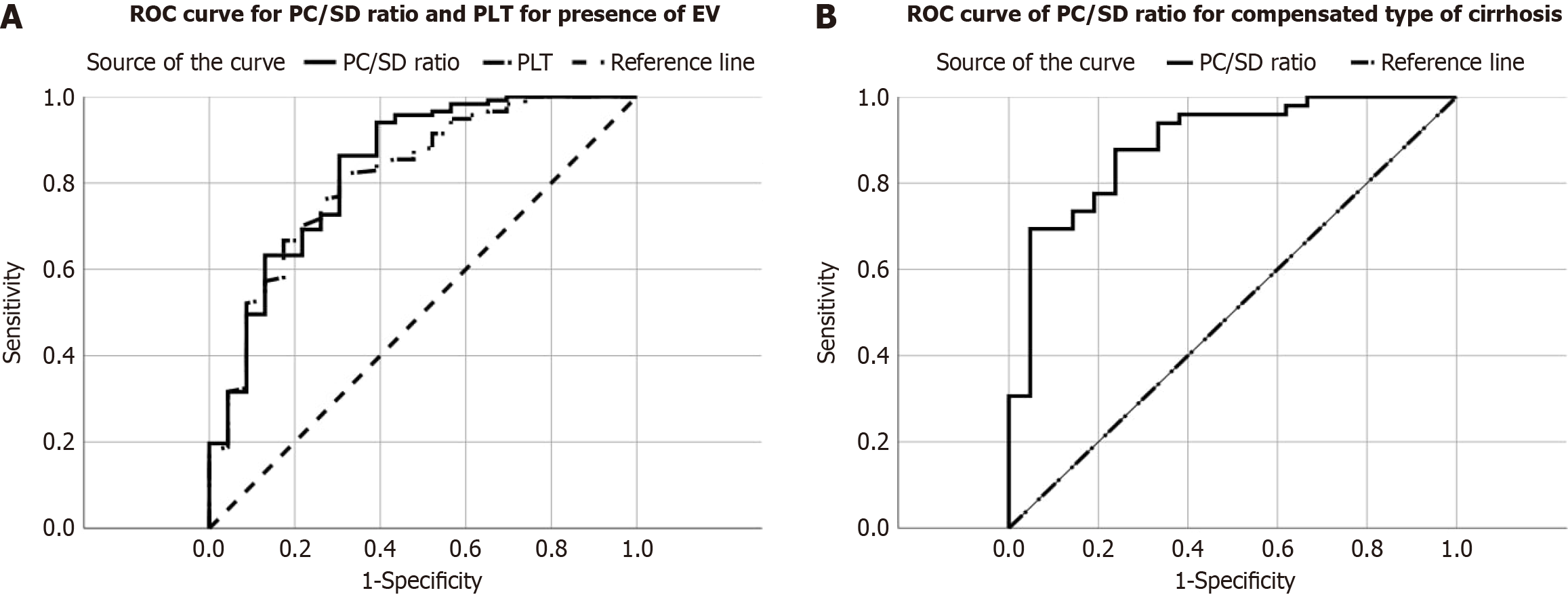
Figure 2 Receiver operating characteristic curve.
A: Receiver operating characteristic (ROC) curve for platelet count and platelet count-spleen diameter ratio (PC/SD) for the presence of esophageal varices; B: ROC curve for PC/SD for the presence of esophageal varices in patients with compensated cirrhosis. Broken line: Platelet count in figure A and reference line in figure B; Solid line: PC/SD; Dashed line: Reference line in figure A. ROC: Receiver operating characteristic curve; PLT: Platelet count; PC/SD: Platelet count-spleen diameter ratio.
- Citation: Mossie GY, Nur AM, Ayalew ZS, Azibte GT, Berhane KA. Platelet counts to spleen diameter ratio: A promising noninvasive tool for predicting esophageal varices in cirrhosis patients. World J Hepatol 2024; 16(10): 1177-1187
- URL: https://www.wjgnet.com/1948-5182/full/v16/i10/1177.htm
- DOI: https://dx.doi.org/10.4254/wjh.v16.i10.1177


