Copyright
©The Author(s) 2015.
World J Stem Cells. Mar 26, 2015; 7(2): 521-534
Published online Mar 26, 2015. doi: 10.4252/wjsc.v7.i2.521
Published online Mar 26, 2015. doi: 10.4252/wjsc.v7.i2.521
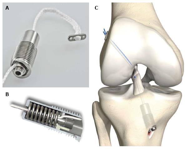
Figure 1 Dynamic intraligamentary stabilization screw called Ligamys© (Mathys, inc.
Bettlach, Switzerland). A: Close-up of the outside of the screw made of titanium and illustrating with a mounted lace which mimics the polyethylene string that is mounted in the real surgery to stabilize the knee joint in case of an ACL rupture; B: Inside of the dynamic fixation screw with the spring that takes the dynamic load of the ACL and stabilizes the joint; C: Illustration of the exact position of the dynamic intraligamentary stabilization in situ in the knee joint. ACL: Anterior cruciate ligament.
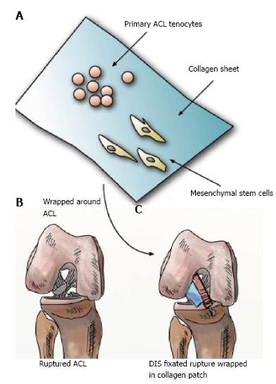
Figure 2 Overview of current regenerative approaches to improve anterior cruciate ligament rupture treatment in combination of the dynamic intraligamentary stabilization approach.
A: First mesenchymal stem cells and/or primary ACL-tenocytes are pre-seeded on collagen patches (shown in blue); B: Ruptured ACL; C: DIS approach takes over major mechanical load and stabilizes the knee, whereas collagen patch wrapped around the ACL provides an ideal solution to improve clinical outcome. ACL: Anterior cruciate ligament; DIS: Dynamic intraligamentary stabilization.
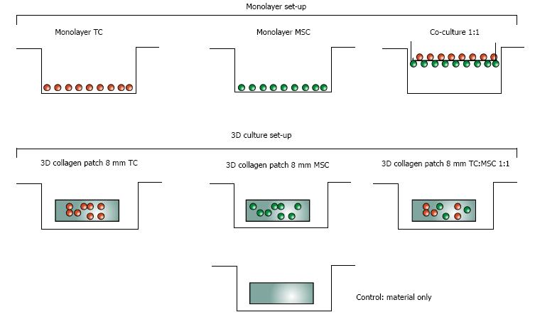
Figure 3 Experimental design of in vitro testing of cyto-compatibility.
All monolayer and collagen patch cultures were run for 7 d under standard culture conditions (37 °C at 100% humidity and 5% CO2) in high glucose Dulbecco’s Modified Eagle Medium and 10% fetal calf serum. MSC: Mesenchymal stem cell; TC: Tenocyte.
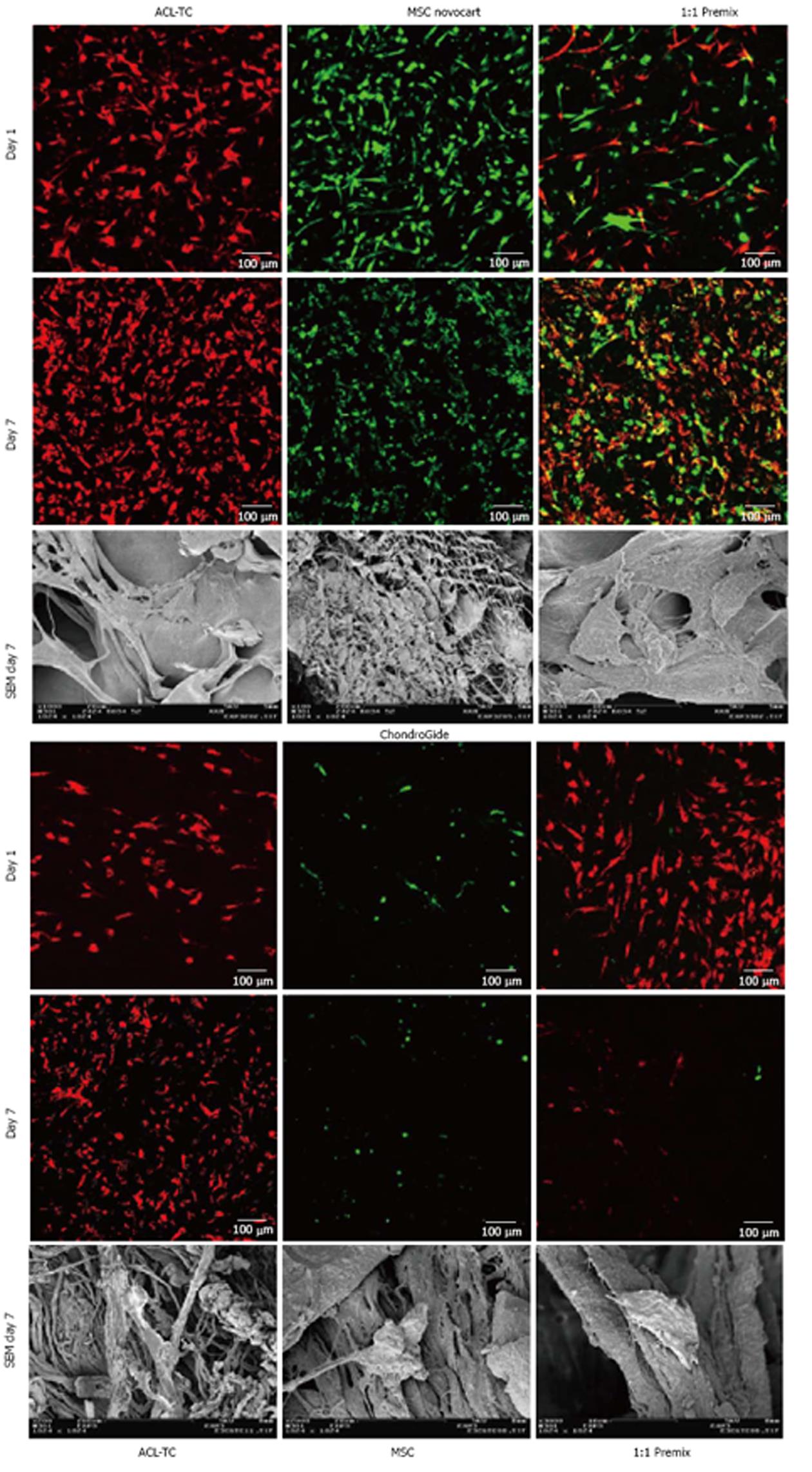
Figure 4 Z-projections of confocal laser scanning microscope images of 200 μm 3D-stacks through the collagen patches of mesenchymal stem cells and anterior cruciate ligament-derived tenocytes on day 1 and after 7 d of culture on Novocart™ and Chondro-Gide™.
Cells were stained with Vybrant™ DiO (DiO = green, MSCs) and DiL membrane tracker dyes (DiL = red, TCs) prior to seeding. Last row are scanning electron microscope pictures of the cell-seeded scaffolds after 7 d of culture showing adherence and shape of the MSCs and tenocytes on the scaffolds. Note that for Chondro-Gide™ patches these are only images from TCs seeded scaffolds. For the Novocart™ patches the right most image is from an experiment containing a premix of cells, and the left most and center SEM image are from experiments with MSCs seeded scaffolds. MSC: Mesenchymal stem cell; ACL-TC: Anterior cruciate ligament-derived tenocyte; SEM: Scanning electron microscope.
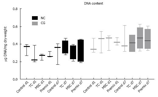
Figure 5 DNA content of collagen patches.
DNA content of collagen patches seeded with anterior cruciate ligament-derived tenocytes (TCs) and/or MSCs on Novocart™ and Chondro-Gide™ normalized per dry weight per carrier for the collagen patches for day 1 and day 7 of the experiment (values are means ± SEM, n = 3 experiments for day 1, 4 experiments for day 7). Note the large amount of DNA present in material controls. MSCs: Mesenchymal stem cells.
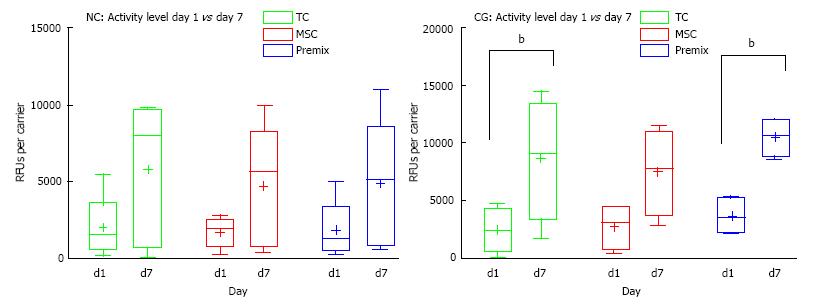
Figure 6 Mitochondrial cell activity.
Mitochondrial cell activity measured by resazurin red assay of collagen patches seeded with anterior cruciate ligament-derived tenocytes (TCs) and/or MSC on NC and CG normalized per dry weight carrier for the collagen patches for day 1 and day 7 of the experiment (values are means ± SEM, n = 4 experiments for CG and n = 5 experiments for NC). b denotes P < 0.01. MSC: Mesenchymal stem cell; NC: Novocart™; CG: Chondro-Gide™; SEM: Scanning electron microscope.
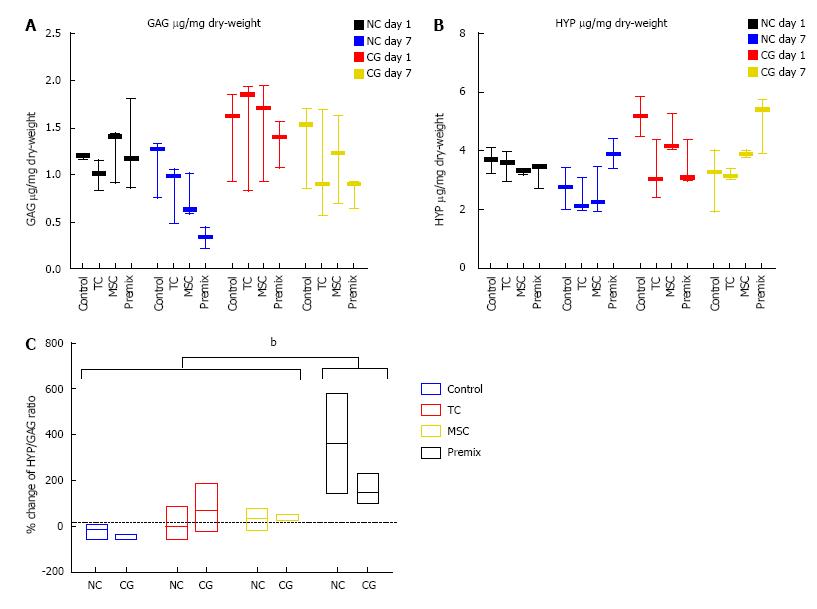
Figure 7 Hydroxy-Proline and glycosaminoglycan content.
A: HYP content assay; B: GAG of collagen patches seeded with anterior cruciate ligament-TCs and/or MSCs on NC and CG normalized per dry weight carrier for the collagen patches for day 1 and day 7 of the experiment (values are means ± SEM, n = 3 experiments for day 1, 4 for day 7); C: Percentage change of HYP to GAG ratio relative to day 1 of NC and CG patches. b denotes a significant P < 0.01. GAG: Glycosaminoglycan; HYP: Hydroxy-proline; MSC: Mesenchymal stem cell; TC: Tenocytes; NC: Novocart™; CG: Chondro-Gide™; SEM: Scanning electron microscope.
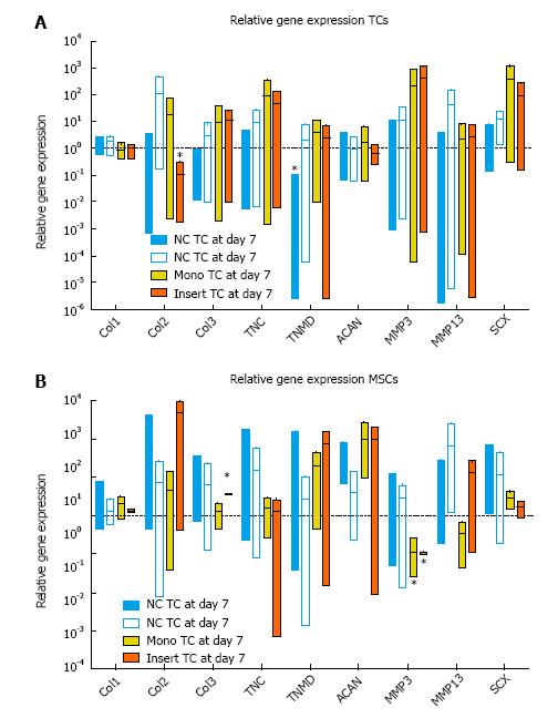
Figure 8 Relative gene expression of major extracellular matrix genes important for anterior cruciate ligament repair and mesenchymal stem cells differentiation grown on 3D collagen patches, i.
e., Novocart™ and ChondroGide™. A: Gene expression of anterior cruciate ligament-derived tenocytes (TCs) relative to day 0 TCs; B: Gene expression of bone marrow-derived MSCs relative to day 0 MSCs (values are means ± SEM, n = 4 experiments for CG and n = 5 for NC). MSC: Mesenchymal stem cell; NC: Novocart™; CG: ChondroGide™; SEM: Scanning electron microscope.
- Citation: Gantenbein B, Gadhari N, Chan SC, Kohl S, Ahmad SS. Mesenchymal stem cells and collagen patches for anterior cruciate ligament repair. World J Stem Cells 2015; 7(2): 521-534
- URL: https://www.wjgnet.com/1948-0210/full/v7/i2/521.htm
- DOI: https://dx.doi.org/10.4252/wjsc.v7.i2.521


