
















Copyright: ©Author(s) 2026.
World J Stem Cells. Jul 26, 2026; 18(7): 119865
Published online Jul 26, 2026. doi: 10.4252/wjsc.119865
Published online Jul 26, 2026. doi: 10.4252/wjsc.119865
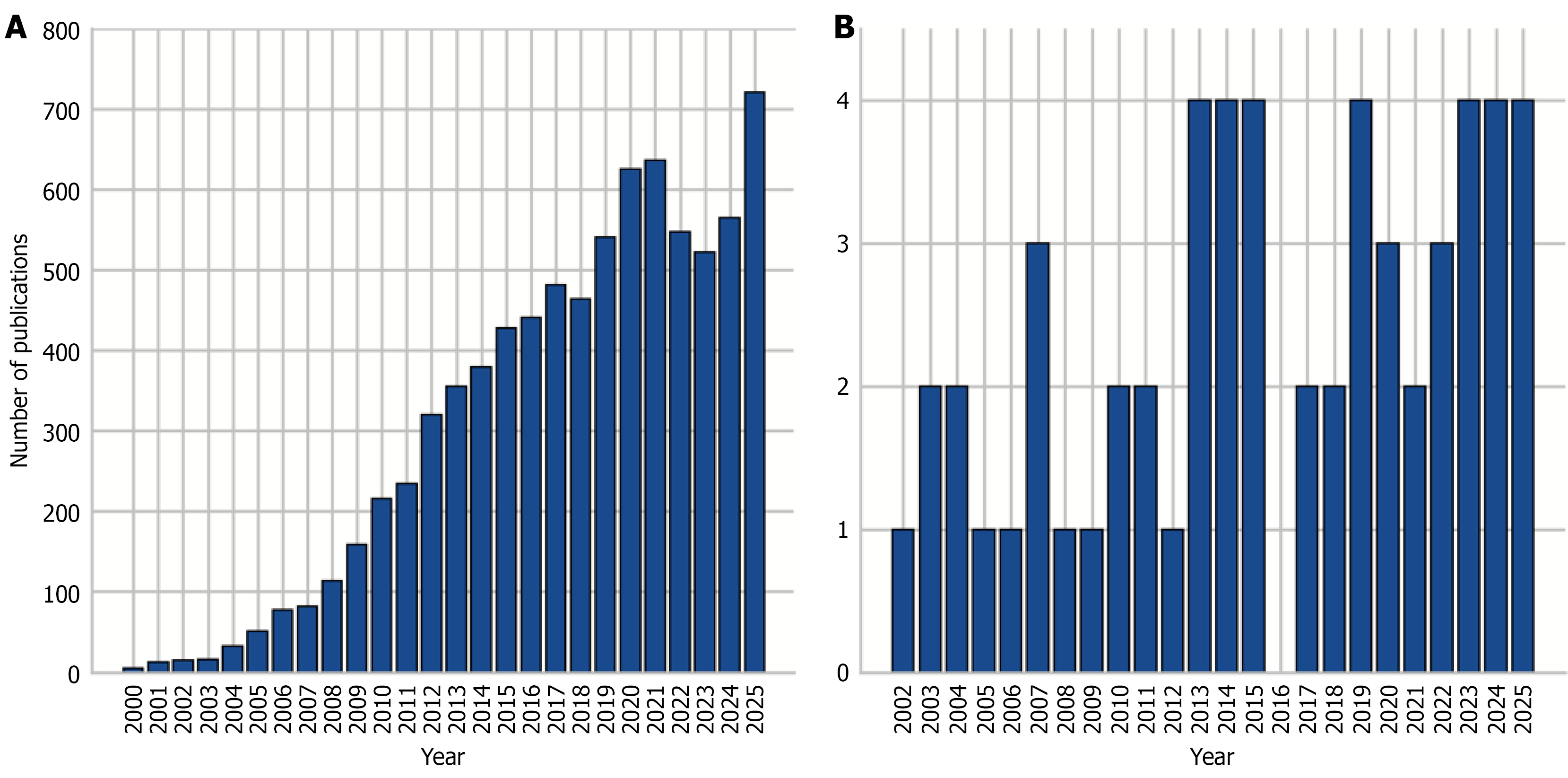
Figure 1 Number of publications indexed in the PubMed database from 2000 to 2025.
A: Publications related to tissue and organ regeneration using mesenchymal stem cells; B: Publications related to tissue and organ regeneration using epiregulin.
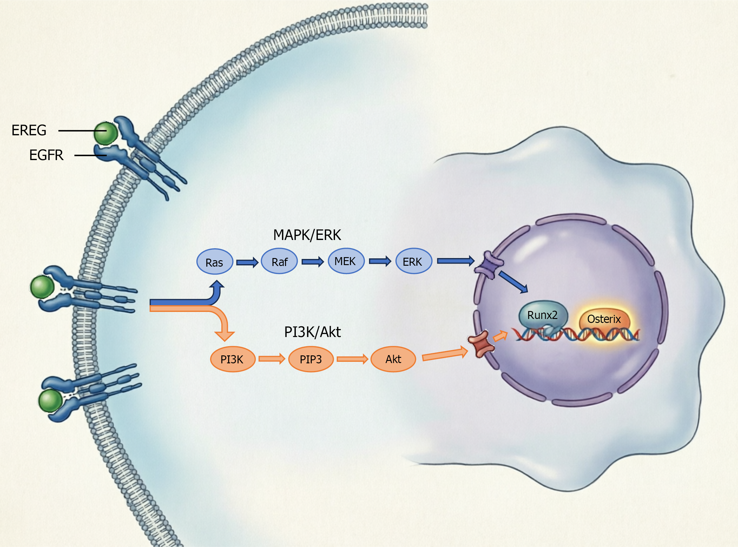
Figure 2 Schematic representation of the molecular mechanisms underlying epiregulin-induced osteogenic differentiation of mesen
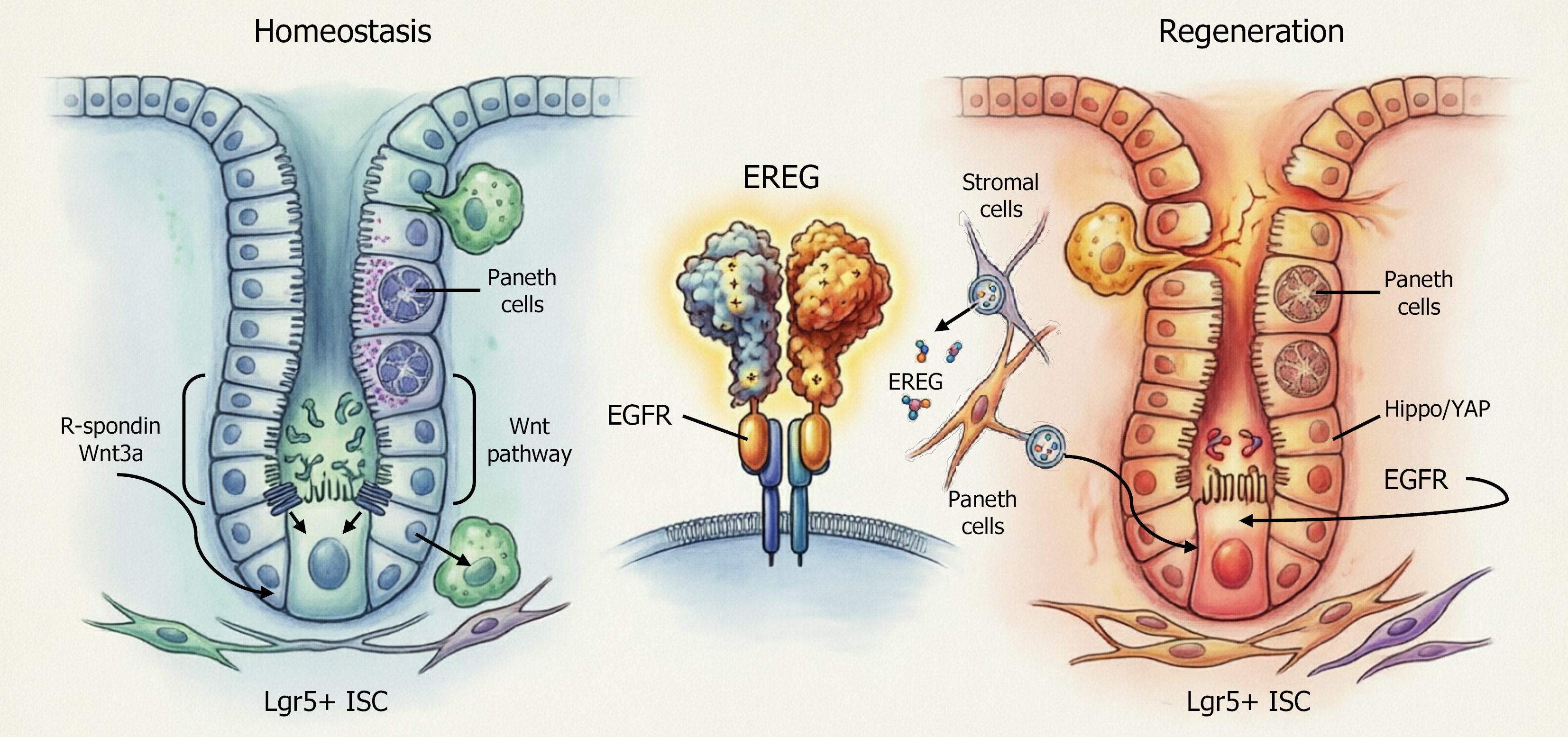
Figure 3 Schematic representation of the mechanisms underlying regeneration of the intestinal epithelium.
Left panel (homeostasis): Intestinal epithelial homeostasis is maintained through canonical Wnt signaling in Lgr5+ intestinal stem cells (ISCs) located at the crypt base. R-spondin and Wnt3a, which are produced by the niche, activate the Wnt pathway, thereby supporting ISC self-renewal and differentiation into epithelial lineages, including Paneth cells. Right panel (regeneration): Following injury, the Hippo pathway effector Yes-associated protein (YAP) transiently reprograms Lgr5+ ISCs. This suppresses excessive Paneth cell differentiation and activates a regenerative program that stimulates proliferation, survival, and epidermal growth factor receptor (EGFR) signaling. Epiregulin (EREG) serves as a critical mediator of YAP-dependent regeneration by binding to EGFR on ISCs. Stromal cells surrounding crypts secrete EREG in response to injury, particularly radiation damage, providing non-cell-autonomous compensation that can rescue organoid formation even in YAP-deficient conditions. Central inset: EREG binds to the EGFR receptors on Lgr5+ ISCs, thereby activating downstream signaling cascades. The ability of EREG to regulate the transition between homeostatic and regenerative states makes it a promising therapeutic target for treating radiation enteropathy, inflammatory bowel disease, and post-surgical intestinal restoration. EGFR: Epidermal growth factor receptor; EREG: Epiregulin; ISC: Intestinal stem cell; YAP: Yes-associated protein.
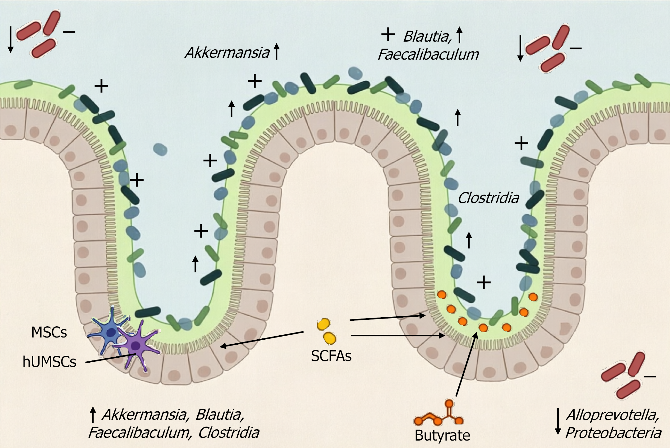
Figure 4 Schematic representation of the effects of human umbilical cord mesenchymal stem cell therapy on the gut microbiota-short-chain fatty acid-immune axis in dextran sodium sulphate-induced colitis.
The administration of human umbilical cord mesenchymal stem cells promotes the enrichment of beneficial bacterial genera, such as Akkermansia, Blautia, Faecalibaculum, and Clostridia, while suppressing pathogenic taxa such as Alloprevotella and Proteobacteria. This restored microbial community enhances the production of short-chain fatty acids, particularly butyrate, thereby strengthening the integrity of the epithelial barrier and exerting anti-inflammatory effects. Adapted from Liu et al[48]. hUMSCs: Human umbilical cord mesenchymal stem cells; MSCs: Mesenchymal stem cell; SCFAs: Short-chain fatty acids.
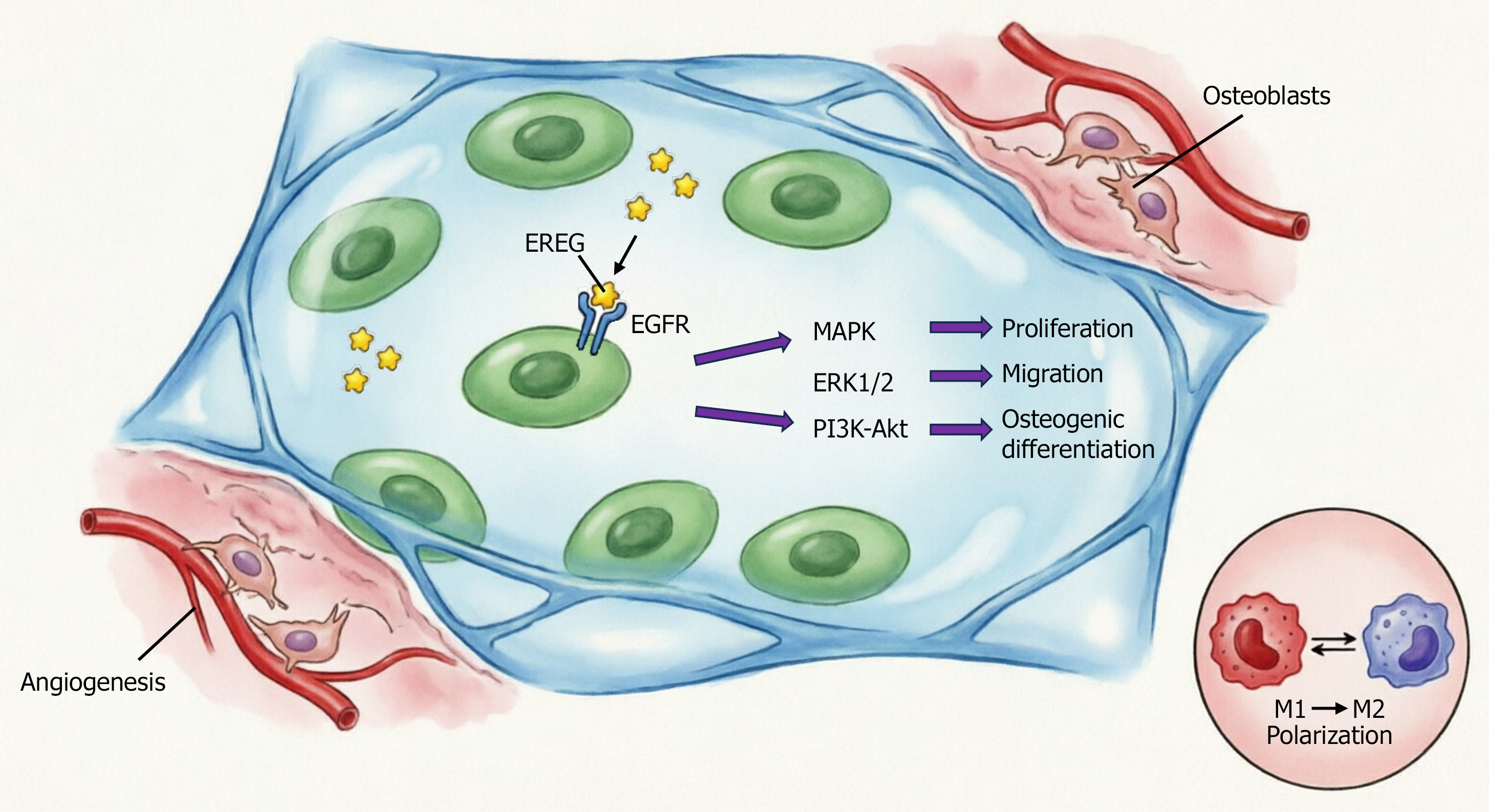
Figure 5 Schematic illustration of the epiregulin-mesenchymal stem cell-hydrogel therapeutic platform for bone/periodontal regeneration.
Biodegradable hydrogel scaffolds serve as a delivery system for mesenchymal stem cells (MSCs) and epiregulin (EREG). EREG binds to epidermal growth factor receptors on MSCs, activating mitogen-activated protein kinase, extracellular signal-regulated kinase 1/2, and phosphatidylinositol 3-kinase/protein kinase B signaling pathways that promote cell proliferation, migration, and osteogenic differentiation. The synergistic actions of the MSC secretome and exogenous EREG modulate the inflammatory microenvironment (M1→M2 macrophage polarization), stimulate angiogenesis, and enhance deposition of mineralized extracellular matrix. This integrated approach enables targeted regeneration of bone and periodontal tissues in inflammatory conditions. Akt: Protein kinase B; EGFR: Epidermal growth factor receptor; EREG: Epiregulin; ERK1/2: Extracellular signal-regulated kinase 1/2; MAPK: Mitogen-activated protein kinase; PI3K: Phosphatidylinositol 3-kinase.
- Citation: Dudun AA, Bonartseva GA, Bonartsev AP. Enhancing mesenchymal stem cell function with epiregulin in tissue engineering: From tissue regeneration to gut microbiota modulation. World J Stem Cells 2026; 18(7): 119865
- URL: https://www.wjgnet.com/1948-0210/full/v18/i7/119865.htm
- DOI: https://dx.doi.org/10.4252/wjsc.119865