©The Author(s) 2025.
World J Stem Cells. Sep 26, 2025; 17(9): 108523
Published online Sep 26, 2025. doi: 10.4252/wjsc.v17.i9.108523
Published online Sep 26, 2025. doi: 10.4252/wjsc.v17.i9.108523
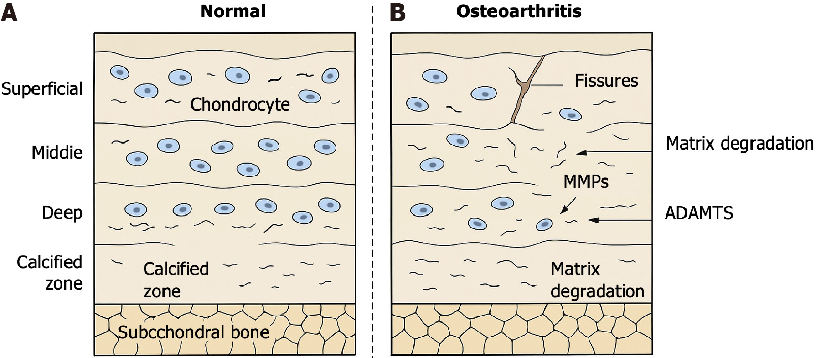
Figure 1 Structural differences between normal and degenerated articular cartilage.
A and B: Schematic representation of healthy (A) and osteoarthritis-affected (B) articular cartilage. Normal cartilage is composed of four zones-superficial, middle, deep, and calcified-organized above the subchondral bone and enriched in collagen type II and aggrecan. In osteoarthritis, progressive matrix degradation, fissures, loss of chondrocytes, and elevated expression of matrix metalloproteinases and aggrecanases (a disintegrin and metalloproteinase with thrombospondin motifs) result in biomechanical failure and tissue degeneration. MMPs: Matrix metalloproteinases; ADAMTs: A disintegrin and metalloproteinase with thrombospondin motifs.
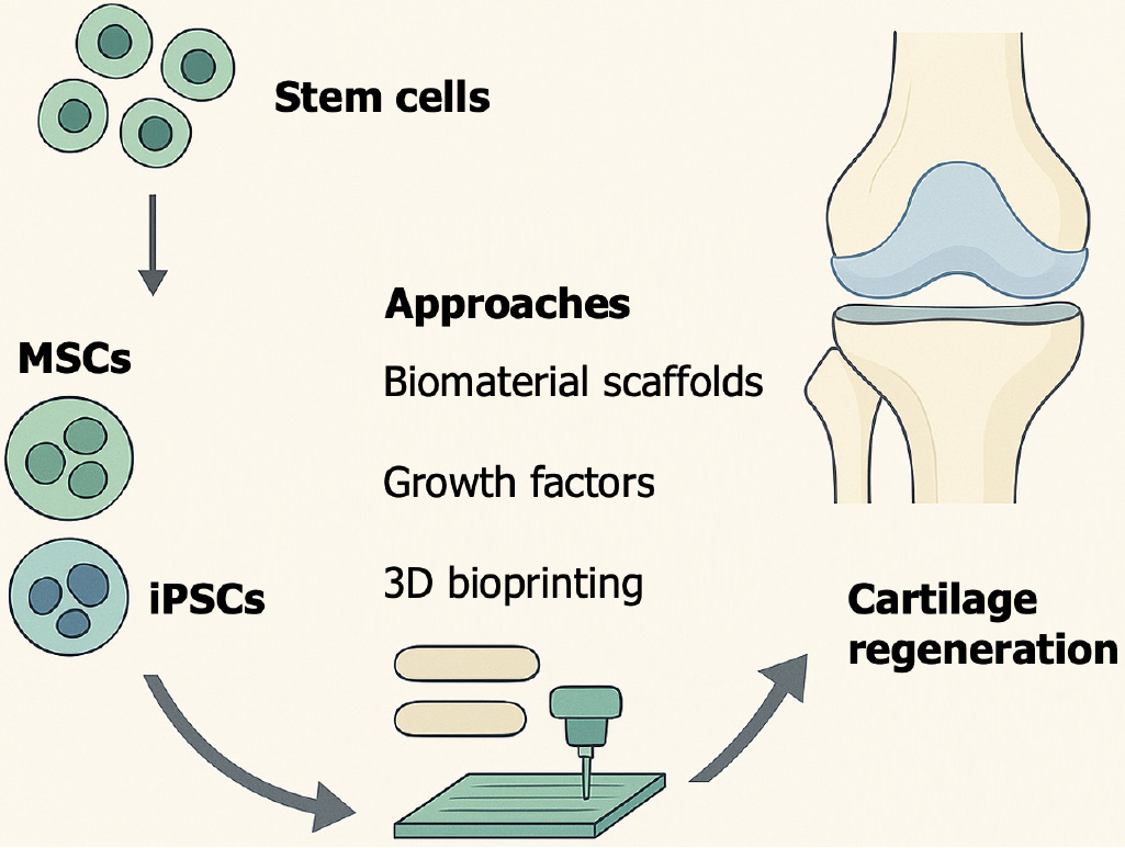
Figure 2 Stem cell-based therapeutic strategies for cartilage repair.
A simplified schematic representing the application of stem cells - including mesenchymal stem cells and induced pluripotent stem cells - in combination with bioengineering tools such as biomaterial scaffolds, growth factors, and three-dimensional bioprinting. These integrated approaches collectively drive chondrogenesis and facilitate functional cartilage regeneration. MSCs: Mesenchymal stem cells; iPSCs: Induced pluripotent stem cells; 3D: Three-dimensional.
- Citation: Cong B, Zhang FH, Zhang HG. Stem cell-based cartilage regeneration: Biological strategies, engineering innovations, and clinical translation. World J Stem Cells 2025; 17(9): 108523
- URL: https://www.wjgnet.com/1948-0210/full/v17/i9/108523.htm
- DOI: https://dx.doi.org/10.4252/wjsc.v17.i9.108523


