Copyright
©The Author(s) 2024.
World J Stem Cells. May 26, 2024; 16(5): 499-511
Published online May 26, 2024. doi: 10.4252/wjsc.v16.i5.499
Published online May 26, 2024. doi: 10.4252/wjsc.v16.i5.499
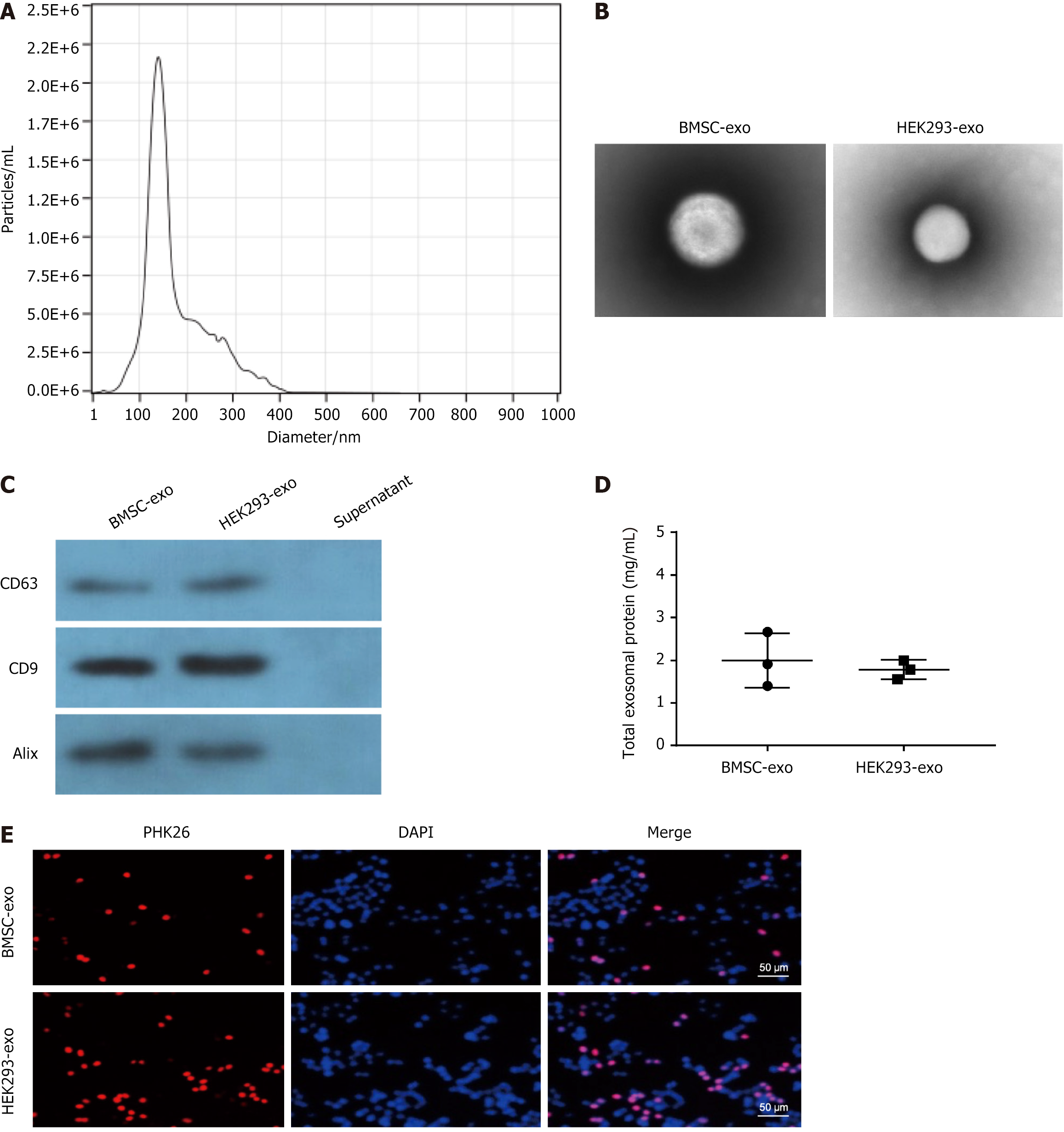
Figure 1 Characterization of bone marrow-derived mesenchymal stem cell- and HEK293-derived exosomes.
A: Particle size distribution of bone marrow-derived mesenchymal stem cell (BMSC)-derived and HEK293-derived exosomes (BMSC-exo and HEK293-exo, respectively) detected by NanoSight; the mean diameter is 150 nm; B: Representative images of the morphology of BMSC-exo and HEK293-exo via transmission electron microscopy; C: Western blot identification of exosome surface markers; D: Expression of BMSC-exo and HEK293-exo protein; E: Internalization of PKH26-labeled exosomes within mouse osteoblast progenitor cells by fluorescence microscopy, Scale bar: 50 μm. BMSC-exo: Bone marrow-derived mesenchymal stem cell-derived exosome.
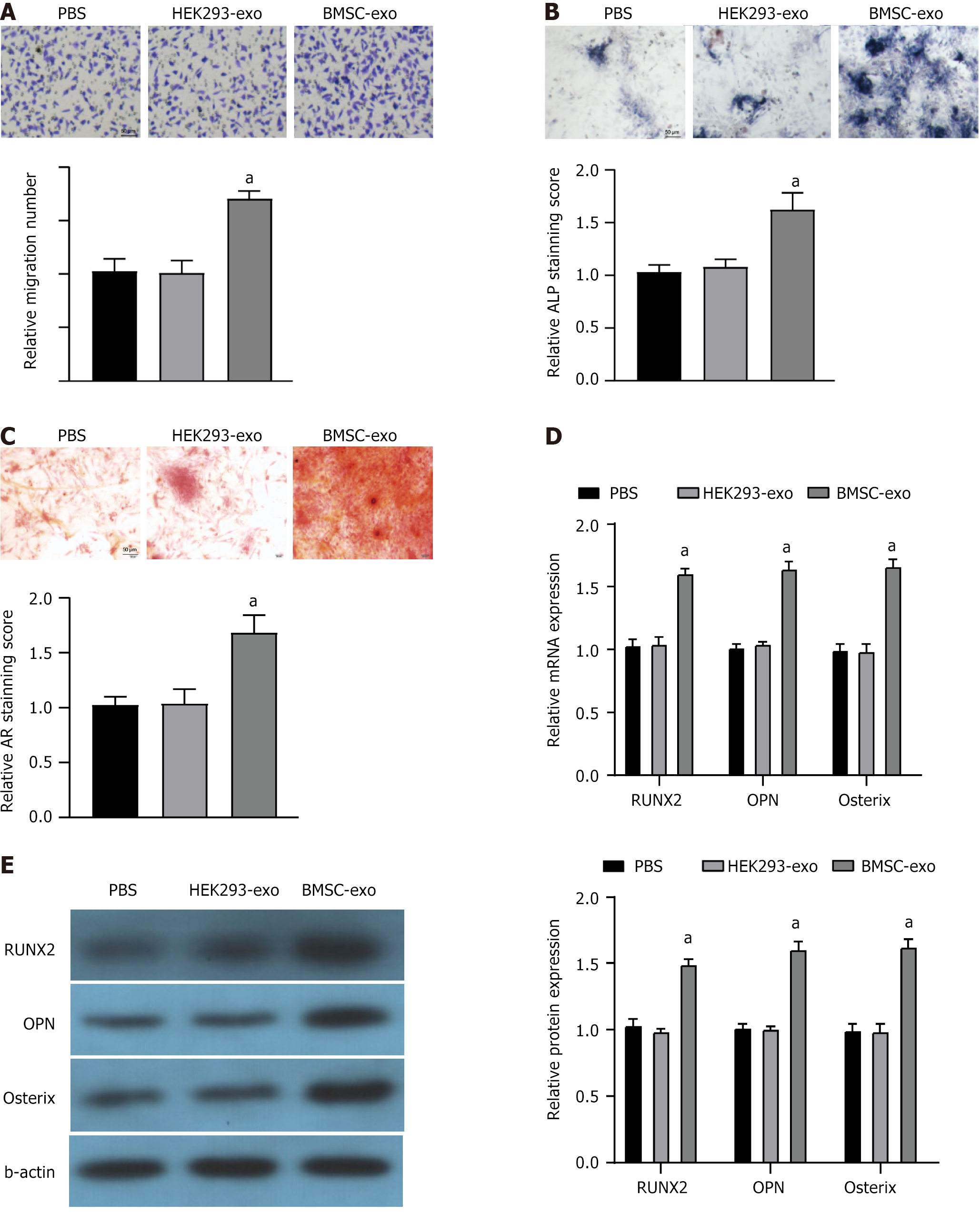
Figure 2 Bone marrow-derived mesenchymal stem cell-derived exosome and HEK293-derived exosome promoted migration and angiogenesis of mouse osteoblast progenitor cells.
A: Mouse osteoblast progenitor cells (mOPCSs) with bone marrow-derived mesenchymal stem cell (BMSC)-derived exosome (BMSC-exo) or HEK293-exo stained by crystal violet in Transwell assays. BMSC-exo enhanced cell migration, Scale bar: 50 μm; B: Alkaline phosphatase staining of mOPCSs with BMSC-exo or HEK293-exo internalization for one week. Scale bar: 50 μm; C: Alizarin red staining of treated mOPCSs after osteogenic induction for two weeks. Scale bar: 50 μm; D and E: mRNA and protein expression of genes associated with osteogenesis (Runx2, OPN, and Osterix) in BMSC-exo and HEK293-exo-treated mOPCSs. BMSC-exo: Bone marrow-derived mesenchymal stem cell-derived exosome; PBS: Phosphate buffered saline; ALP: Alkaline phosphatase.
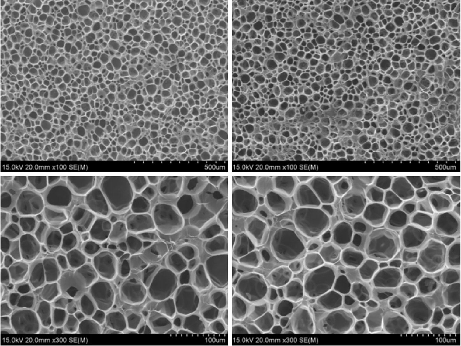
Figure 3 Morphology and characterization of chondroitin-6-sulfate/silk fibroin/glycol chitosan/dialysis-purified maleic anhydride-modified polyethylene glycol hydrogel.
The analysis of scanning electron microscopy images revealed that the pores in the chondroitin-6-sulfate/silk fibroin/glycol chitosan/dialysis-purified maleic anhydride-modified polyethylene glycol hydrogel exhibited predominantly circular or elliptical shapes with uniform sizes and were well interconnected.
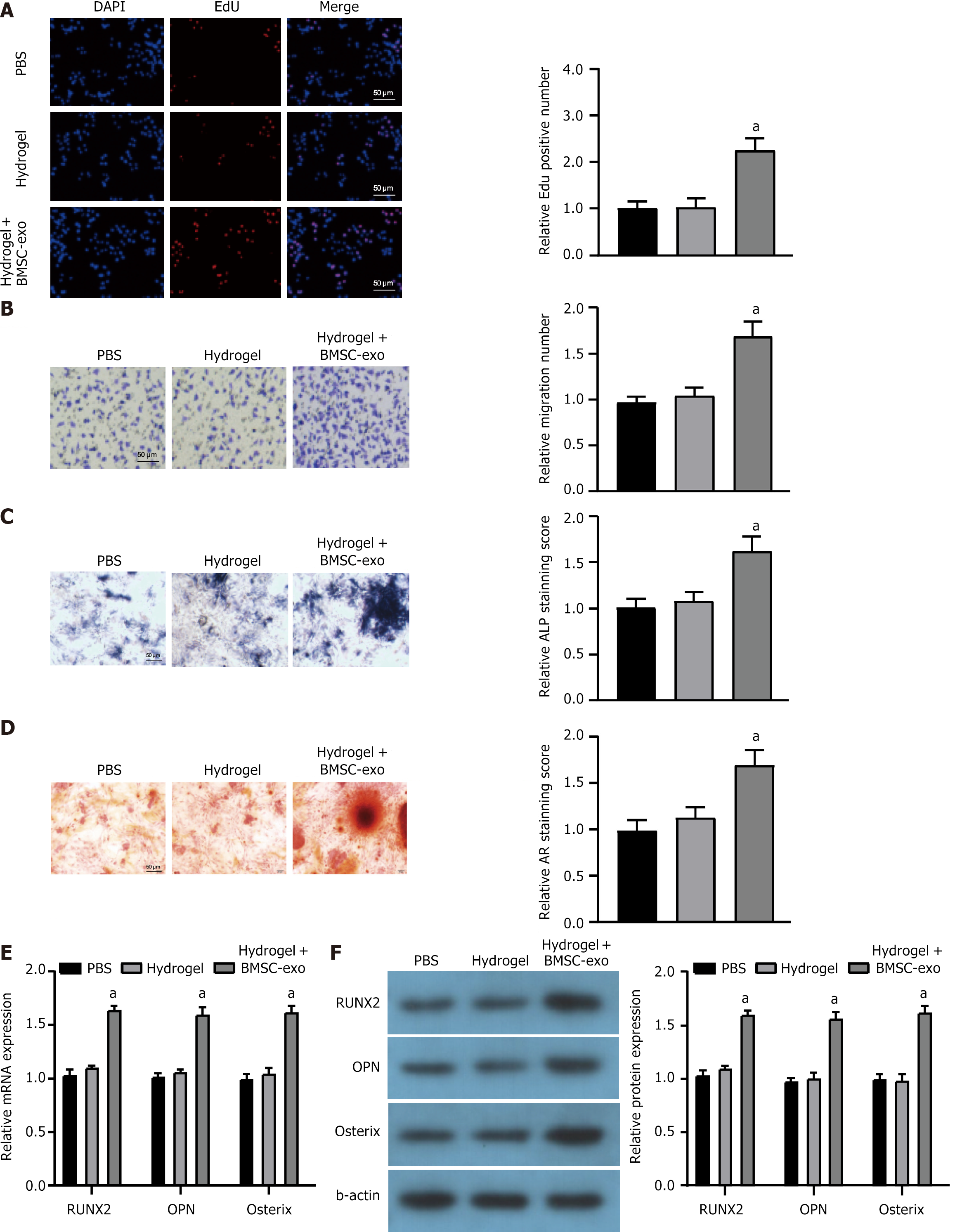
Figure 4 Fabrication and efficacy of bone marrow-derived mesenchymal stem cell-derived exosome-treated hydrogel.
A: Cells proliferation of mouse osteoblast progenitor cells (mOPCSs) with different treatments by fluorescence microscopy; B: mOPCSs with hydrogel or bone marrow-derived mesenchymal stem cell (BMSC)-derived exosome (BMSC-exo)-treated hydrogel stained by crystal violet in Transwell assays. Hydrogel + BMSC-exo enhanced cell migration, Scale bar: 50 μm; C: Alkaline phosphatase staining of mOPCSs treated with hydrogel or hydrogel + BMSC-exo for one week. Scale bar: 50 μm; D: Alizarin red staining of mOPCSs with different treatments after osteogenic induction for two weeks. Scale bar: 50 μm; E and F: mRNA and protein expression of genes associated with osteogenesis (Runx2, OPN, and Osterix) in mOPCSs with different treatments. aP < 0.05. BMSC-exo: Bone marrow-derived mesenchymal stem cell-derived exosome; PBS: Phosphate buffered saline; DAPI: 4’,6’-diaminido-2-phenylindole; ALP: Alkaline phosphatase.
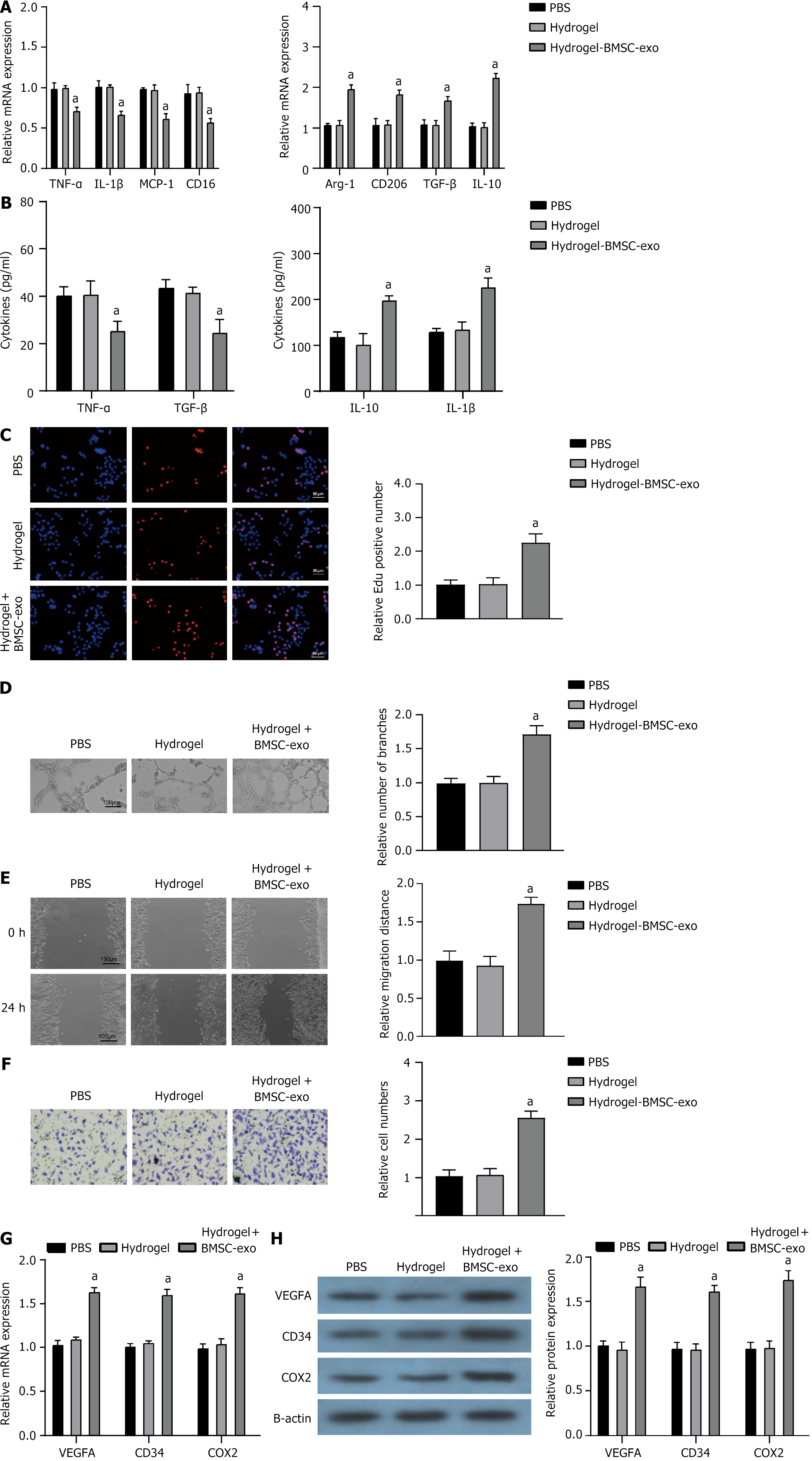
Figure 5 Bone marrow-derived mesenchymal stem cell-derived exosome hydrogels effectively suppressed the inflammatory response of macrophages while facilitating angiogenesis.
A: Quantitative real-time polymerase chain reaction assessment of mRNA expression of tumor necrosis factor (TNF)-α, interleukin (IL)-1β, monocyte chemoattractant protein-1, CD16, arginase-1, CD206, transforming growth factor (TGF)-β, and IL-10 in RAW264.7 cells cultured on hydrogels with or without bone marrow-derived mesenchymal stem cell (BMSC)-derived exosome (BMSC-exo) for 24 h; B: Expression of TNF-α, IL-1β, TGF-β, and IL-10 cytokines was assessed in RAW264.7 macrophages across different experimental groups; C: Cells proliferation of mouse osteoblast progenitor cells (mOPCSs) co-cultured with human umbilical vein endothelial cell in different groups by fluorescence microscopy; D: Tube formation assay of mOPCs in different groups. Scale bar: 50 μm; E and F: Scratch wound healing assay and Transwell migration assay of mOPCSs in different groups. The hydrogel + BMSC-exo group demonstrated enhanced migration, Scale bar: 50 μm; G and H: mRNA and protein expression of angiogenesis markers (vascular endothelial growth factor A, CD34, and cyclooxygenase-2) in mOPCSs in different groups. aP < 0.05. TNF: Tumor necrosis factor; IL: Interleukin; MCP-1: Monocyte chemoattractant protein-1; Arg-1: Arginase-1; TGF: Transforming growth factor; VEGFA: Vascular endothelial growth factor A; COX-2: Cyclooxygenase-2; BMSC-exo: Bone marrow-derived mesenchymal stem cell-derived exosome; PBS: Phosphate buffered saline.
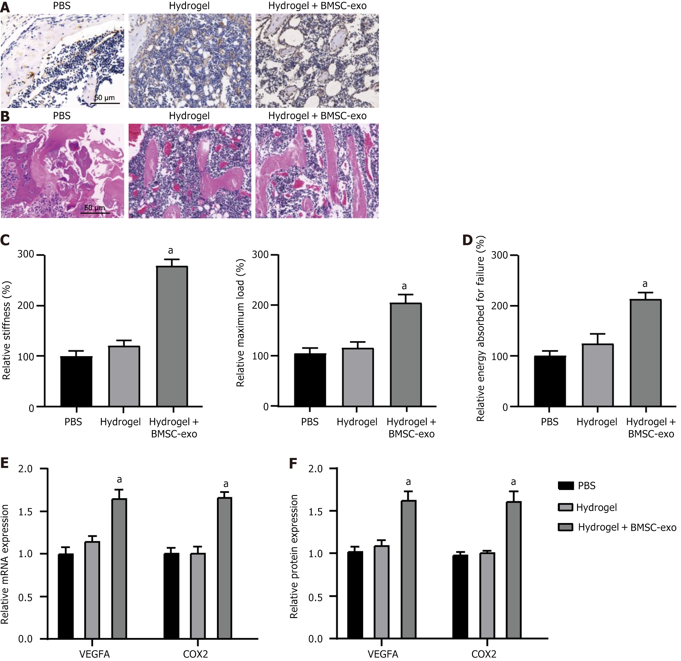
Figure 6 Bone marrow-derived mesenchymal stem cell-derived exosome hydrogel promoted fracture healing and angiogenesis in vivo.
A: Alcian blue and orange G staining of regenerated bone sections treated by hydrogels with or without bone marrow-derived mesenchymal stem cell (BMSC)-derived exosome (BMSC-exo) on week 2 and week 4 after fracture; B: Hematoxylin-eosin staining of regenerated bone tissues; C and D: Maximum load, stiffness, and energy absorbed for failure or repaired bone; E and F: mRNA and protein expression of angiogenesis markers (vascular endothelial growth factor A and cyclooxygenase-2) in fracture healing model treated by hydrogels with or without BMSC-exo. n = 6, aP < 0.05. VEGFA: Vascular endothelial growth factor A; COX-2: Cyclooxygenase-2; BMSC-exo: Bone marrow-derived mesenchymal stem cell-derived exosome; PBS: Phosphate buffered saline.
- Citation: Zhang S, Lu C, Zheng S, Hong G. Hydrogel loaded with bone marrow stromal cell-derived exosomes promotes bone regeneration by inhibiting inflammatory responses and angiogenesis. World J Stem Cells 2024; 16(5): 499-511
- URL: https://www.wjgnet.com/1948-0210/full/v16/i5/499.htm
- DOI: https://dx.doi.org/10.4252/wjsc.v16.i5.499


