Copyright
©The Author(s) 2023.
World J Stem Cells. May 26, 2023; 15(5): 453-465
Published online May 26, 2023. doi: 10.4252/wjsc.v15.i5.453
Published online May 26, 2023. doi: 10.4252/wjsc.v15.i5.453
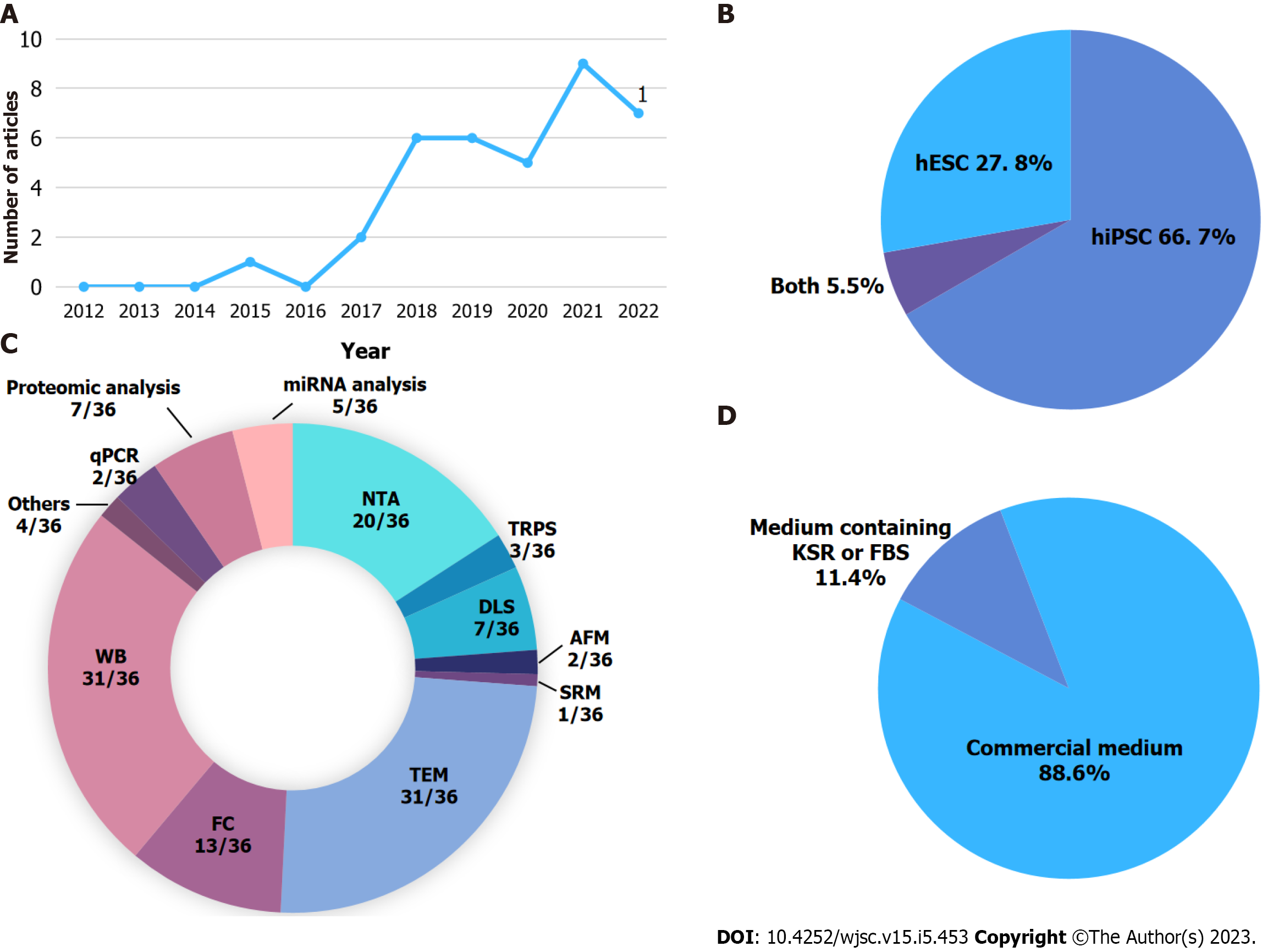
Figure 1 Overview of studies on human pluripotent stem cell-derived extracellular vesicles published between 2012 and 2022.
A: Timeline of published articles on human pluripotent stem cell-derived extracellular vesicles (hPSC-EVs). 1Two articles were published online in 2022 but published in print in 2023; B: Analysis of the percentage of articles that use human embryonic stem cells, human induced pluripotent stem cell, or both cell types to isolate EVs; C: Methods used in the studies to characterize hPSC-EVs. The graphic depicts the number of articles using certain techniques/total number of articles included in the analysis; D: Analysis of media used to culture hPSC to isolate the EVs. AFM: Atomic force microscopy; DLS: Dynamic light scattering; FC: Flow cytometry; NTA: Nanoparticle tracking analysis; qPCR: Quantitative polymerase chain reaction; SRM: Super-resolution microscopy; TEM: Transmission electron microscopy; TRPS: Tunable resistive pulse sensing; WB: Western blot; hESC: Human embryonic stem cells; hIPSC: Human induced pluripotent stem cell.
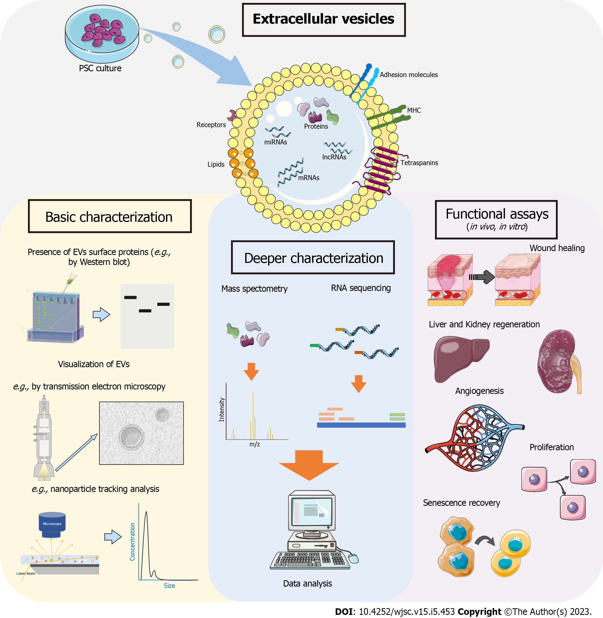
Figure 2 Diagram of pluripotent stem cell-derived extracellular vesicle isolation, its most common characterizations, and the applications described or indicated for these extracellular vesicles.
EV: Extracellular vesicle; miRNA: MicroRNA; lncRNA: Long noncoding RNA; PSC: Pluripotent stem cell. The images were obtained from Servier Medical Art (http://smart.servier.com), licensed under a Creative Commons Attribution 3.0 Unported License.
- Citation: Matos BM, Stimamiglio MA, Correa A, Robert AW. Human pluripotent stem cell-derived extracellular vesicles: From now to the future. World J Stem Cells 2023; 15(5): 453-465
- URL: https://www.wjgnet.com/1948-0210/full/v15/i5/453.htm
- DOI: https://dx.doi.org/10.4252/wjsc.v15.i5.453


