©The Author(s) 2025.
World J Gastroenterol. Nov 7, 2025; 31(41): 111449
Published online Nov 7, 2025. doi: 10.3748/wjg.v31.i41.111449
Published online Nov 7, 2025. doi: 10.3748/wjg.v31.i41.111449
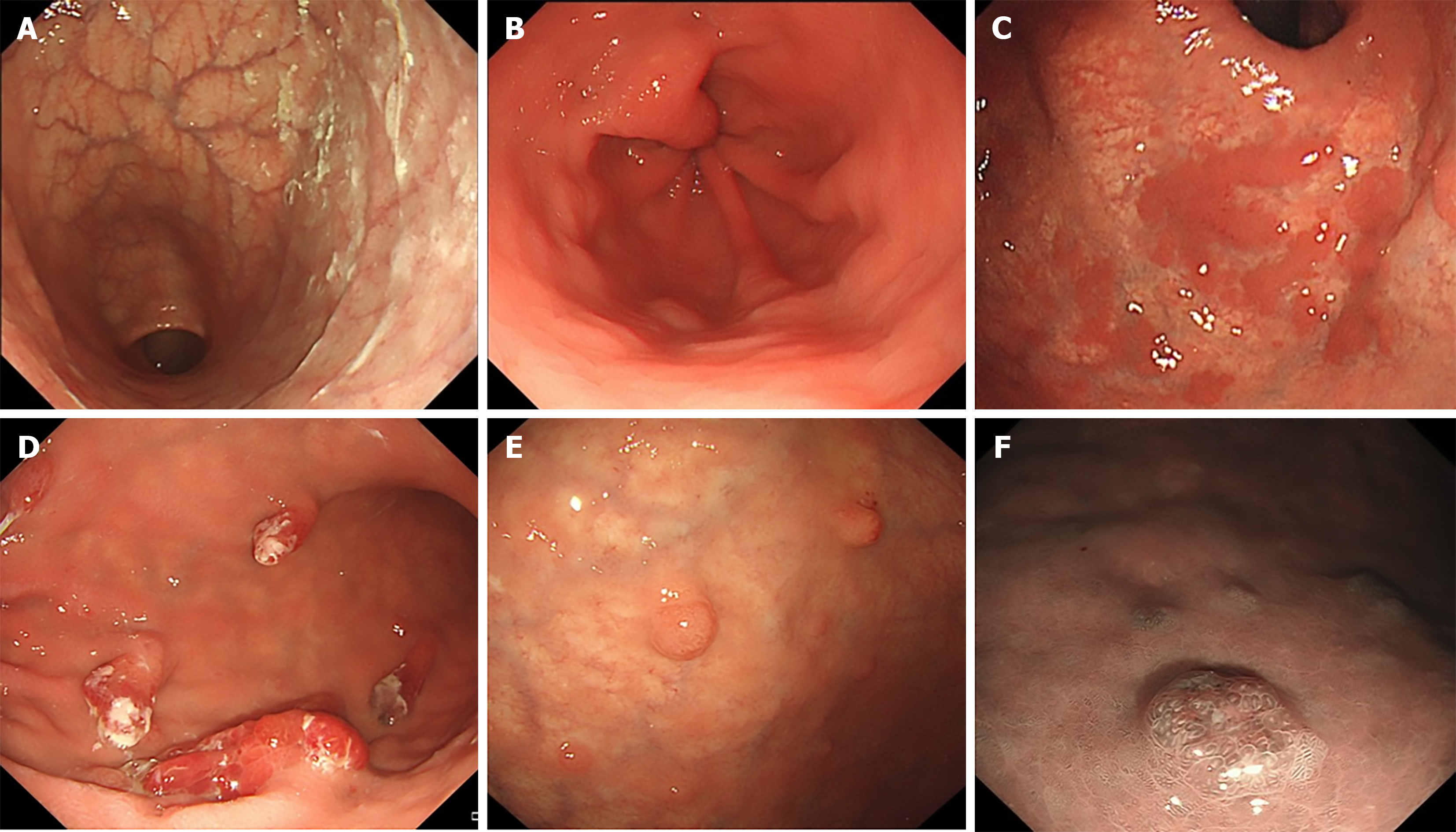
Figure 1 Some endoscopic findings in patients with autoimmune gastritis.
A: Mucosal atrophy of the gastric fundus and corpus (reverse atrophy) with sticky adherent dense mucus; B: Circular wrinkle-like pattern of the mucosa with no atrophy in antrum; C: Remnants of oxyntic mucosa, presenting as flat localized type; D: Hyperplastic polyps; E: Scattered type I gastric neuroendocrine tumors in the atrophic gastric fundus and body; F: Narrow band imaging of gastric neuroendocrine tumors.
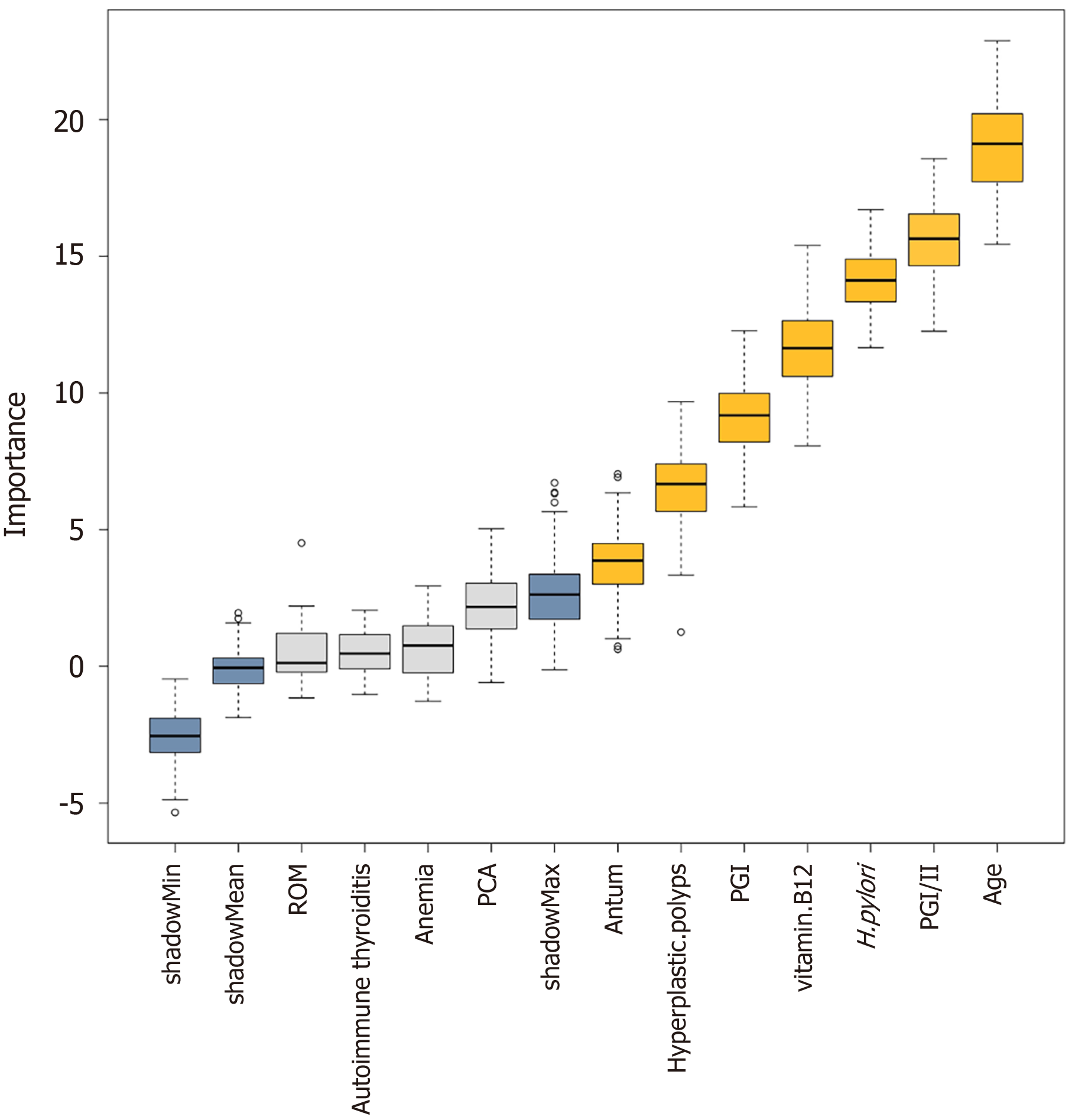
Figure 2 Feature selection for gastric neuroendocrine tumors in patients with autoimmune gastritis with Boruta algorithm.
Variables with yellow box plots are considered important, while those with grey box plots are rejected, and the blue plot represents the minimum, average, and max shadow scores. ROM: Remnants of oxyntic mucosa; PCA: Parietal cell antibody; PGI: Pepsinogen I; H. pylori: Helicobacter pylori; PGI/II: Ratio of pepsinogen I to II.
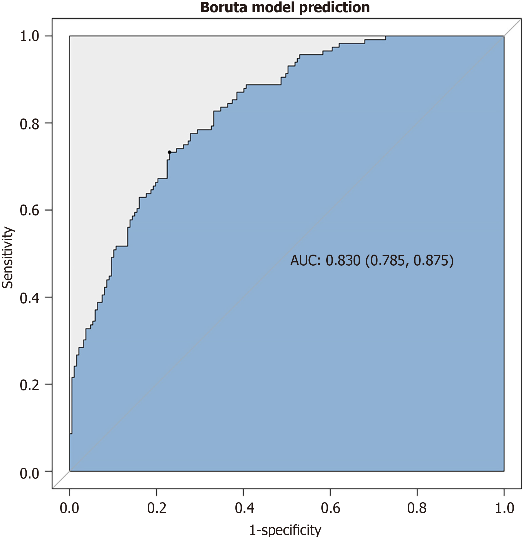
Figure 3 The receiver operating characteristic curve of the discriminative ability for predicting neuroendocrine tumor by the associated variables screened with the Boruta method.
AUC: Area under curve.
- Citation: Li YM, Guo WJ, Deng C, Luo J, Shi YF, Zhu D, Wei QL, Zhang MG, Du SY, Tan HY. Risk assessment of type I gastric neuroendocrine tumors based on endoscopic and clinical features of autoimmune gastritis. World J Gastroenterol 2025; 31(41): 111449
- URL: https://www.wjgnet.com/1007-9327/full/v31/i41/111449.htm
- DOI: https://dx.doi.org/10.3748/wjg.v31.i41.111449


