©The Author(s) 2025.
World J Gastroenterol. Sep 14, 2025; 31(34): 109825
Published online Sep 14, 2025. doi: 10.3748/wjg.v31.i34.109825
Published online Sep 14, 2025. doi: 10.3748/wjg.v31.i34.109825
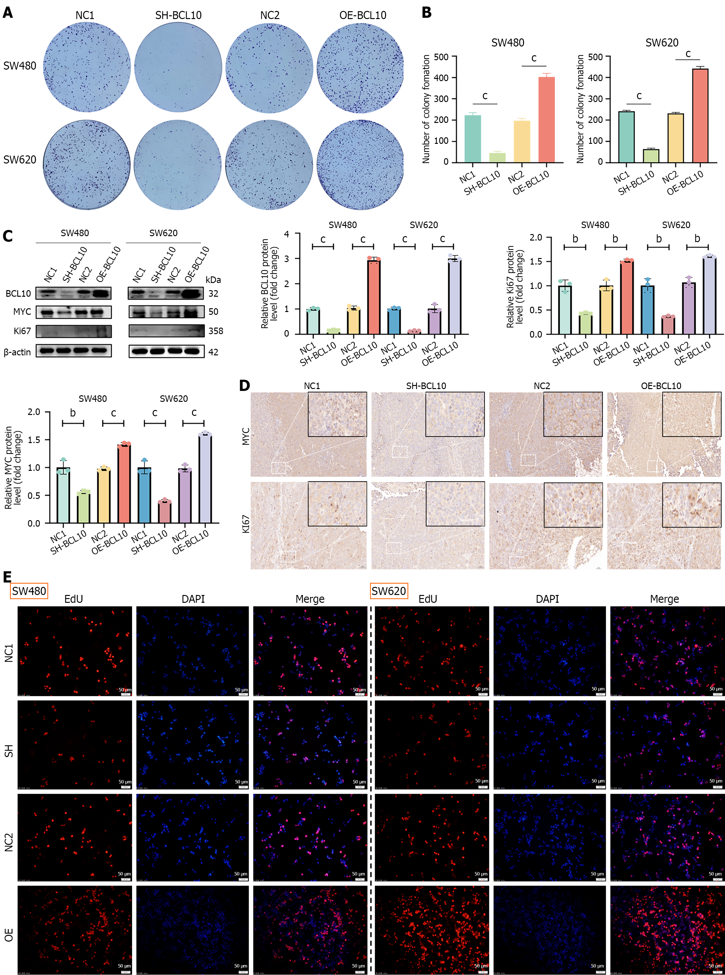
Figure 1 B-cell CLL/Lymphoma 10 promotes the proliferation of colorectal cancer cells in vitro and in vivo.
A and B: Colony formation assays showing proliferation ability in SW480 and SW620 cell lines with BCL10 knockdown (SH-BCL10) or overexpression (OE-BCL10). NC1: Scrambled shRNA control; NC2: Empty vector control. cP < 0.001; C: Western blot analysis of MYC and Ki67 expression. bP < 0.01; cP < 0.001; D: Immunohistochemical staining of MYC and Ki67 in xenograft tumors. Scale bars: 50 μm (main panels, 20 × magnification); 20 μm (insets, 40 × magnification); E: EdU assay for cell proliferation detection. Scale bars: 50 μm.
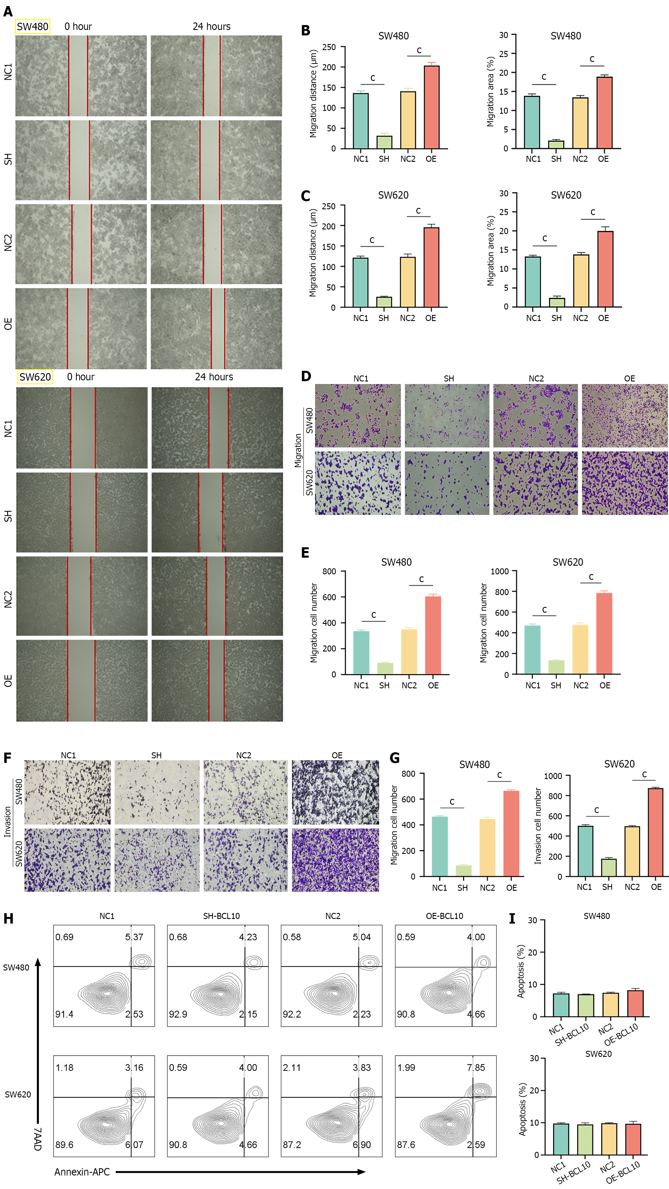
Figure 2 B-cell CLL/Lymphoma 10 promotes invasion and migration without affecting baseline apoptosis.
A-C: Wound healing assays showing migration ability in SW480 and SW620 cells. cP < 0.001; D-G: Transwell invasion assays with representative images (D and F) and quantification (E and G). cP < 0.001; H and I: Flow cytometry analysis of apoptosis using Annexin V/PI staining.
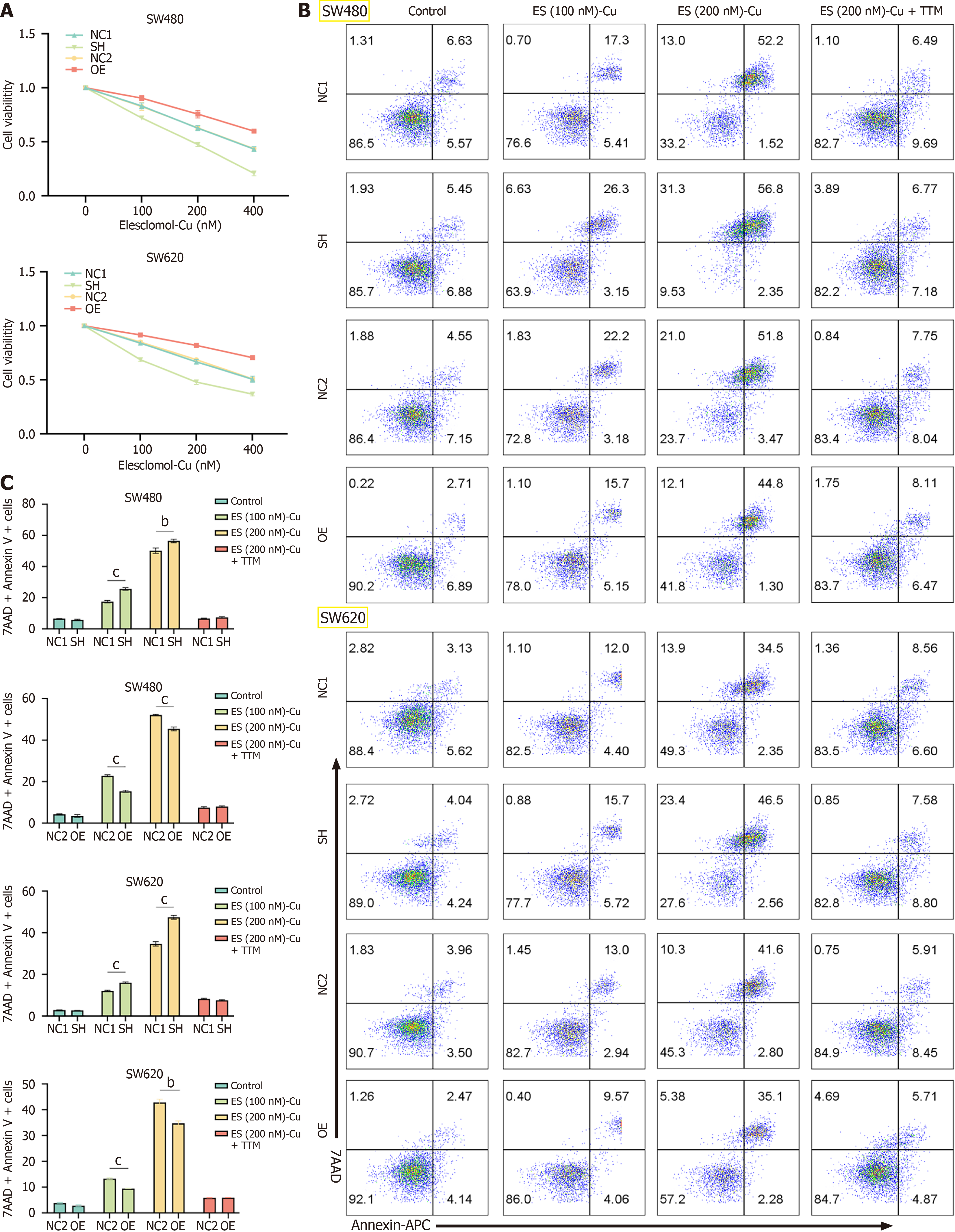
Figure 3 B-cell CLL/Lymphoma 10 regulates sensitivity to cuproptosis.
A: Cell viability after elesclomol-Cu treatment (CCK-8 assay); B and C: Flow cytometry analysis of cell death using 7-AAD and Annexin V staining. TTM: Tetrathiomolybdate. bP < 0.01; cP < 0.001.
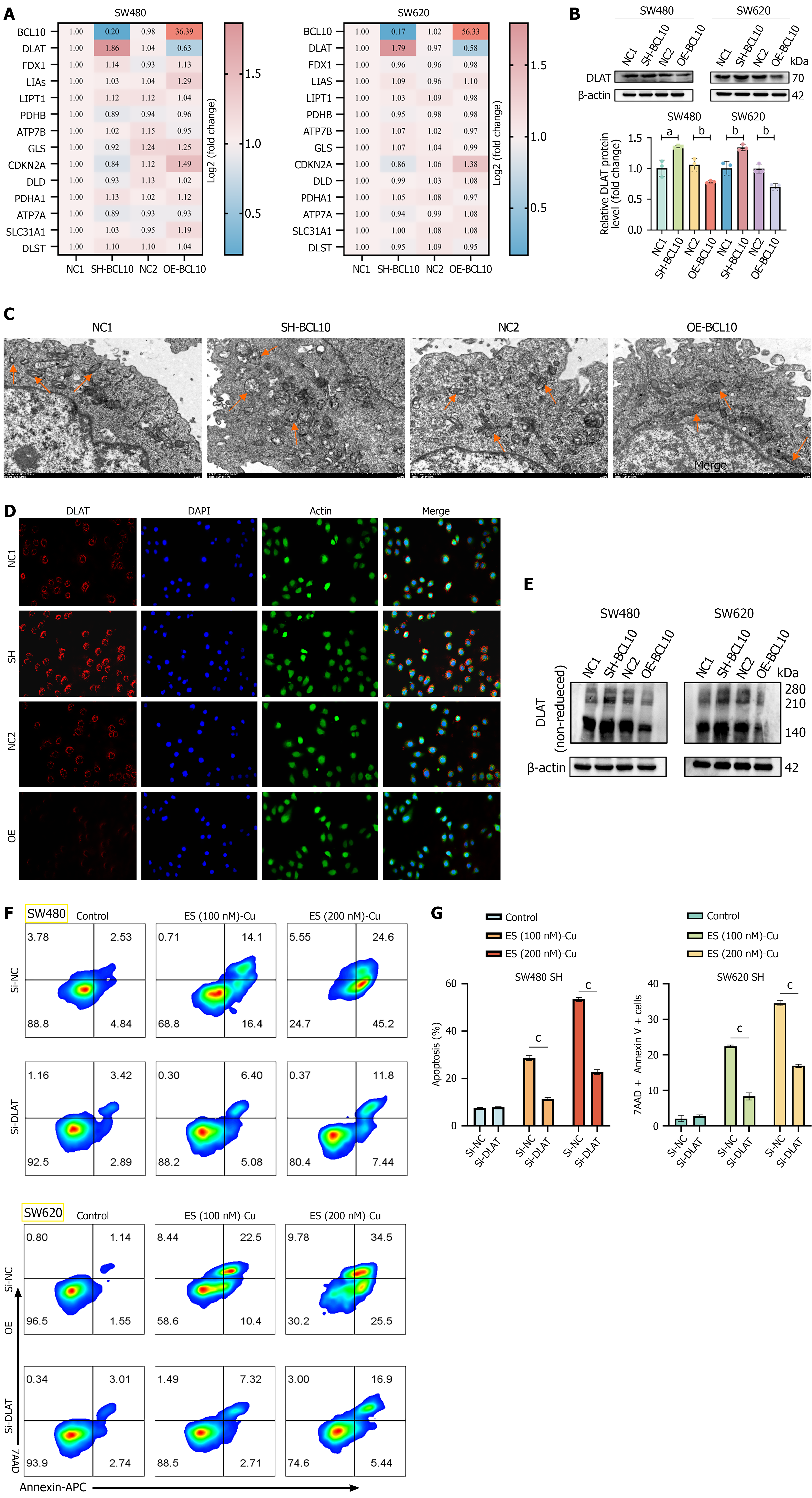
Figure 4 B-cell CLL/Lymphoma 10 affects cuproptosis sensitivity through DLAT regulation.
A: Quantitative PCR analysis of cuproptosis-related genes; B: Western blot of B-cell CLL/Lymphoma 10 (BCL10) and DLAT expression. aP < 0.05; bP < 0.01; C: TEM images of mitochondrial morphology (orange arrows: Damaged mitochondria with cristae disruption); D: Immunofluorescence of indicated proteins after elesclomol-Cu treatment; E: Non-reducing Western blot of DLAT oligomers; F and G: Flow cytometry after DLAT knockdown. cP < 0.001.
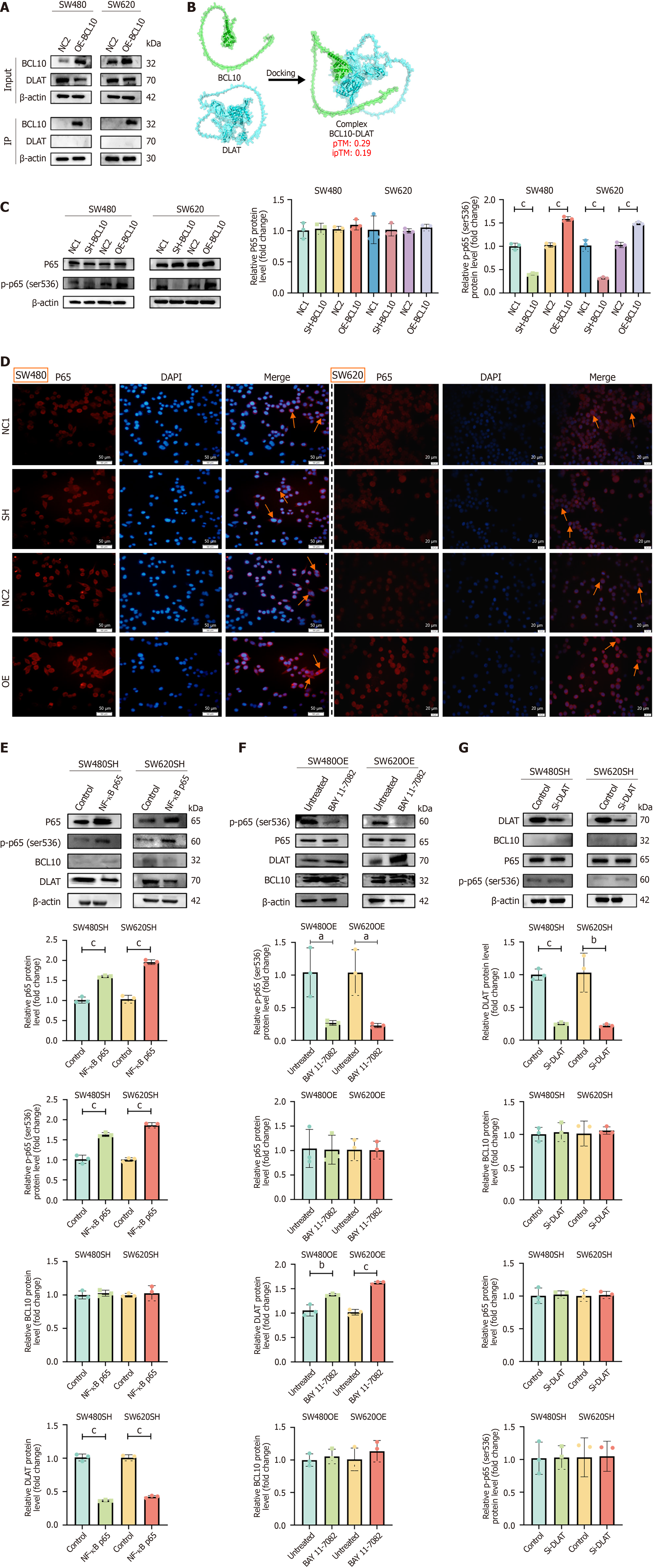
Figure 5 B-cell CLL/Lymphoma 10 regulates DLAT through NF-κB p65 phosphorylation.
A: Co-IP of DLAT and B-cell CLL/Lymphoma 10 (BCL10); B: AlphaFold3 predicted binding model (ipTM < 0.6); C: Western blot of p65 and p-p65 (Ser536). cP < 0.001; D: Immunofluorescence of p65 (red) and DAPI (blue); orange arrows indicate nuclear translocation; E: Western blot after p65 transfection in BCL10-knockdown cells. cP < 0.001; F: Western blot after BAY 11-7082 treatment. aP < 0.05. bP < 0.01. cP < 0.001; G: Western blot after DLAT knockout. cP < 0.001.
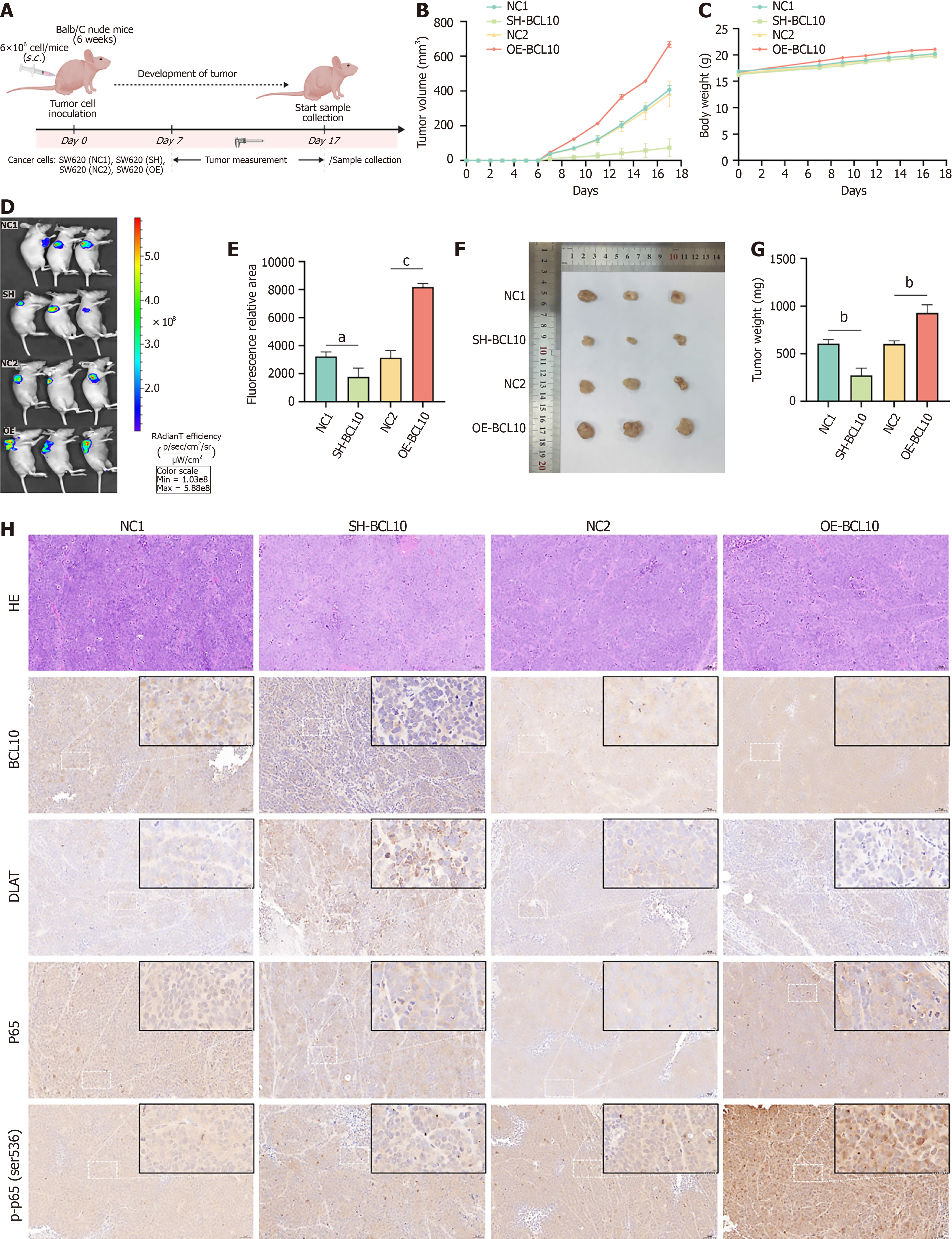
Figure 6 In vivo effects of B-cell CLL/Lymphoma 10 on tumor growth and DLAT/p65 signaling.
A: Experimental timeline; B-G: Tumor volume (B, D, E) and weight (C, F, G) measurements. aP < 0.05; bP < 0.01; cP < 0.001; H: Hematoxylin and eosin staining and IHC for BCL10, DLAT, p65, and p-p65 (Ser536). Scale bars: 50 μm (main panels, 20 × magnification); 20 μm (insets, 40 × magnification).
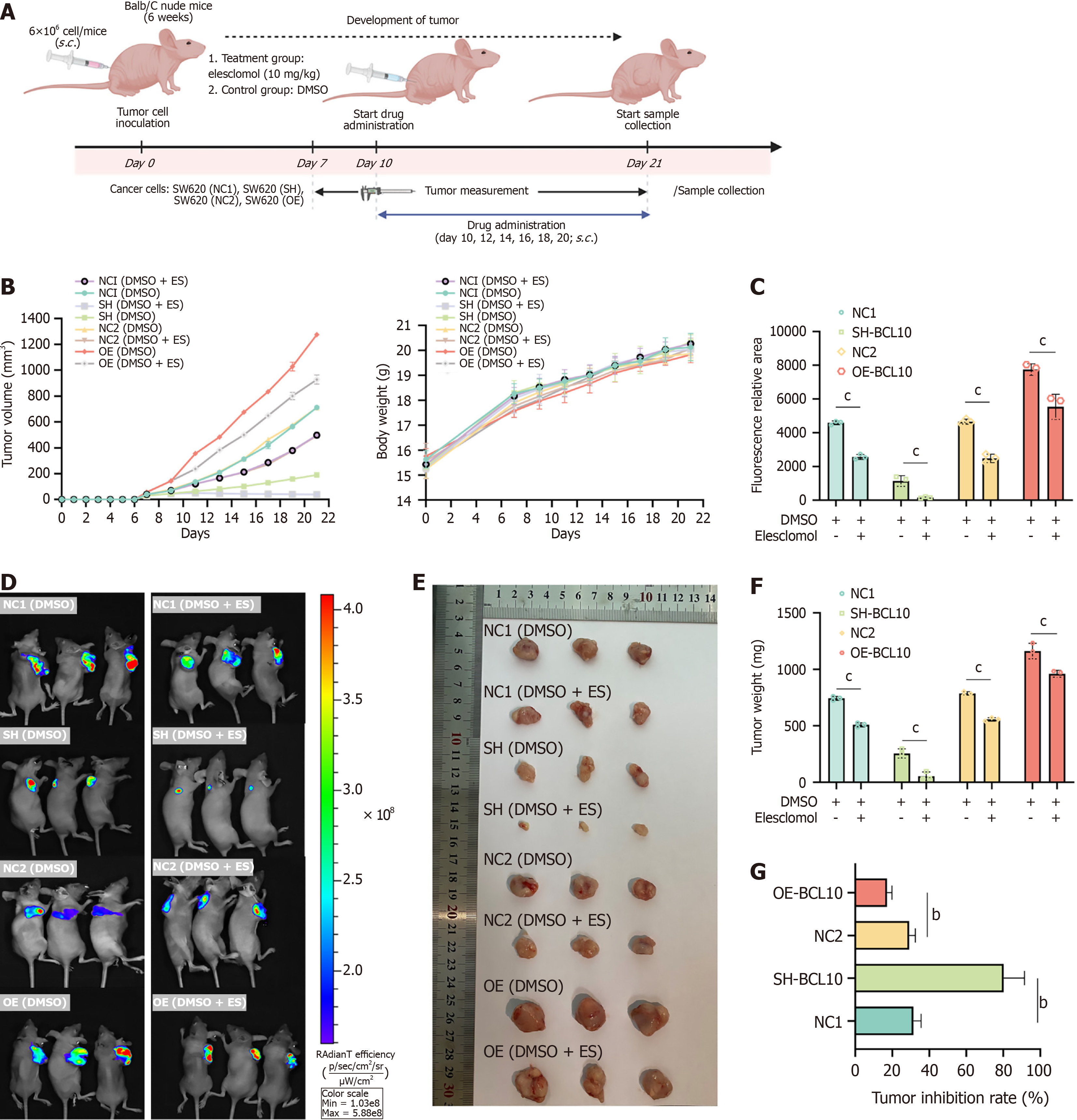
Figure 7 B-cell CLL/Lymphoma 10 modulates copper treatment sensitivity in vivo.
A: Experimental timeline; B: Tumor volume and mouse weight changes; C-F: Quantification of tumor volume (C and D) and weight (E and F). cP < 0.001; G: Tumor growth inhibition rate. bP < 0.01.
- Citation: Xiao PT, Li CF, Liu YD, Zhong J, Cui XL, Liu C, Yang W. B cell CLL/lymphoma 10 promotes colorectal cancer cell proliferation and regulates cuproptosis sensitivity through the NF-κB signaling pathway. World J Gastroenterol 2025; 31(34): 109825
- URL: https://www.wjgnet.com/1007-9327/full/v31/i34/109825.htm
- DOI: https://dx.doi.org/10.3748/wjg.v31.i34.109825


