©The Author(s) 2023.
World J Gastroenterol. May 28, 2023; 29(20): 3157-3167
Published online May 28, 2023. doi: 10.3748/wjg.v29.i20.3157
Published online May 28, 2023. doi: 10.3748/wjg.v29.i20.3157
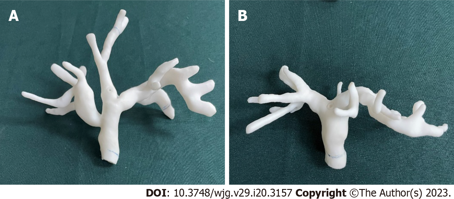
Figure 1 Biliary phantom setup.
A: Biliary model based on patient A; B: Biliary model based on patient B.
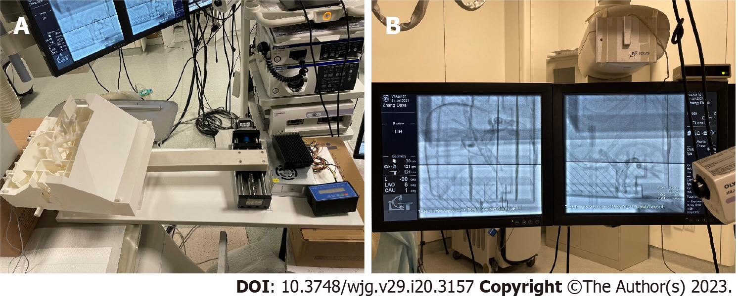
Figure 2 Surgical environment for endoscopic retrograde cholangiopancreatography phantom.
A: Respiratory motion simulator; B: X-ray fluoroscopy guidance in navigation.
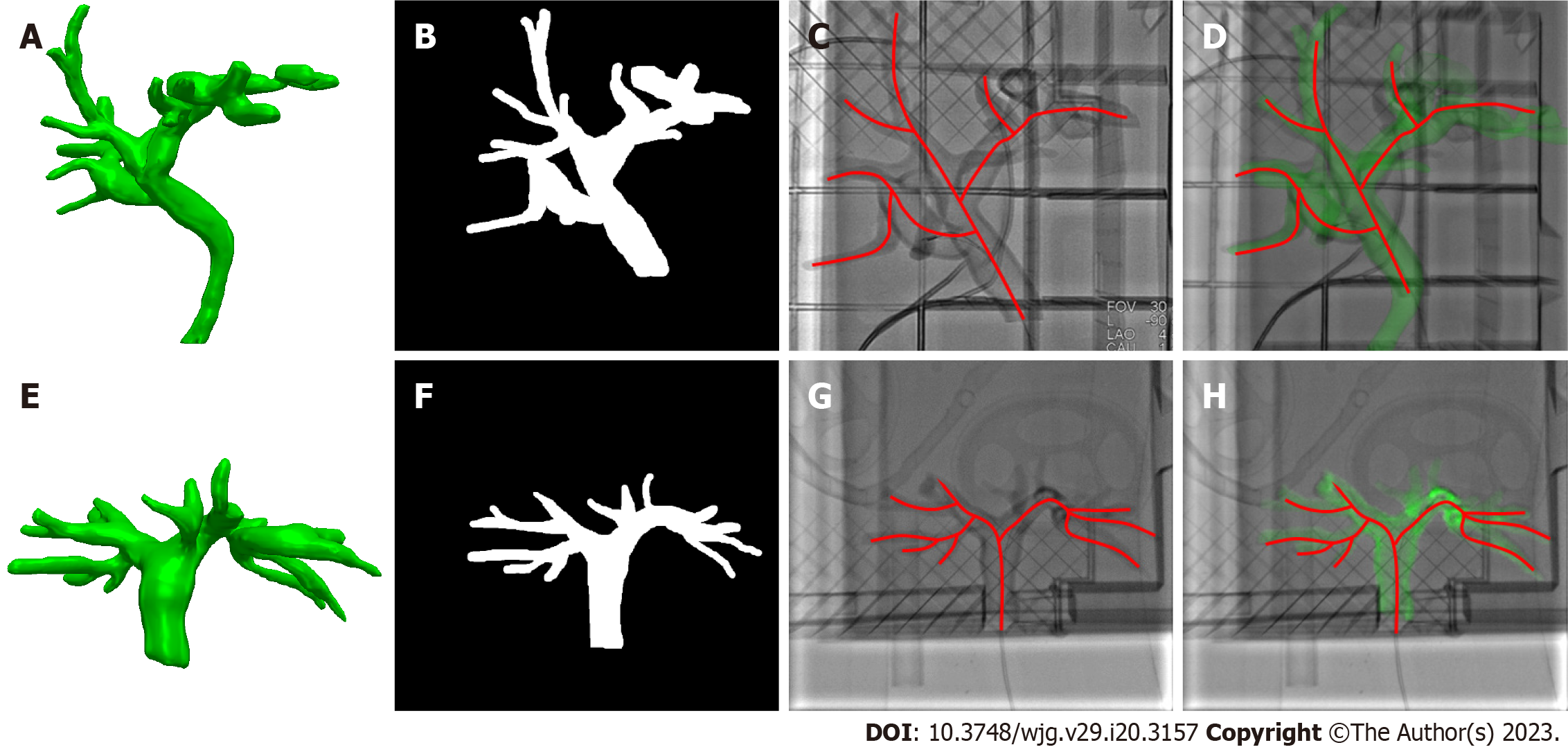
Figure 3 Phantom study of registration and fusion.
A and E: Biliary phantom segmentation using computed tomography images; B and F: Biliary phantom segmentation using X-ray fluoroscopy; C and G: Biliary lumen centerline extraction; D and H: Three-dimensional biliary model registered with the best matching frame using X-ray fluoroscopy.
Figure 4 Automatic segmentation of structures based on preoperative computed tomography.
A and B: Biliary and skin phantoms of different patients; C: Bone, liver, biliary tract, and gallbladder segmented by a three-dimensional UNet network based on preoperative computed tomography data.
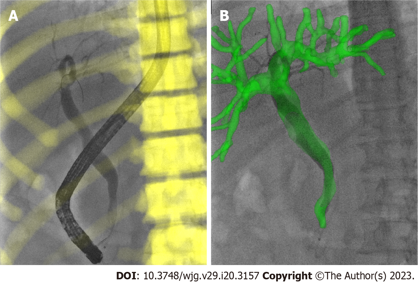
Figure 5 Registration and fusion on the best matching frame of the patient.
A: Three- dimensional/two-dimensional registration based on bone during endoscopic retrograde cholangiopancreatography; B: Overlay the biliary model on X-ray fluoroscopy through the transformation from bone registration.
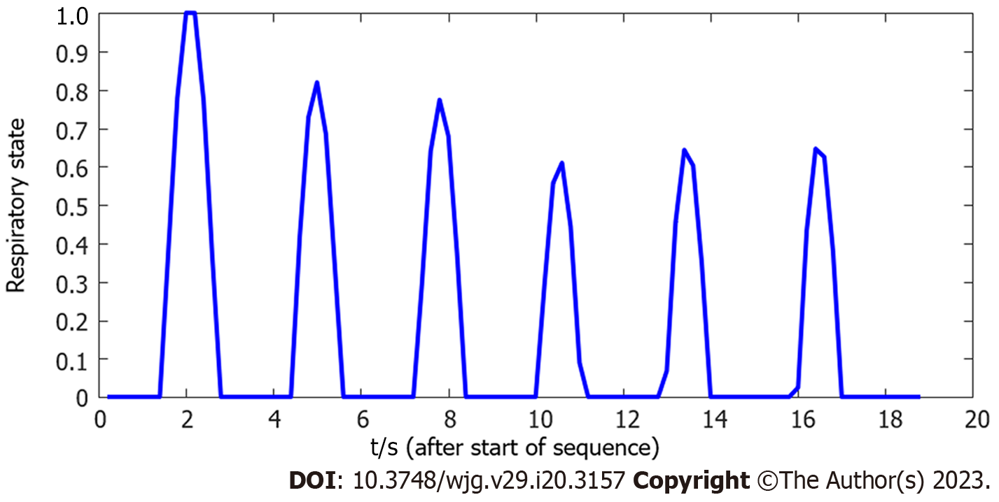
Figure 6 Example of the estimated respiratory state over an 18.
8 s X-ray fluoroscopic image sequence.
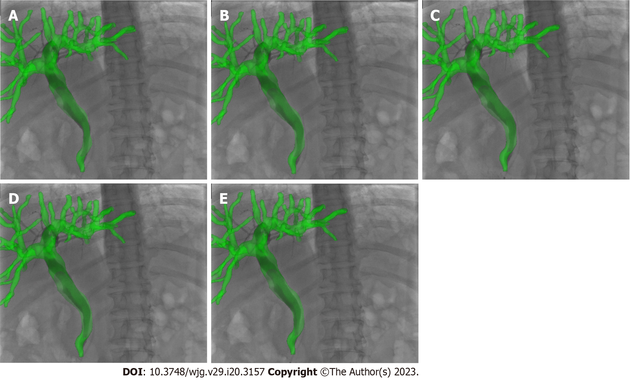
Figure 7 Real-time navigation of image-guided endoscopic retrograde cholangiopancreatography.
A: Beginning of inhalation; B: During inhalation; C: End of inhalation; D: During exhalation; E: End of exhalation.
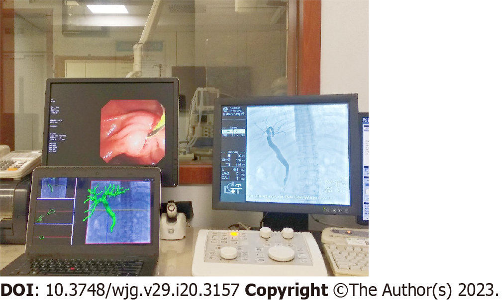
Figure 8 Image-guided endoscopic retrograde cholangiopancreatography navigation system.
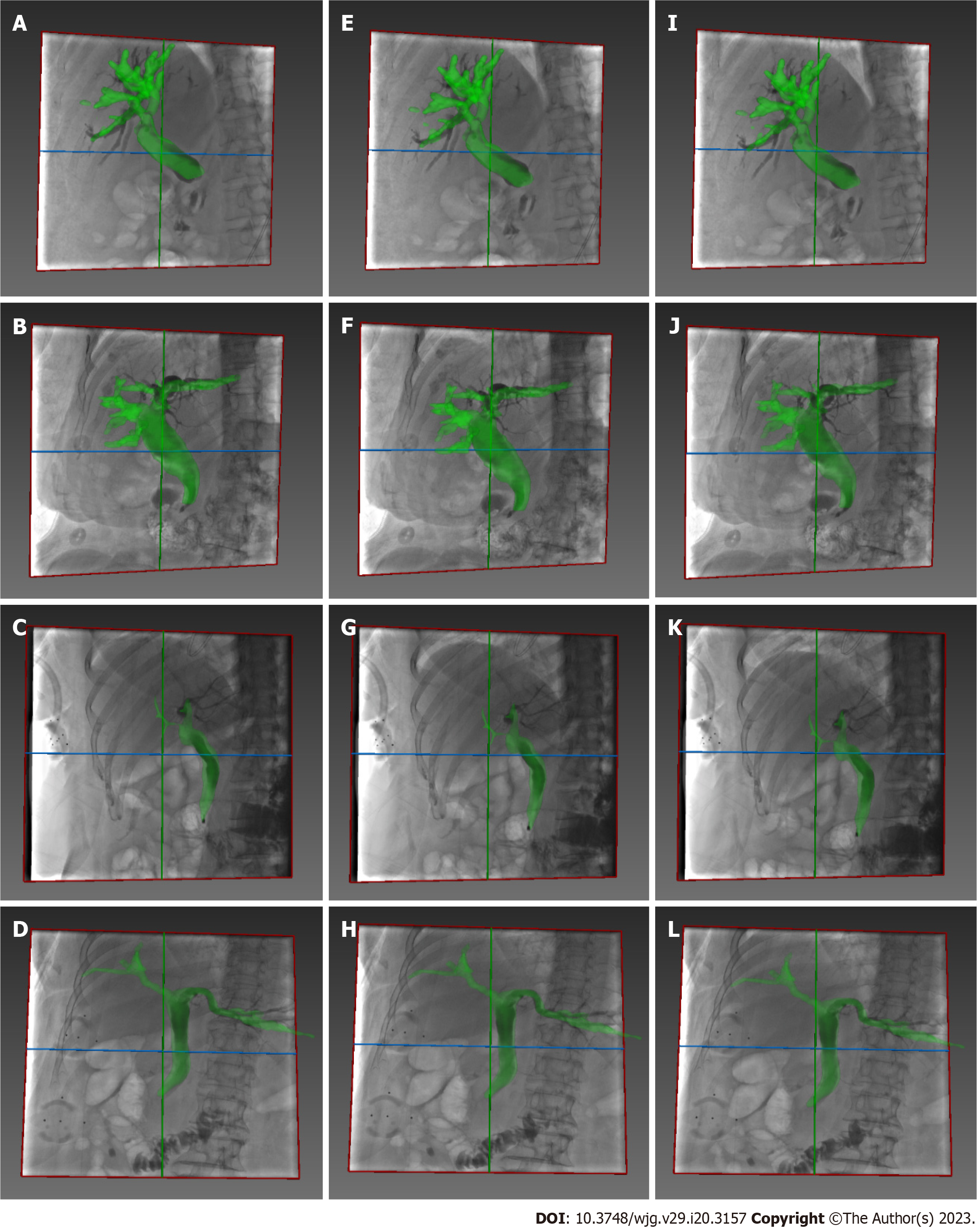
Figure 9 Additional examples of real-time image guidance navigation.
A-D: Beginning of the inhalation; E-H: During the inhalation; I-L: End of the inhalation.
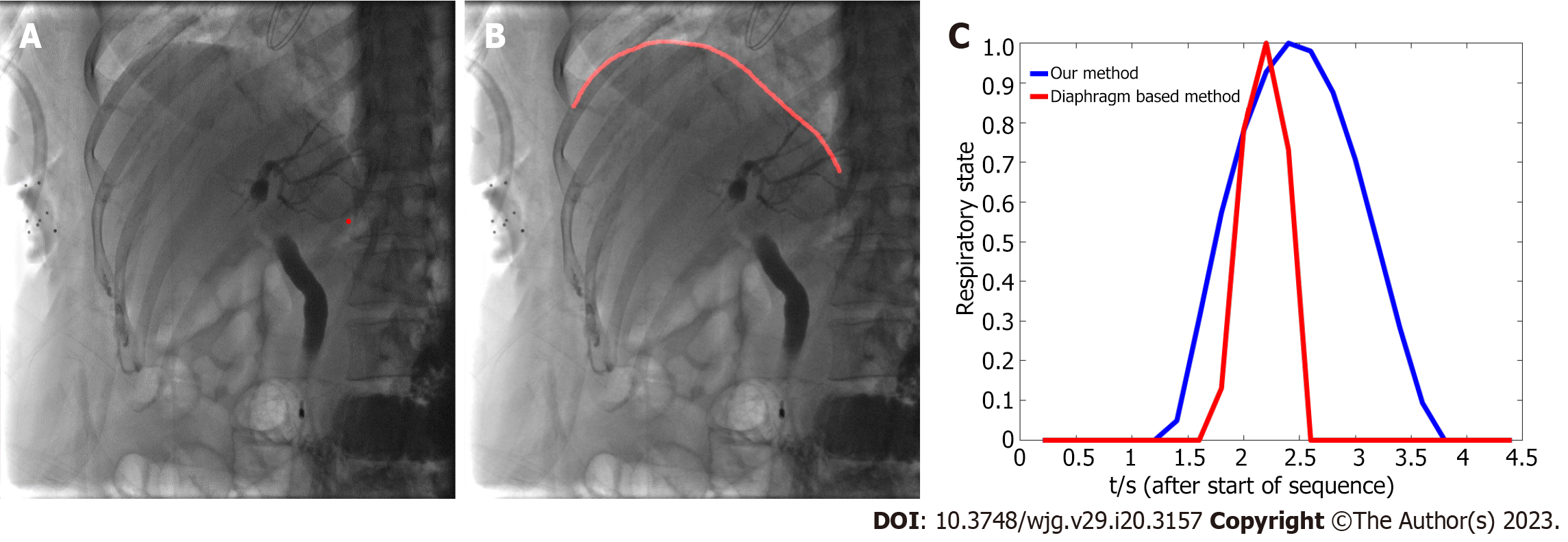
Figure 10 Comparison between two methods with respect to respiratory state estimation.
A: Centroid of the X-ray image; B: Diaphragm of the X-ray image; C: Example of the estimated respiratory state over a 4.4 s X-ray image sequence.
- Citation: Zhang DY, Yang S, Geng HX, Yuan YJ, Ding CJ, Yang J, Li MY. Real-time continuous image guidance for endoscopic retrograde cholangiopancreatography based on 3D/2D registration and respiratory compensation. World J Gastroenterol 2023; 29(20): 3157-3167
- URL: https://www.wjgnet.com/1007-9327/full/v29/i20/3157.htm
- DOI: https://dx.doi.org/10.3748/wjg.v29.i20.3157


